Chapter 4 Tissues Anatomy 32 A tissue is


 or develop fibrous")

- Slides: 49
Chapter 4: Tissues Anatomy 32
A tissue is composed of a group of cells with similar structure that perform related functions and are surrounded by extracellular material (non-living). The four different tissue types can be associated with a general function: epithelial for covering, connective for support, muscle for movement, and nervous for control.
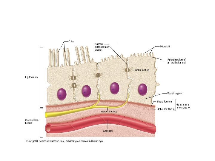
I. Epithelia and Glands- Epithelial tissue serves as a lining for organs that have an opening to the environment. It has several functions: boundary layer, interface layer, protection, transport such as secretion and absorption. A. Special Characteristics of Epithelia- because every tissue type has a specific function and structure, each as distinguishing characteristics 1. Cellularity- dense cell areas, multiple points of connection between cells, few extracellular matrix material. 2. Specialized contacts- multiple cell junctions tightly connect neighboring cells. 3. Polarity- each cell has a distinguishable apical (top) and basal (bottom) side The nuclei tend to be towards the basal side. The cells are anchored at the basal side by basal lamina which is part of the basement membrane. 4. Support by connective tissue- an underlying layer of connective tissue supports epithelial tissue. 5. Avascular but innervated- blood vessels to not cross into epithelial tissue but nerve ending do. 6. Regenetration- because they line organs epithelial cells are exposed to many conditions that destroy the cells, thus they have a great ability to regenerate quickly.
Lateral Cell Junctions
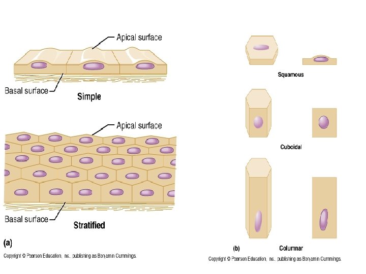
b. Classification of Epithelia- this tissue is identified by two names one describes cell layers and the other the shape.
1. Simple epithelia- a single layer of epithelia is called simple epithelia, it is design to allow molecules to cross the membrane quickly, such as in the capillaries and lungs. i. Simple squamous epithelia- a flat layer of cells. Endothelium produces a slick slippery surface. Mesothelium serves as a middle covering as part of serosa found in body cavities.
• Simple cuboidal epithelia- a layer of cube like cells, their height and width are just about equal. It form gland ducts and tubules.
• iii. Simple columnar epithelium- a layer of tall cells that line the digestive tract. It functions in absorption, secretion, and ion movement, some as cilia in their apical side.
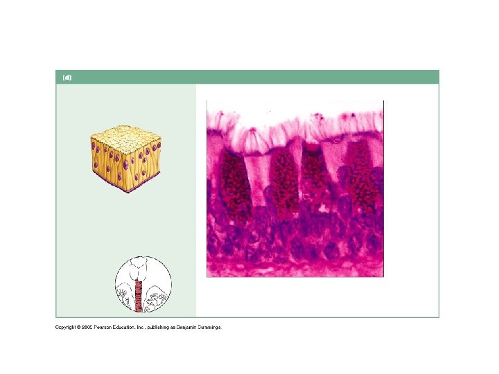
iiii. Pseudostratified Columnar Epithelium- a single layer with cells of different heights which makes it appearing as if there are different layers. Short cells are undifferentiated and give rise to tall cells. Tall cells function in secretion and absorption.
2. Stratified epithelia- two or more layers of cells, cells rise from basal side, main function is protection. i. Stratified squamous epithelium- many layers of cuboidal or columnar cells with squamous cells at the surface. It is the best for protection and lines areas that are often-abraded. They make up the skin and MAY OR MAY NOT have keratin.
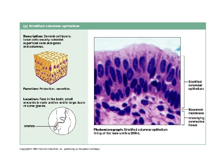
ii. Stratified cuboidal and columnar epithelium- rare and forms only large ducts of glands.
iii Transitional epithelium- lines hollow urinary organs and has the ability to stretch from six to three cells thick.
What type of tissue is found in endothelium?
c. Epithelial Surface Features- each side of the cells has a special feature. 1. Apical Surface Features: Microvilli and Cilia- are extensions on the apical side that increase surface areas. They can move to push substances in a certain direction. 2. Lateral Surface Features: Cell Junctions-cells are held together by proteins that attach lateral cell surfaces. Gap junctions are special openings that allow substances to flow from cell to cell. Desmosomes are proteins that support and link two different cell walls. Tight junctions also attach cell surfaces but use a different structure than desmosomes. 3. Basal Surface Features: The Basal Lamina- thin noncellular support sheet made up of proteins, controls what enters epithelium, assist in regeneration. Below basal lamina is the basal membrane which is thicker.
Lateral Cell Junctions
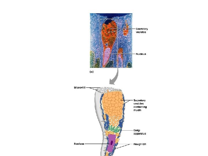
d. Glands- groups of epithelial cells that make and secrete a substance. Glands may be unicellular or multicellulaer. 1. Exocrine Glands- substances like mucus, sweat, and saliva are secreted onto body surfaces or cavities. Multicellular exocrine glands have ducts and can be classified by the number and arrangement of the ducts (see page 79). 2. Endocrine Glands- have no ducts, secrete hormones into the blood stream.
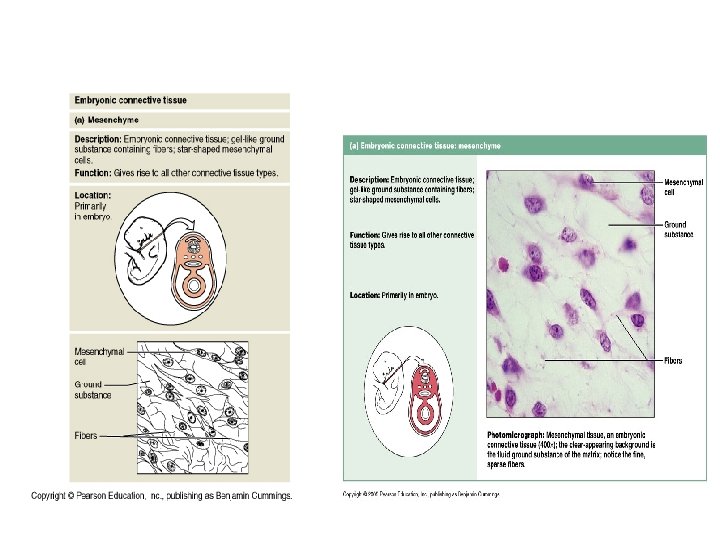
II. Connective Tissue- this tissue type is the most abundant and diverse. It has cells separated by a large amount of nonliving extracellular matrix. All cells originate from embryonic tissue called mesenchyme and do not have an apical or basal side. There are four main types of tissue: connective tissue proper, cartilage, bone, and blood. Table 4. 2 summarizes and compares the different types of connective tissue.
Mesenchyme gives rise to all types of connective tissue. The four basic functions of connective tissue are: 1. Support and bind other tissues- ligaments and tendon support joints and attach bone to bone and muscle to bone 2. Hold body fluids- blood contains plasma which holds a high percentage of water 3. Defends the body against infection- immune system cells arise from mesenchyme and are found in many different types of connective tissue. 4. Store nutrients- adipose cells store fat, bone cells store calcium.
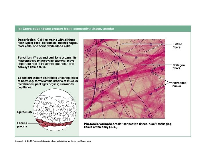
a. Connective Tissue Proper- forms the supportive framework of different organs and is made up of the following main components fibers, ground substance, immune cells, and adipose cells. A great example or connective tissue proper is areolar connective tissue. Areolar Tissue: A Model Connective Tissue- most abundant type, supports and binds other tissues, holds fluids, stores nutrients and defends against infection (see page 85). The fibers are created by cells called fibroblast. 1. Fibers: Collagen fibers are the strongest and withstand pulling. Reticular fibers are bundles of collagen fibrils forming a network for support. Elastic fibers are long and thin and form wide networks within extracellular matrix, allow tissues to reshape when stretched 2. Ground substance: this is a jelly-like substance that holds interstitia fluid (tissue fluid). It allows materials to cross to and from capillaries because areolar connective tissues lies between capillaries and other tissues. 3. Defense cells: these are immune system cells that help in preventing disease causing organisms to enter the blood stream. Know the cells in general: Macrophages are “big eaters” that engulf (phagocytic) foreign materials, Plasma cells secrete antibodies (proteins that destroy cells), mast cells cause inflammation, neurtophils, lymphocytes, and eosinophils go to infected areas and phagocytoze bacteria 4. Fat cells: also called adipose cells hold fat (nutrient), they are large cells and doesn’t divide.
• Connective Tissue Proper
b. Other Loose Connective Tissues- these are connective tissues that resemble areolar connective tissue. 1. Adipose Tissue- 90% fat cells (white adipose), highly vascularized, found below skin and mesenteries, cushions kidneys and eyes. In babies brown adipose keeps them warm.
2. Reticular connective- only has reticular fibers in the matrix that forms a supportive mesh. Found in bone marrow, spleen, and lymph nodes
c. Dense Connective Tissue- contains a high percentage of collagen. 1. Irregular dense connective tissue has thick collagen fibers running in different planes, it is found in the dermis and capsules around organs.
2. Regular dense connective tissue have fibers that run parallel to the pull direction, poorly vascularized, makes up tendons and ligaments, high amount of elastic fibers.
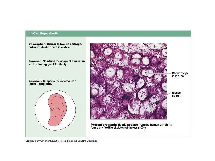
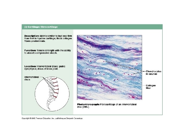
d. Other Connective Tissues: Cartilage, Bone, and Blood- connective tissue types that resist compression. 1. Cartilage- firm but flexible, high tissue fluid, no blood or nerves, has chondrocytes and chondroblasts. There are three types of cartilage: Hyaline, Elastic, and Fribrocartilage.
2. Bone Tissue- highly supportive and protective, matrix has inorganic salts, high collagen fiber content, has osteocytes
3. Blood- most unusual connective tissue, develops from mesenchyme cells and has plasma (nonliving matrix). It transport nutrients and cell
III. Muscle Tissue- involved in body movement because cells are capable of contraction, contains myofilaments (muscle filaments), there are three types. a. Skeletal muscle- attatches to bones, striated, multiple nucleic cells
III. Muscle Tissue- involved in body movement because cells are capable of contraction, contains myofilaments (muscle filaments), there are three types. a. Skeletal muscle- attatches to bones, striated, multiple nucleic cells
b. Cardiac muscle-makes the heart, striated, single nucleated cells, involuntary, pumps blood.
b. Cardiac musclemakes the heart, striated, single nucleated cells, involuntary, pumps blood.
• c. Smooth muscle- lines organs, no striations, spindle shaped cells, involuntary, moves substances inside the body.
• c. Smooth muscle- lines organs, no striations, spindle shaped cells, involuntary, moves substances inside the body.
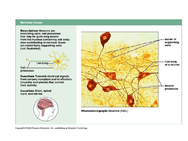
IV. Nervous Tissue- makes up the brain, spinal cord, and nerves that innervate the body. Some nervous system cells have the ability to create electrical impulses and stimulate other tissue. a. Neurons- nerve cells that extend long distances and can send an impulse to activate a cell. These control body function. b. Glial- nerve cells that form supportive structures, insulate, and nourish neurons. These cells come in direct contact with neurons but do not control body function
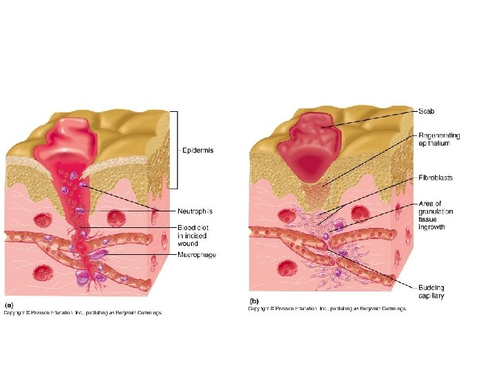
V. Tissue Response- Mechanisms such as a skin layer, secretion of mucus, and tears assist in protection against invading organism. However, when an injury occurs that breaks the skin, the physical barrier is removed. The tissue reacts by using inflammation and a immune response to protect the body. a. b. Inflammation- when tissue is injured the damaged cells release inflammatory chemicals that signal the blood vessels to bring more blood. The excess blood flow causes the area to become red and hot. The heat can serve as a general mechanism to destroy bacteria. As fluid from blood accumulates the area swells and the swelling causes pain because it puts pressure on pain receptors. An immune response takes place at this site. The chemicals released during the injury call immune sytem cells to fight the infection. Some cells specialize in detecting and killing bacteria, others specialize in phagocytosis to clear off dead cells.
b. Repair-after a scab forms healing tissues may replace themselves (regeneration) or develop fibrous tissues also known as a scar (fibrosis). 1. Tissues have different abilities to heal: epithelia, areolar connective tissue, fat tissue, dense irregular connective tissue (fascia), and blood forming tissue regerenate readily, smooth muscle and dense regular tissue (tendons) regenerates moderately, skeletal muscle and cartilage regenerate poorly, and cardiac muscle and nervous tissue do not regenerate.
VI The Tissue Throughout Life Connective, endothelium and muscle = mesenchyme mesoderm Epithelial tissues, brain and spinal cord = embryonic epithelium ectoderm and endoderm Primary tissues all formed by the 2 nd month and rapid growth continues in prenatal period. Nerve cells stop by fetal period and cell division slows down in adult hood. Stem cells are maintained throughout life for regeneration. Good nutrition and circulation helps in tissue regeneration. Tissue repair becomes less efficient with age and bone, muscle, and nervous tissues deregenerate.