CHAPTER 4 SKELETAL SYSTEM Skeletal system is composed

")
 n n Bone matrix is non-living, but it changes constantly,")


 anatomy of a Long Bone 1 -Diaphysis · Shaft · Composed of")

 · Cavity of the shaft · Contains yellow marrow (mostly")
 is the unit of bone structure")

 -Arranged in concentric rings (lamellae) around the")


")

 Mandible (1) Palatine (2) Maxillae (2) Zygomatic")


 Mandible( n n The only freely movable bone of the skull(with auditory")


 n n n Forms the posterior wall of")


 Consists of: -2 clavicles -2 scapulae Connects with the")


 -Functions Holds bones together n Allows bones to move n All bones")


 and convex posteriorly")
- Slides: 75
CHAPTER 4 SKELETAL SYSTEM Skeletal system is composed of: -Bones -Cartilages -Joints -Ligaments Functions of Skeletal System n SUPPORT: Hard framework that supports and anchors the soft organs of the body. n PROTECTION: Surrounds organs such as the brain and spinal cord. n MOVEMENT: Allows for muscle attachment therefore the bones are used as levers. n STORAGE: Minerals and lipids are stored within bone material. n BLOOD CELL FORMATION: The bone marrow is responsible for blood cell production.
Structure of Bone n n n Bone is a tissue (? ? ? ) Bone cells are called osteocytes (osteoblasts), and bone matrix is made of calcium salts and collagen. The calcium salts are calcium carbonate (Ca. CO 3) and calcium phosphate (Ca 3(PO 4)2), which give bone the strength required to perform its supportive & protective functions.
Structure of Bone (Cont’) n n Bone matrix is non-living, but it changes constantly, with calcium release & deposition. The function of osteocytes, is to regulate the amount of Ca that is deposited in, or removed from, the bone matrix.
Classification of bones 1 -According to structure 1 -Compact bone; looks solid & made of osteon & haversian system n Outer layer of bone, very hard and dense. n Organized in structural units called Haversian systems n Matrix is composed of Ca salts (Ca carbonate and Ca phosphate) n Osteocytes – living bone cells that live in matrix. 2 - Spongy (Porous) bone with visible holes & cavities n Located in the ends of long bones. n Many spaces that are filled with red bone marrow which produces bone cells.
2 -According to the shape 1 -Long bones · Typically longer than wide · Have a shaft with heads at both ends · Contain mostly compact bone, with a canal (medullary cavity) Examples: femur, humerus, limbs without wrists & ankles 2 -Short bones · Generally cube-shape · Contain mostly spongy bone Examples: carpals, tarsals, Slide 5. 4 a
3 -Flat bones · Thin and flattened · Usually curved · Thin layers of compact bone around a layer of spongy bone Examples: Skull, ribs, sternum, scapula 4 -Irregular bones · Irregular shape · Do not fit into previous bone categories Examples: Vertebrae and facial bones ** Short, flat, & irregular are made of spongy bone, covered by a thin layer of compact one. Slide 5. 5 a
Classification of bones according to Shape
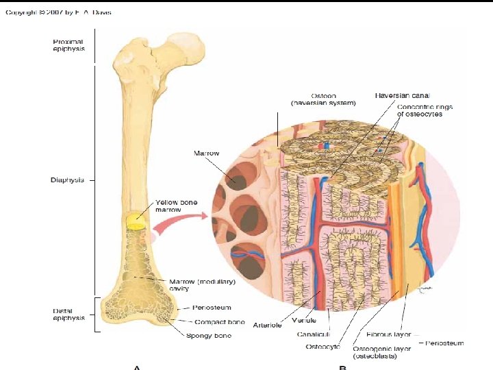
Macroscopic (gross) anatomy of a Long Bone 1 -Diaphysis · Shaft · Composed of compact bone 2 -Epiphysis · Ends of the long bone · Composed mostly of spongy bone Figure 5. 2 a Slide 5. 6
3 -Periosteum · Outside covering of the diaphysis · Fibrous connective tissue membrane secured to underlying bone by Sharpey`s fibres 4 -Articular cartilage · Covers the external surface of the epiphyses · Made of hyaline cartilage · Decreases friction at joint surfaces. Copyright © 2003 Pearson Education, Inc. publishing as Benjamin Cummings Slide 5. 7
5 -Medullary cavity (canal) · Cavity of the shaft · Contains yellow marrow (mostly fat) in adults · Contains red marrow (for blood cell formation) in infants and children. Figure 5. 2 a Copyright © 2003 Pearson Education, Inc. publishing as Benjamin Cummings Slide 5. 8 b
Microscopic anatomy of Bone · Osteon (Haversian System) is the unit of bone structure , it consists of: - Central (Haversian) canal · Runs in the center of an osteon · Carries blood vessels and nerves - Perforating (Volkman’s) canal · Perpendicular to the central canal · Carries blood vessels and nerves Copyright © 2003 Pearson Education, Inc. publishing as Benjamin Cummings Slide 5. 10 a
Figure 5. 3 Copyright © 2003 Pearson Education, Inc. publishing as Benjamin Cummings Slide 5. 10 b
· Lacunae -Cavities containing bone cells (osteocytes) -Arranged in concentric rings (lamellae) around the central canal called · Canaliculi -Tiny canals radiate from the central canal to the lacunae. -Form a transport system. Copyright © 2003 Pearson Education, Inc. publishing as Benjamin Cummings Figure 5. 3 Slide 5. 11 a
Changes in the Human Skeleton · In embryos, the skeleton is primarily hyaline cartilage. · During development, this cartilage is gradually replaced by bone through three centers of ossifications in the diaphsis and the two epiphyses. After puperty , cartilage remains in some areas as: · Bridge of the nose · Parts of ribs · Joints Copyright © 2003 Pearson Education, Inc. publishing as Benjamin Cummings Slide 5. 12
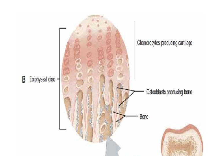
Bone Growth -Growth in length of long bones during childhood is achieved by epiphyseal (cartilage) plates which lie between epiphysis and diaphysis. New cartilage is added while older cartilage becomes ossified so bone become longer. -Bones grow in width through periosteum. · Bones change shape continuously by pull of gravity &muscle exercise. Slide 5. 13 a
Long Bone Formation and Growth Figure 5. 4 a Copyright © 2003 Pearson Education, Inc. publishing as Benjamin Cummings Slide 5. 14 a
Factors affecting bone growth n n Hereditary Nutrition Hormones; growth hormon, thyroxin, parathyroid, and insulin Exercises
Types of Bone Cells · Osteocytes · Mature bone cells · Osteoblasts · Bone-forming cells · Osteoclasts · Bone-destroying cells that break down bone matrix to: - release of calcium to the blood if needed. - Remodel the bone after fracture. · Bone remodeling is a process by both osteoblasts and osteoclasts. Copyright © 2003 Pearson Education, Inc. publishing as Benjamin Cummings Slide 5. 15
Fractures Closed fracture : skin is intact n Open fracture : skin is open Fracture reduction : 1 -closed reduction , no surgery is needed 2 -open reduction , surgery is needed Healing time for simple fracture is 6 -8 weeks (longer in elderly people), it occurs in FOUR major events: n 1 -hematoma formation n 2 -fibrocartilage callus formation n 3 -bony callus formation n 4 -bone remodeling by osteoclasts. n
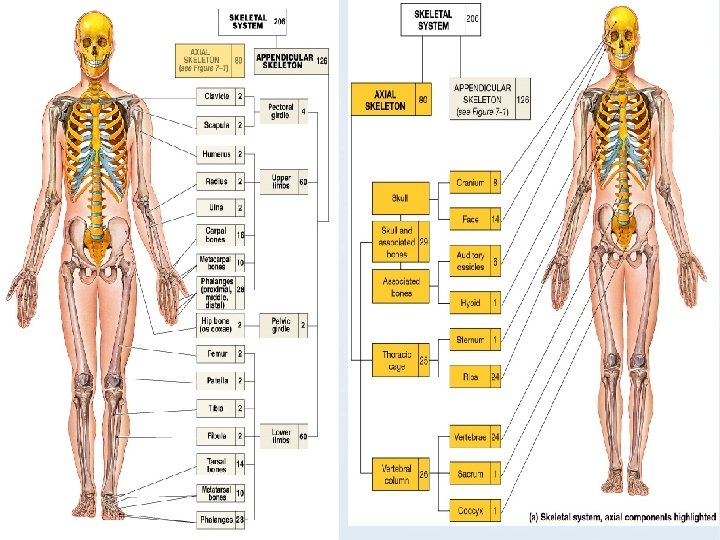
Divisions of the Skeletal system n Axial skeleton n Skull and associated bones(Auditory ossicles and Hyoid bones). n Vertebral column n Thoracic cage (Ribs+ sternum) § Appendicular skeleton -Pectoral girdle +upper limb -Pelvic girdle +lower limb
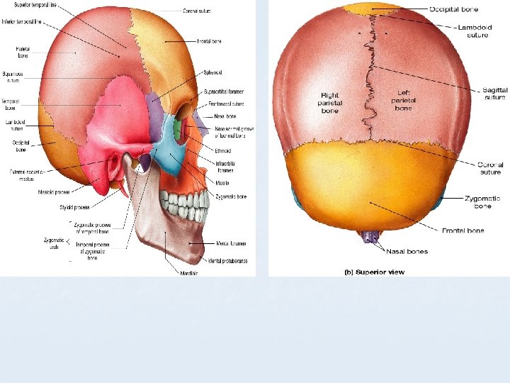
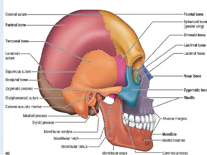
Sutures n Most skull bones are flat bones. Except for the mandible, which is connected to the rest of the skull by a freely movable joint, all bones of the adult skull are firmly united by interlocking joints called sutures (soo′cherz). The suture lines have a saw-toothed or serrated appearance. (except: malleus, incus, and stapes) The major skull sutures are: n Coronal – between parietal and frontal n Sagittal– between parietal bones n Lambdoid – between the parietal and occipital n Squamous – between the parietal and temporal
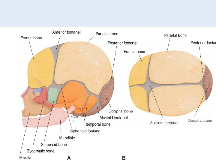
Fontanels: From each center of ossification, bone growth and radiates outward as calcium salts are deposited in the fibrous model of the bone. This process is not complete at birth; a baby has areas of fibrous connective tissue remaining between the bones of the skull. These are called fontanels , which permit: -compression of the baby’s head during birth without breaking the still thin cranial bones. -the growth of the brain after birth. *By the age of 2 years, all the fontanels have become ossified, and the skull becomes a more effective protective covering for the brain.
The Adult Skull • skull has 22 bones 1 -cranium = 8 bones: frontal, occipital, 2 temporals, 2 parietals, sphenoid and ethmoid 2 -facial bones = 14 bones: nasals, maxillae, zygomatics, mandible, lacrimals, palatines, inferior nasal conchae, vomer. • skull has a larger cranial cavity. It also has the nasal cavity, the orbits and paranasal sinuses. • outer surface provides large areas for muscle attachment that move the head or provide facial expressions. • Attached to its inner surfaces are membranes called meninges that stabilize the brain.
The Skull and Associated Bones
Frontal bone n n Forms the forehead Roof of the orbit articulates with parietal (coronal suture) Has frontal sinuses
• Parietal bones -Part of the superior and lateral surfaces of the cranium -articulate with each other – sagittal suture -articulate with occipital (lambdoid suture), and frontal(coronal), and temporal(squamous suture).
• Temporal bone -houses the inner ear -mastoid process (air spaces that communicate with the middle ear)and is an attachment of sternocleidomastoid muscle -anterior to mastoid process is external acoustic meatus -inferior and medial to the mastoid process is styloid process (muscle attachment) -stylomastoid foramen (7 th cranial nerve)
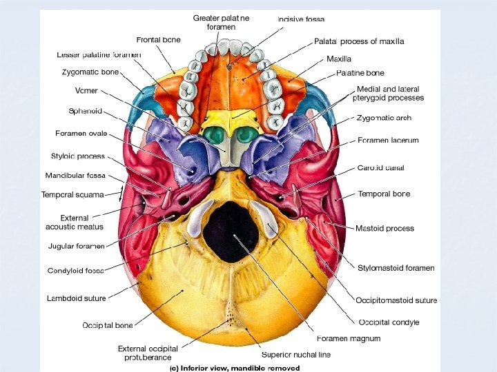
n n n Occipital bone part of the base of the skull articulates with parietal and temporal. foramen magnum for spinal cord. occipital condyles to sit on atlas (first) vertebra.
Sphenoid bone - Contributes to floor of cranium - Has sella turcica (turk`s saddle) that house the pituitary gland.
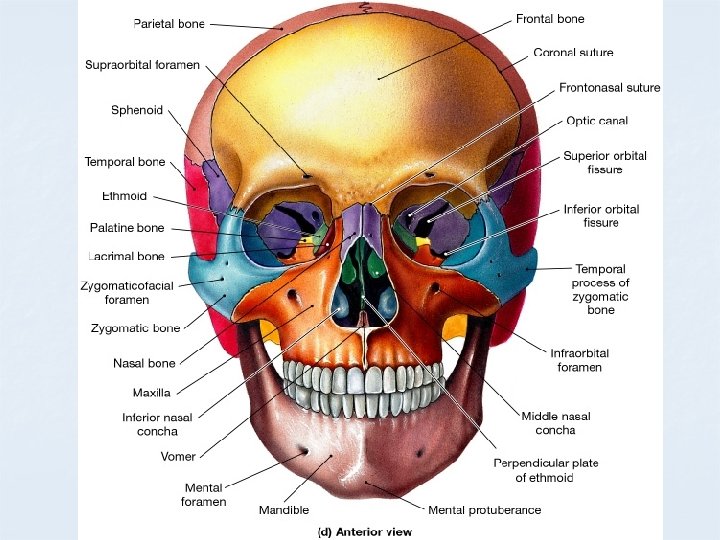
Ethmoid bone n n n Forms part of orbital wall Forms roof of nasal cavity connects with the vomer contain the ethmoid sinuses Cribiform plate: perforations for olfactory nerve, midline is the crista galli
Facial Bones : 14 bones Nasal (2) Mandible (1) Palatine (2) Maxillae (2) Zygomatic (2) Lacrimal (2) Inferior nasal conchae (2) Vomer (1)
Bones of the Face n Maxillae n n Paired bone Largest of facial bones Form upper jaw body contains the maxillary sinuses
n Palatine bones Form posterior portion of hard palate *The anterior portion of the hard palate is made by the palatine process of the maxillae n
Mandible(lower jaw) Mandible( n n The only freely movable bone of the skull(with auditory ausscicles) articulations with temporal bone in temperomandibular joint.
The Hyoid Bone n n The only bone in our body with no articulation with other bone. Base for muscles of the tongue and larynx
Paranasal Sinuses -Lined with mucous membranes and open into nasal cavity though openings called ostia -Resonating chambers for voice, lighten the skull - Four sinuses: n frontal sinus: n sphenoid sinus: n ethmoid sinus: n maxillary sinus which is: n The largest of the sinuses n close to the upper alveolar margin -Sinusitis is inflammation of the sinuses. It can easily spread from the nose to any sinus and from one sinus to the other as mucous membrane is continuous.
Adult Vertebral Column n 26 vertebrae n n n 24 individual vertebrae Sacrum Coccyx Seven cervical vertebrae n Twelve thoracic vertebrae n Five lumbar vertebrae n Sacrum and coccyx are Fused together. n
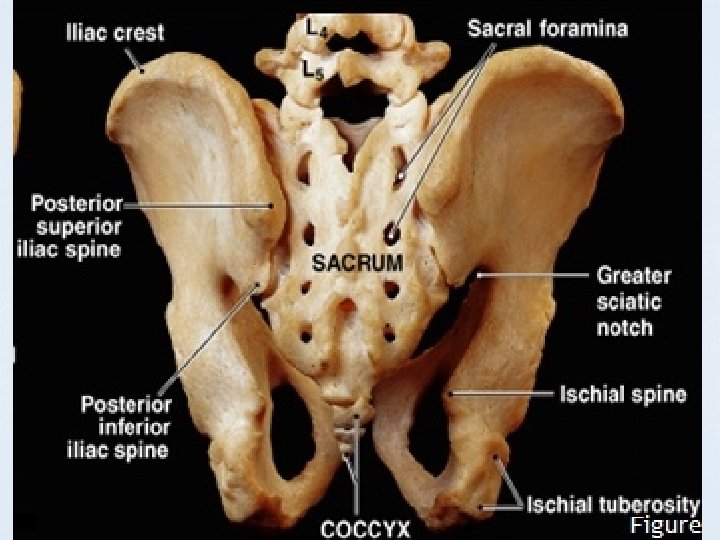
Sacrum (S 1 – S 5) n n n Forms the posterior wall of pelvis Formed from 5 fused vertebrae Superior surface articulates with L 5 Inferiorly articulates with coccyx Sacral promontory n Where the first sacral vertebrae bulges into pelvic cavity Center of gravity is 1 cm posterior to sacral promontory Coccyx Is the “tailbone” n Formed from 3 – 5 fused vertebrae n Offers only slight support to pelvic organs n
Sacrum Figure 7. 18 a, b
Bony Thorax n n Forms the framework of the chest Components of the bony thorax Thoracic vertebrae – posteriorly n Ribs – laterally n Sternum and costal cartilage – anteriorly n n Protects thoracic organs Supports shoulder girdle and upper limbs Provides attachment sites for muscles
The Bony Thorax Figure 7. 19 a
Sternum n n Formed from three parts : n Manubrium – superior part n Articulates with medial end of clavicles n Body – bulk of sternum n Sides are notched as articulations for costal cartilage of ribs 2– 7 n Xiphoid process – inferior end of sternum n Ossifies around age 40 Anatomical landmarks n Jugular notch n Central indentation at superior border of the manubrium n Sternal angle n A horizontal ridge where the manubrium joins the body, it is at the level of the second rib.
Ribs n All ribs attach to vertebral column posteriorly n True ribs - superior seven pairs of ribs n n Attach to sternum by costal cartilage False ribs – inferior five pairs of ribs , attatch indirectly to the sternum nfloating ribs 11– 12 are short and free anteriuorly.
Ribs Figure 7. 20 a
Ribs Figure 7. 20 b
Disorders of the Axial Skeleton n Abnormal spinal curvatures n n Scoliosis – an abnormal lateral curvature Kyphosis – an exaggerated thoracic curvature Lordosis – an accentuated lumbar curvature – “swayback” Stenosis of the lumbar spine n A narrowing of the vertebral canal
The Appendicular Skeleton Allows us to move and manipulate objects n Includes all bones other than axial skeleton, it includes: n n n the supportive girdles (pectoral &pelvic girdles) the limbs (upper & lower limbs)
th Figure 8– 1
The Pectoral Girdle (Shoulder girdle) Consists of: -2 clavicles -2 scapulae Connects with the axial skeleton only at the manubrium (claviculosternal joint) Figure 8– 2 a
The Wrist Figure 8– 6
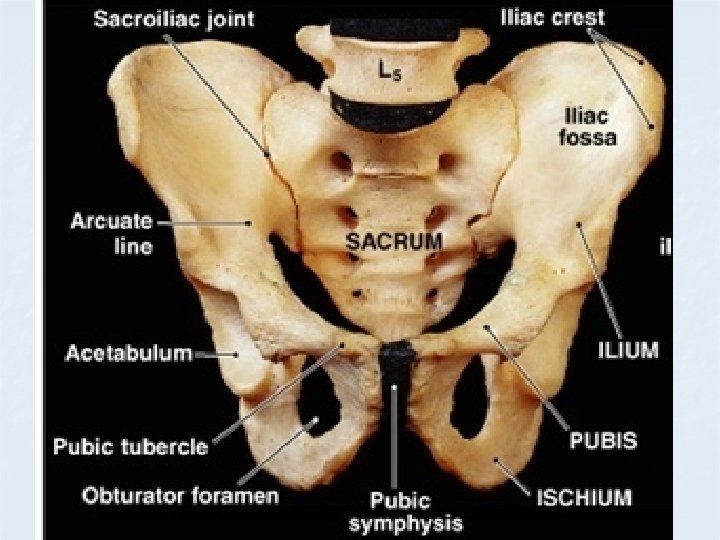
Pelvic girdle: two ossa coxae, sacrum and coccyx
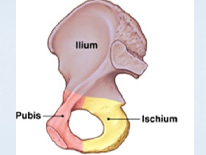
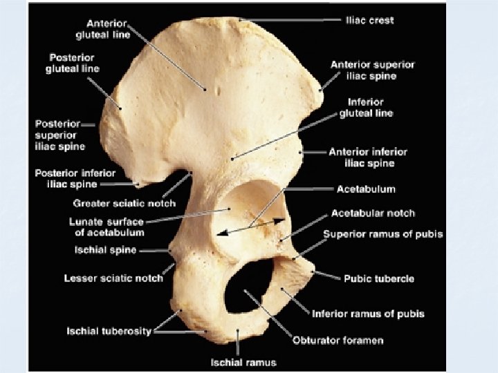
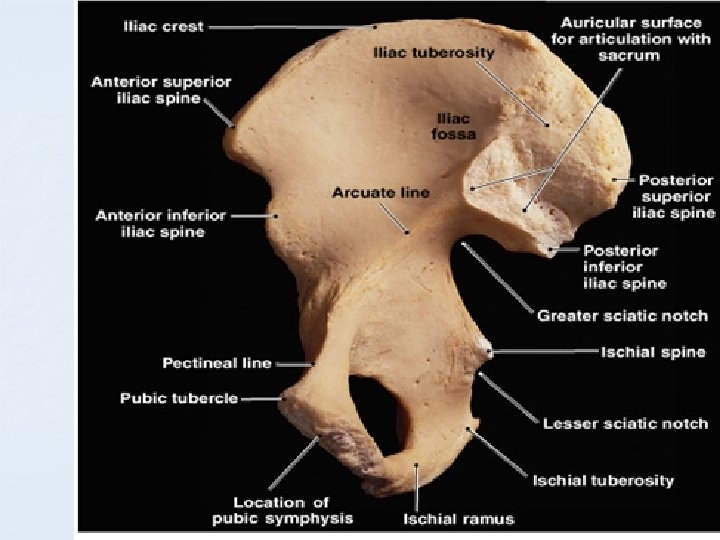
The OSSA COXAE n Also called hipbones Strong to bear body weight &stress of movement Each is made up of 3 fused bones: n ilium (articulates with sacrum) n ischium n Pubis The Acetabulum (vinegar cup) Also called the hip n n n socket n n Is the meeting point of the ilium, ischium, and pubis Articulates with head of the femur (Hip joint).
Landmarks of the Pubis n Pubic symphysis: gap between pubic tubercles n padded with fibrocartilage n Landmarks of the Ischium • Ischial tuberosity: posterior projection you sit on • Ischeal spine, superior to the tuberosity , important during labour
Divisions of the Pelvis Figure 8– 9
Comparing the Male and Female Pelves n Female pelvis is: n smoother n lighter n less prominent muscle and ligament attachments Pelvis Modifications for Childbearing n n n Enlarged pelvic outlet Broad pubic angle (> 100°) Less curvature of sacrum and coccyx Wide, circular pelvic inlet Ilia project laterally.
Comparing the Male and Female Pelvis Figure 8– 10
The Ankle n n n Also called the tarsus: consists of 7 tarsal bones. Calcaneus (heel bone): n n transfers weight to ground attaches Achilles tendon Figure 8– 14 a
Arches of the foot : Bones are arranged to form -THREE strong arches, 2 longitudinal (medial & lateral & 1 transverse -Ligaments & tendons help to hold the bones firmly in the arched position but still allow a certain amount of spriginess. Week arches are referred to as flat foot Figure 8– 14 b
Articulations (Joints) -Functions Holds bones together n Allows bones to move n All bones articulate with at least one other bone except the hyoid. -Synovial Joints – Structure 1. Articular cartilage: hyaline 2. Joint Cavity: space filled with lubricating fluid 3. Fibrous Capsule: fibrous CT lined with a smooth synovial membrane 4. Reinforcing Ligament: can be inside or outside the joint capsule n
Ball and socket joints A spherical head of one bone fits into a round socket in another These Multiaxial joints allow movements in all axes including rotation Shoulder and Hip are examples
Developmental aspects n n n n Long bones are formed on top of Hyaline cartilage Flat bones of skull are formed on top of fibrous membranes At birth, some fontanels still remain By end of adolescence, the epiphysial plates are fully ossified Adult skull is 1/8 & infant skull is 1/4 of the total body length. At birth, cranium is huge relative to face, it is related to rapid growth of brain By 2 years skull is 3/4 adult size By 9 years, skull become nearly of adult size
n n n n At birth, the spine is arched (primary) and convex posteriorly Secondary curvature are convex anteriorly, cervical with raising the head & lumber with start of walking S-shaped spine in adult At birth the UL ratio is 1. 7 to 1 At 10, UL is 1 to 1 Bones become stronger with pull of gravity &muscle contractions Osteoporosis (thin and fragile bones) occurs in totally inactive persons. It occurs in half women after 65 and in 20% of men after 70(estrogen maintain healthy bones)