Chapter 4 Protein 3 Dimensional Structure and Function


 polypeptides arranged in long strands or sheets 2) water")


 has mostly hydrophobic AA’s Two")


- Slides: 35
Chapter 4 Protein 3 -Dimensional Structure and Function
Terminology • Conformation – spatial arrangement of atoms in a protein • Native conformation – conformation of functional protein
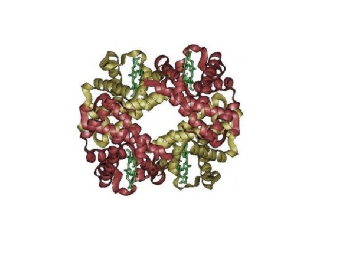
Protein Classification • Simple – composed only of amino acid residues • Conjugated – contain prosthetic groups (metal ions, co-factors, lipids, carbohydrates) Example: Hemoglobin – Heme
Protein Classification • • One polypeptide chain - monomeric protein More than one - multimeric protein Homomultimer - one kind of chain Heteromultimer - two or more different chains (e. g. Hemoglobin is a heterotetramer. It has two alpha chains and two beta chains. )
Protein Classification Fibrous – 1) polypeptides arranged in long strands or sheets 2) water insoluble (lots of hydrophobic AA’s) 3) strong but flexible 4) Structural (keratin, collagen) Globular 1) 2) 3) 4) – polypeptide chains folded into spherical or globular form water soluble contain several types of secondary structure diverse functions (enzymes, regulatory proteins)
catalase keratin collagen
Protein Function • • • Catalysis – enzymes Structural – keratin Transport – hemoglobin Trans-membrane transport – Na+/K+ ATPases Toxins – rattle snake venom, ricin Contractile function – actin, myosin Hormones – insulin Storage Proteins – seeds and eggs Defensive proteins – antibodies
4 Levels of Protein Structure
Non-covalent forces important in determining protein structure • • van der Waals hydrogen bonds ionic bonds hydrophobic interactions
1 o Structure Determines 2 o, 3 o, 4 o Structure • Sickle Cell Anemia – single amino acid change in hemoglobin related to disease • Osteoarthritis – single amino acid change in collagen protein causes joint damage
Classes of • Alpha helix • B-sheet • Loops and turns o 2 Structure
Alpha-Helix • First proposed by Linus Pauling and Robert Corey in 1951 • Identified in keratin by Max Perutz • A ubiquitous component of proteins • Stabilized by H-bonds
Alpha-Helix • Residues per Right handed turn: 3. 6 helix • Rise per residue: 1. 5 Angstroms • Rise per turn (pitch): 3. 6 x 1. 5 A = 5. 4 Angstroms • amino hydrogen H -bonds with carbonyl oxygen located 4 AA’s away forms 13 atom loop
Alpha-Helix All H-bonds in the alpha-helix are oriented in the same direction giving the helix a dipole with the Nterminus being positive and the C -terminus being negative
Alpha-Helix • Side chain groups point outwards from the helix • AA’s with bulky side chains less common in alpha-helix • Glycine and proline destabilizes alphahelix
Amphipathic Alpha-Helices + One side of the helix (dark) has mostly hydrophobic AA’s Two amphipathic helices can associate through hydrophobic interactions
Beta-Strands and Beta-Sheets • Also first postulated by Pauling and Corey, 1951 • Strands may be parallel or antiparallel • Rise per residue: • – 3. 47 Angstroms for antiparallel strands – 3. 25 Angstroms for parallel strands – Each strand of a beta sheet may be pictured as a helix with two residues per turn
Beta-Sheets • Beta-sheets formed from multiple side-byside beta-strands. • Can be in parallel or anti-parallel configuration • Anti-parallel betasheets more stable
Beta-Sheets • Side chains point alternately above and below the plane of the beta-sheet • 2 - to 15 beta-strands/beta-sheet • Each strand made of ~ 6 amino acids
Loops and turns Loops • Loops usually contain hydrophillic residues. • Found on surfaces of proteins • Connect alpha-helices and beta-sheets Turns • Loops with < 5 AA’s are called turns • Beta-turns are common
Beta-turns • allows the peptide chain to reverse direction • carbonyl C of one residue is H-bonded to the amide proton of a residue three residues away • proline and glycine are prevalent in beta turns
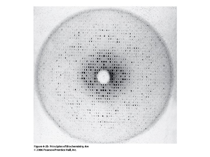
Protein 3 -D structure: 3 o and 4 o structure and protein folding.
o 3 Structure • third level of protein organization • folding of polypeptide chain causes 2 o structures to interact • formation of motifs and domains
Proteins with similar 1 o structure also have similar 3 o structure tuna yeast rice 1 GDVAKGKKTFVQKCAQCHTVENGGKHKVGPNLWGLFGRKTGQAEGYSYTDANKSKGIVWN 1 GSAKKGATLFKTRCLQCHTVEKGGPHKVGPNLHGIFGRHSGQAEGYSYTDANIKKNVWDE 1 GNPKAGEKIFKTKCAQCHTVDKGAGHKQGPNLNGLFGRQSGTTPGYSYSTANKMAVIWEE 61 ETLMEYLENPKKYIPGTKMIFAGIKKKGERQDLVAYLKSATS 61 NNMSEYLTNPKKYIPGTKMAFGGLKKEKDRNDLITYLKKACE 61 NTLYDYLLNPKKYIPGTKMVFPGLKKPQERADLISYLKEATS
Common Motifs
Motifs Combine to form Domains • Domains are independent folding units in a 3 o structure of a protein • Individual domains have specific function Parallel twisted sheet • Hydrophobic interactions are the major driving force in folding domains Alpha/beta barrel
Protein family members share common domain structures lactate dehydrogenase malate dehydrogenase COO- COOO C + NADH CH 3 COOHO CH + NAD+ CH 3 O C H 2 C HO + NADH COO- CH + NAD+ CH 2 COO-
o 4 Structure • Quaternary structure describes the organization of subunits in a protein with multiple subunits (oligomeric protein) • Can have homo-multimers or hetero-multimers a 2 b 2 a 2 bg
4 o Structure • Determine molecular weight of native protein by gel permeation chromatography • Determine molecular weight of individual subunits by SDS-PAGE • Can use the information to determine subunit composition If……. Native protein – 160, 000 daltons and a-Subunit – 50, 000 daltons b-Subunit – 30, 000 daltons Then…… Protein can have a 2 b 2 structure
4 o Structure • Subunits held together by non-covalent interactions • Oligomeric protein is more stable than disassociated subunits • Active site often made up of AA residues from different subunits • 4 o and 3 o structure is often affected by ligand (substrate or inhibitor) binding. Important in enzyme regulation
Protein denaturation • Denaturation – disruption of native conformation • Heat commonly used to denature proteins • Tm = temperature where 50% folded/50% unfolded. • Typical Tm = 40 -60 o. C • Tm for thermophiles >100 o. C (Taq DNA polymerase) • Chemical denaturants Chaotrophic agents = Urea, KCN detergents = SDS Tm
Disulfides Bonds • Stabilize native structure • Formed after native conformation achieved • Abundant in secreted proteins but not in intracellular proteins • Protein disulfide isomerase catalyzes reduction of incorrect disulfide linkages