Chapter 4 Principles of Neural and Hormonal Communication


 changes in membrane potential during")

 – Voltage-gated")


 Dendrites Cell body of postsynaptic neuron Axon hillock Myelinated")


 Postsynaptic neuron")


- Slides: 59
Chapter 4 Principles of Neural and Hormonal Communication Human Physiology by Lauralee Sherwood © 2007 Brooks/Cole-Thomson Learning
Outline • • Graded Potentials Action Potentials Synapses and integration Intracellular communication Signal Transduction Hormonal Communication Nervous vs. Endocrine System
Communication is critical for the survival of the cells that compose the body. Two major regulatory systems of the body – nervous and endocrine - communicate with the cells/tissues/organs/systems they control.
Neural Communication • Nerve and muscle are excitable tissues • Can undergo rapid changes in their membrane potentials • Can change their resting potentials into electrical signals – Electrical signals are critical to the function of the nervous system and all muscles
Neural Communication • Membrane electrical states – Polarization • Any state when the membrane potential is other than 0 m. V – Depolarization • Membrane becomes less polarized than at resting potential – Repolarization • Membrane returns to resting potential after having been depolarized – Hyperpolarization • Membrane becomes more polarized than at resting potential
Types of Changes in Membrane Potential
Voltage clamp • • • The technique allows an experimenter to "clamp" the cell potential at a chosen value. This makes it possible to measure how much ionic current crosses a cell's membrane at any given voltage. This is important because many of the ion channels in the membrane of a neuron are voltage gated ion channels, which open only when the membrane voltage is within a certain range. Kenneth Cole[2] and George Marmount
Patch clamp • • • A patch-clamp microelectrode is a micropipette with a relatively large tip diameter. The microelectrode is placed next to a cell, and gentle suction is applied through the microelectrode to draw a piece of the cell membrane (the 'patch') into the microelectrode tip; the glass tip forms a high resistance 'seal' with the cell membrane. This can be used for studying the activity of the ion channels that are present in the patch of membrane. If more suction is now applied, the small patch of membrane in the electrode tip can be displaced, leaving the electrode sealed to the rest of the cell. This "whole-cell" mode allows very stable intracellular recording. This technique was developed by Erwin Neher and Bert Sakmann who received the Nobel Prize in 1991.
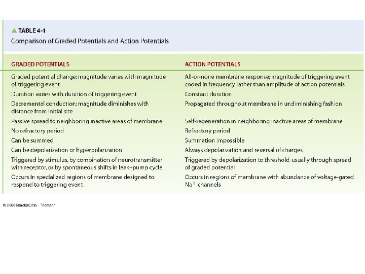
Channels • Leak channels – Unregulated passage of ions • Gated channels – – Voltage gated Chemically gated Mechanically gated Thermally gated • These channels create and alter membrane potentials • Two kinds of potential change – Graded potentials • Serve as short-distance signals – Action potentials • Serve as long-distance signals
Graded Potential • Occurs in small, specialized region of excitable cell membranes • Magnitude of graded potential varies directly with the magnitude of the triggering event • Die out over short distances
Fig. 4 -2, p. 87
Current Flow During a Graded Potential
Portion of excitable cell Initial site of potential change Loss of charge Direction of current flow from initial site * Numbers refer to the local potential in m. V at various points along the membrane. Loss of charge Direction of current flow from initial site Fig. 4 -4, p. 89
Graded Potentials Examples of graded potentials: • • • Postsynaptic potentials Receptor potentials End-plate potentials Pacemaker potentials Slow-wave potentials
Action Potentials • Brief, rapid, large (100 m. V) changes in membrane potential during which potential actually reverses • Involves only a small portion of the total excitable cell membrane • Do not decrease in strength as they travel from their site of initiation throughout remainder of cell membrane
Fig. 4 -7, p. 91
Action Potentials • When membrane reaches threshold potential – (-50 to-55 mv) – Voltage-gated channels in the membrane undergo conformational changes – Flow of sodium ions into the ICF reverses the membrane potential from -70 m. V to +30 m. V – Flow of potassium ions into the ECF restores the membrane potential to the resting state
Action Potentials • Additional characteristics – Sodium channels open during depolarization by positive feedback. – When the sodium channels become inactive, the channels for potassium open. This repolarizes the membrane. – As the action potential develops at one point in the plasma membrane, it regenerates an identical action potential at the next point in the membrane. – Therefore, it travels along the plasma membrane undiminished.
Action Potentials Permeability Changes and Ion Fluxes During an Action Potential
Fig. 4 -3 b, p. 88
Action Potentials The Na+/K+ pump gradually restores the concentration gradients disrupted by action potentials. • Sodium is pumped into the ECF • Potassium is pumped into the ICF • Refractory period keeps the action potential going in one direction and limits the Ap frequency • All or none • Frequency and line coding
Neuron • Once initiated, action potentials are conducted throughout a nerve fiber • Action potentials are propagated from the axon hillock to the axon terminals • Basic parts of neuron (nerve cell) – Cell body – Dendrites – Axon
Neuron
Neuron • Cell body – Houses the nucleus and organelles • Dendrites – Project from cell body and increase surface area available for receiving signals from other nerve cells – Signal toward the cell body Dendrite and cell body serve as the neurons input zone.
Neuron • Axon – Nerve fiber – Single, elongated tubular extension that conducts action potentials away from the cell body – Conducting zone of the neuron – Collaterals • Side branches of axon – Axon hillock • First portion of the axon plus the region of the cell body fro m which the axon leaves • Neuron’s trigger zone – Axon terminals • Release chemical messengers that simultaneously influence other cells with which they come into close association • Output zone of the neuron
Action Potentials • Two types of propagation – Contiguous conduction • Conduction in unmyelinated fibers • Action potential spreads along every portion of the membrane – Saltatory conduction • Rapid conduction in myelinated fibers • Impulse jumps over sections of the fiber covered with insulating myelin
Contiguous Conduction
Saltatory Conduction
Saltatory Conduction • Propagates action potential faster than contiguous conduction because action potential does not have to be regenerated at myelinated section • Myelinated fibers conduct impulses about 50 times faster than unmyelinated fibers of comparable size • Myelin – Primarily composed of lipids – Formed by oligodendrocytes in CNS – Formed by Schwann cells in PNS
Regeneration of Nerve Fibers • Regeneration of nerve fibers depends on its location • Schwann cells in PNS guide the regeneration of cut axons • Fibers in CNS myelinated by oligodendrocytes do not have regenerative ability – Oligodendrocytes inhibit regeneration of cut central axons
Synapses • Junction between two neurons • Primary means by which one neuron directly interacts with another neuron (muscle cells or glands as well) • Anatomy of a synapse – Presynaptic neuron – conducts action potential toward synapse – Synaptic knob – contains synaptic vesicles – Synaptic vesicles – stores neurotransmitter (carries signal across a synapse) – Postsynaptic neuron – neuron whose action potentials are propagated away from the synapse – Synaptic cleft – space between the presynaptic and postsynaptic neurons
Fig. 4 -16, p. 103
Synapses Signal at synapse either excites or inhibits the postsynaptic neuron • Two types of synapses – Excitatory synapses – Inhibitory synapses
Neurotransmitters • Vary from synapse to synapse • Same neurotransmitter is always released at a particular synapse • Quickly removed from the synaptic cleft • Some common neurotransmitters – – – – – Acetylcholine Dopamine Norepinephrine Epinephrine Serotonin Histamine Glycine Glutamate Aspartate Gamma-aminobutyric acid (GABA)
Neuropeptides • Large molecules consisting of from 2 to 40 amino acids • Synthesized in neuronal cell body in the endoplasmic reticulum and Golgi complex • Packaged in large, dense-core vesicles present in axon terminal • Act as neuromodulators, not creating potentials but altering the membranes Norepinephrine : Galanin Enkephalin Neuropeptide Y GABA Somatostatin (in the hippocampus) Cholecystokinin Neuropeptide Y (in the arcuate nucleus) Acetylcholine VIP Substance P Dopamine Cholecystokinin Neurotensin Epinephrine (adrenaline) Neuropeptide Y Neurotensin Serotonin (5 -HT) Substance P TRH Enkephalin
Comparison of Classical Neurotransmitters and Neuropeptides Characteristic Classical Neurotransmitters Neuropeptides Size Small, one amino acid or similar chemical Large, 2 to 40 amino acids in length Site of Synthesis Cytosol of synaptic knob Endoplasmic reticulum and Golgi complex in cell body, travel to synaptic knob by axonal transport Site of Storage In small synaptic vesicles in axon terminal In large dense-core vesicles in axon terminal Site of Release Axon terminal, may be cosecreted with neurotransmitter Speed and Duration of Action Rapid, brief response Slow, prolonged response Site of Action Subsynaptic membrane of postsynaptic cell Nonsynaptic sites on either presynaptic or postsynaptic cell at much lower concentrations than classical neurotransmitters Effect Usually alter potential of postsynaptic cell by opening specific ion channels Usually enhance or suppress synaptic effectiveness by long-term changes in neurotransmitter synthesis or postsynaptic receptor sits (act as neuromodulators)
Synaptic inputs (presynaptic axon terminals) Dendrites Cell body of postsynaptic neuron Axon hillock Myelinated axon Fig. 4 -15, p. 102
Fig. 4 -17, p. 104
Neuronal Integration • Multiple EPSP and IPSP’s from numerous synapses converge on one neuron. • These signals can cause different changes in the postsynaptic neuron – Cancellation – Spatial summation – Temporal summation
Threshold = approx -55 mv Fig. 4 -18, p. 106
Fig. 4 -19, p. 109
Presynaptic inputs Convergence of input (one cell is influenced by many others) Postsynaptic neuron Presynaptic inputs Divergence of output (one cell influences many others) Postsynaptic neurons Arrows indicate direction in which information is being conveyed. Fig. 4 -20, p. 111
The Retina Example of convergence and divergence
Synaptic Drug Interactions • Possible drug actions – Altering the synthesis, axonal transport, storage, or release of a neurotransmitter – Modifying neurotransmitter interaction with the postsynaptic receptor – Influencing neurotransmitter reuptake or destruction – Replacing a deficient neurotransmitter with a substitute transmitter
Examples of drugs that alter synaptic transmission • Cocaine – Blocks reuptake of neurotransmitter dopamine at presynaptic terminals • Strychnine – Competes with inhibitory neurotransmitter glycine at postsynaptic receptor site • Tetanus toxin – Prevents release of inhibitory neurotransmitter GABA, affecting skeletal muscles
Chemical Messengers • Four types of chemical messengers – Paracrines • Local chemical messengers • Exert effect only on neighboring cells in immediate environment of secretion site – Neurotransmitters • Short-range chemical messengers • Diffuse across narrow space to act locally on adjoining target cell (another neuron, a muscle, or a gland)
Chemical Messengers – Hormones • Long-range messengers • Secreted into blood by endocrine glands in response to appropriate signal • Exert effect on target cells some distance away from release site – Neurohormones • Hormones released into blood by neurosecretory neurons • Distributed through blood to distant target cells
Fig. 4 -21, p. 112
Chemical Messengers • Extracellular chemical messengers bring about cell responses primarily by signal transduction – Process by which incoming signals are conveyed to target cell’s interior • Binding of extracellular messenger (first messenger) to matching receptor brings about desired intracellular response by either – Opening or closing channels – Activating second-messenger systems • Activated by first messenger • Relays message to intracellular proteins that carry out dictated response
Hormones • Endocrinology – Study of homeostatic activities accomplished by hormones • Two distinct groups of hormones based on their solubility properties – Hydrophilic hormones (Proteins, peptides) • Highly water soluble • Low lipid solubility – Lipophilic hormones (Steroids) • High lipid solubility • Poorly soluble in water
Fig. 4 -21, p. 112
Table 4 -4, p. 114
Fig. 4 -22, p. 115
Fig. 4 -23, p. 116
Fig. 4 -24, p. 118
Fig. 4 -25, p. 119
Fig. 4 -26, p. 122
Comparison of Nervous System and Endocrine System