CHAPTER 4 Origins of Life and Cells THE


 Small cells No membrane-bound organelles � No nucleus Circular chromosome")
 Pores in nuclear")
 – intracellular transport � Membrane channels that extend throughout the")


 carbohydrate receptor protein recognition protein cholesterol phospholipid binding site cytosol (inside)")
 head (hydrophilic) Figure 5 -2 Biology: Life on Earth 8/e © 2008")
 hydrophilic heads phospholipid hydrophobic tails bilayer hydrophilic heads cytosol (watery")
 (inside) reactions Figure 5 -5 Biology: Life on Earth 8/e © 2008 Pearson")

 � Does not require the input")
 Simple diffusion through the phospholipid bilayer lipid-soluble molecules (extracellular fluid) and O 2,")
 Isotonic solution Equal movement of water into and out of cells. (b) Hypertonic")


 Facilitated diffusion through a carrier protein amino acids, sugars, small proteins (extracellular fluid)")


 Pinocytosis (extracellular fluid) 1 3 2 (cytosol) vesicle containing extracellular fluid 1 A")
 Pinocytosis in a smooth muscle cell. 1 extracellular fluid 2 cytosol Figure 5")
 receptors coated pit 1 2 3 (cytosol) 4 coated")
 (cytosol) 1 2 protein coating")
 Phagocytosis food particle pseudopods 1 (cytosol) (extracellular fluid) 2 food vacuole 3 1")
 Amoeba An Amoeba (a freshwater protist), engulfs a Paramecium using phagocytosis. Figure 5")
 White blood cell A white blood cell ingests bacteria using phagocytosis. Figure 5")
 vesicle (cytosol) Material is enclosed in a vesicle")
 Gap junctions (b) Plasmodesmata root liver cells root cells plasma membrane Gap junctions:")
 Desmosome small intestine cells lining small intestine (b) Tight junction urinary bladder cells")
- Slides: 56
CHAPTER 4 Origins of Life and Cells
THE ORIGINS OF CELLULAR LIFE Primitive Earth provided inorganic precursors from which organic molecules could have been synthesized � Large amounts of free energy present � Reducing atmosphere – absence of oxygen (no oxidation) Carbon dioxide, nitrogen, water vapor, ammonia, methane These molecules served as monomers for the formation of more complex molecules � Amino acids and nucleotides The joining of these monomers produced polymers with the ability to replicate, store, and transfer information. These complex reactions could have occurred in solution (organic soup model) or as reactions on solid reactive surfaces. � The RNA World hypothesis proposes that RNA could have been the earliest genetic material.
SCIENTIFIC EVIDENCE SUPPORTS MODELS OF THE ORIGIN OF LIFE Geological evidence provides support for models of the origin of life on Earth. � The Earth formed approximately 4. 6 billion years ago (bya), � The the earliest fossil evidence for life dates to 3. 5 bya. Taken together, this evidence provides a plausible range of dates when the origin of life could have occurred. Chemical evidence shows it is possible to form complex organic molecules from inorganic molecules in the absence of life. Molecular and genetic evidence indicates that all organisms on Earth share a common ancestral origin of life. Molecular building blocks that are common to all life forms. Scientific evidence includes a common genetic code.
ORGANISMS SHARE MANY CONSERVED CORE PROCESSES AND FEATURES THAT ARE WIDELY DISTRIBUTED AMONG ORGANISMS TODAY. FOUR types of structural and functional evidence support the relatedness of all domains of life. � DNA and RNA are carriers of genetic information through transcription, translation and replication. ALL life on earth uses DNA and/or RNA � Major features of the genetic code are shared by all modern living systems. Code is universal to all life and determines amino acid sequences � Metabolic pathways are conserved across all currently recognized domains. Cell respiration; transport; regulation � Structural evidence supports the relatedness of all eukaryotes. Cellular structures; organelles; cell membranes;
MILLER AND UREY EXPERIMENT Miller and Urey's experiment reproduced a model of earth's possible reducing atmosphere and produce complex organic molecules. Simple organic compounds were spontaneously produced, including amino acids and other organic acids
THE ORIGINS OF CELLS Many different versions of bubble hypotheses have been proposed. Current hypotheses encompass chemical evolution within bubbles, but whether early bubbles were lipid or protein remains unsolved. Documented microfossils have been found that are as old as 2. 5 billion years. � Archaeobacteria – anaerobic ancient bacteria that are alive Live in extreme environments Hydrothermal vents, the Black Sea, hot springs Examples include: Thermophiles – hot temperature Acidophiles – strong acid Halophiles – high salt Methanogens – methane producing bacteria Cell membranes contain unusual lipids not found in other cells Cell wall components differ from other bacteria now
THE FIRST BACTERIA Cyanobacteria � Photosynthetic Produced oxygen Increased oxygen levels promoted ozone formation Decrease in UV radiation – helped form stable atmosphere
THE FIRST EUKARYOTIC CELLS Appeared about 1. 5 billion years ago. Larger than prokaryotes and contain internal membranes and thicker cell walls Endosymbiotic theory � Suggests that energy-producing bacteria may have come to reside within larger bacteria � Eventually evolving into mitochondria and chloroplasts These organelles contain separate circular DNA Sexual reproduction � Frequent genetic recombination � Serves as the raw material for evolution � Great source of VARIATION Multicellularity promoted the development of diversity.
CHAPTER 5 Cell Structure
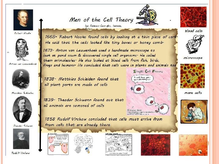
THE CELL THEORY Proposed by Schwann, Schleiden, and Virchow Three major tenets of the cell theory: 1. 2. 3. All organisms are composed of one or more cells Cells are the smallest fundamental life form Cells arise from pre-existing cells Exceptions to cell theory Viruses – are they living? What are they really? Where did first cell come from?
WHY ARE CELLS SMALL? Surface Area-to-Volume Ratio � Cells remain small to maintain a large surface area relative to the volume � Larger cells contain a greater volume � As cells grow larger cellular processes become inefficient Homeostasis is maintained by slow processes Diffusion, osmosis, energy acquisition
STRUCTURES COMMON TO ALL CELLS Four Structures found in ALL cell types � Nucleic Acids – DNA and RNA � Ribosomes – Protein synthesis � Plasma Membranes – Transport, Signaling, Regulation � Cytoplasm – reservoir of ions, inorganic and organic molecules
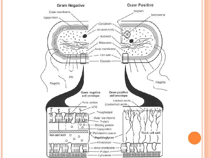
TYPES Prokaryote (Primitive Cell) Small cells No membrane-bound organelles � No nucleus Circular chromosome � No proteins on DNA OF CELLS Characteristic Size Eukaryote (True Cell) Organelles DNA No chromatin DNA is in cytoplasm Plasmid – small ring of DNA in addition to chromosome � Contain extra genes Cell Wall contains peptidoglycan (amino sugar) Small ribosomes in cytoplasm No Cytoskeleton � Cell Walls Ribosomes Cytoskeleton Larger than prokaryote Many membrane bound organelles � Some contain DNA Multiple chromosomes � Surrounded by proteins called histones which regulate genes � DNA & Protein is called chromatin No Plasmids Cell Walls contain chitin or cellulose (carbohydrates) Large ribosomes in cytoplasm AND attached to membranes Cytoplasm is called cytosol Cytoskeleton for support � Protein filaments
EUKARYOTIC ORGANELLES Nucleus - Regulation � Surrounded by nuclear envelope (membrane) Pores in nuclear envelope regulate RNA and ribosome transport Nucleolus synthesizes ribosomes Contains CHROMATIN (DNA + histone protein) Ribosomes- protein synthesis � Made of two subunits of r. RNA � Reads m. RNA and assembles amino acids to form polypeptides. � Located in cytoplasm and bound to endoplasmic reticulum
Endoplasmic Reticulum (ER) – intracellular transport � Membrane channels that extend throughout the cell Transport proteins to Golgi or other areas of cell Form transport vesicles � Two Types of ER Rough ER – contains ribosomes on outer surface Ribosomes secrete proteins into the empty space called a lumen Smooth ER – no ribosomes on surface Lipid synthesis Pinches to form transport vesicles Forms plasma membranes Golgi Apparatus – protein modification � Flat membrane sacs called cisternae with enzymes � Modify proteins to form: Receptors, enzymes, hormones, neurotransmitters, antibodies � Form secretory vesicles for exocytosis � Produce lysosomes
Lysosomes – Intracellular digestion � Membrane-enclosed sacs that contain hydrolytic enzymes � Recycling of a cell’s organic materials and programmed cell death (apoptosis). Vacuole � Membrane-bound sac that plays roles in intracellular digestion and the release of cellular waste products. � In plants, store pigments or poisonous substances � Allows for a large surface area to volume ratio Cell Wall � Chitin or cellulose � Permeable to water and gases. � Provide structure and support for cells
Plant central vacuoles used in several ways: • Maintain water balance • Store hazardous wastes, nutrients, or pigments • Provide turgor pressure on cytoplasm to keep cells rigid Don’t forget to water the plants!
VACUOLES SERVE MANY FUNCTIONS Fluid-filled sacs with a single membrane Functions of vacuoles � Contractile vacuoles in freshwater organisms used to collect and pump water out
Mitochondria - energy capture and transformation � Double membrane that allows compartmentalization � Smooth outer membrane - Inner membrane is highly convoluted Form folds called cristae Cristae contain enzymes important to ATP production Folded membrane increases the surface area for ATP production. � Matrix – central area of mitochondria contains enzymes � Contain circular DNA similar to prokaryotes Chloroplasts - capture energy through photosynthesis � Found in algae and higher plants. � Contain chlorophylls Green pigmented light-trapping molecules in photosynthesis � Double outer membrane that allows compartmentalization � Within the chloroplasts are membrane-bound structures called thylakoids. � Thylakoids are organized in stacks, called “grana” Produce ATP and NADPH 2 � Stroma – gel of enzymes surrounding grana Carbon fixation occurs in the stroma CO 2 converted to carbohydrates. � Contain circular DNA similar to prokaryotes
Cytoskeleton � Network of structural proteins � Facilitate cell movement � Morphological integrity (cell shape) � Organelle movement within the cell � Three types of protein filaments Microtubules Thin, hollow tubes Form the spindle fibers of mitosis to distribute chromosomes equally � Form cilia and flagella Form centrioles (in animal cells ONLY) – form spindle fibers Intermediate Filaments Insoluble Structural support, cell shape, organelle movement Microfilaments Cell movement, endocytosis and exocytosis Contain actin and mysoin (muscle proteins)
THE CYTOSKELETON Cytoskeleton forms a network of protein fibers within the cytoplasm � Composed of microfilaments, intermediate filaments, and microtubules
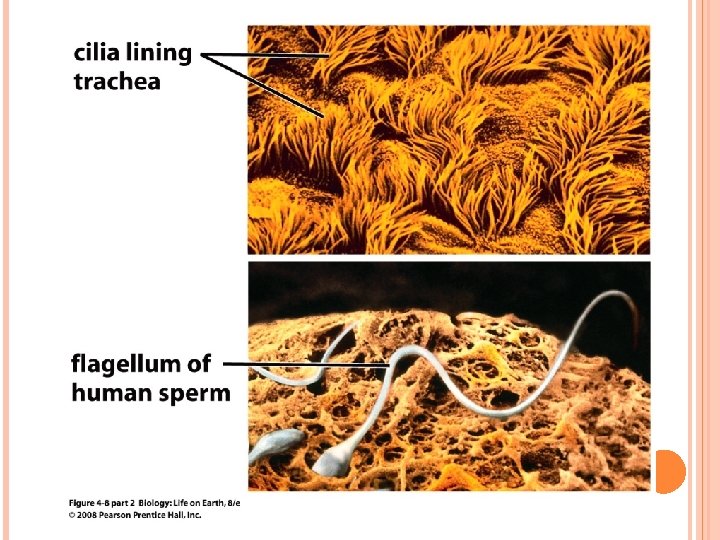
CILIA AND FLAGELLA Cilia and flagella are extensions of the plasma membrane Cilia are short and numerous while flagella are long but few in any cell
EUKARYOTE RELATEDNESS Structural evidence supports the relatedness of all eukaryotes. Cytoskeleton Membrane-bound organelles � mitochondria and/or chloroplasts WITH circular DNA Linear chromosomes � Chromatin (DNA + Protein) Endomembrane systems, including the nuclear envelope
ENDOMEMBRANE SYSTEM The interaction between the following: Nuclear membrane Endoplasmic reticulum Golgi Apparatus Secretory Vesicles Lysosomes The products of protein synthesis are modifies and processed through the membrane bound system that results in secretion of protein products, or the use of materials within the cell
Secretion Endocytosis
CHAPTER 6 Membranes and Cellular Transport
FOUR FUNCTIONS 1. 2. 3. OF THE PLASMA MEMBRANE Containment and separation of cell interior Material exchange – selectively permeable Information detection � Receptors � Glycoproteins � Glycolipids 4. Movement � Proteins pull or pinch membrane for cells to move Pseudopods – cytoplasmic extensions for movement/ingestion in white blood cells
STRUCTURE OF BIOLOGICAL Phospholipids � � Form a semipermeable barrier Outer surfaces are hydrophilic � Peripheral – surface layer proteins � � Function as receptors or attachment sites Integral – proteins that extend across the entire width of the membrane Function as transport proteins or motor proteins Embedded proteins can be hydrophilic, with charged and polar side groups, or hydrophobic, with nonpolar side groups. Glycolipids – sugar + lipid � Hydrophobic fatty acid portions face each other within the interior of the membrane itself. Proteins � The hydrophilic phosphate portions of the phospholipids are oriented toward the aqueous external or internal environments Inner component is hydrophibic MEMBRANES Cell recognition sites and provide energy Glycoprotein – sugar + protein � Cell recognition and regulate cell transport as “gateways”
extracellular fluid (outside) carbohydrate receptor protein recognition protein cholesterol phospholipid binding site cytosol (inside) Figure 5 -1 Biology: Life on Earth 8/e © 2008 Pearson Prentice Hall, Inc. glycoprotein phospholipid bilayer protein transport pore protein filaments
tails (hydrophobic) head (hydrophilic) Figure 5 -2 Biology: Life on Earth 8/e © 2008 Pearson Prentice Hall, Inc.
extracellular fluid (watery environment) hydrophilic heads phospholipid hydrophobic tails bilayer hydrophilic heads cytosol (watery environment) Figure 5 -3 Biology: Life on Earth 8/e © 2008 Pearson Prentice Hall, Inc.
(outside) (inside) reactions Figure 5 -5 Biology: Life on Earth 8/e © 2008 Pearson Prentice Hall, Inc.
PERMEABILITY OF THE PLASMA MEMBRANE Selective permeability is a direct consequence of membrane structure � Substances that freely pass across the lipid bilayer: Small uncharged polar molecules (urea) Small nonpolar molecules, such as N 2 , O 2, and CO 2 across the. � Substances that require embedded channel proteins Hydrophilic substances such as large polar molecules, proteins, and all ions (H+ Na + OH- ) Ions attract water and become hydrated (and large) Water moves across membranes and through channel proteins called aquaporins. Membrane transport proteins � Channel protein � Form a channel or pore to permit transport of substances based upon size and charge Carrier protein Bind to molecules and change shape to transport across the membrane Specific to shape and chemical properties of molecules being transported
TRANSPORT ACROSS THE MEMBRANE Passive Transport (NO ENERGY) � Does not require the input of metabolic energy; the net movement of molecules is from high concentration to low concentration. � Plays a primary role in the import of resources and the export of wastes. � Diffusion: movement of material from high to low concentration across the lipid bilayer Facilitated Diffusion: diffusion of charged and polar molecules through a membrane Uses protein channels or carrier proteins without energy expenditure Hydrogen ion channels, potassium ion channels, glucose carriers Osmosis: Diffusion of water from high concentration to low concentration Water molecules move DOWN their osmotic potential gradient Water potential is expressed as a negative value Lower potential (more negative) is more concentrated � Water flows from a high potential to a lower (less negative) potential � solution “A” Ψ = -2. 55 solution “B” Ψ = -7. 82 Which Solution will GAIN water from the other solution? “B” �
(a) Simple diffusion through the phospholipid bilayer lipid-soluble molecules (extracellular fluid) and O 2, CO 2, H 2 O O 2 (cytosol) Figure 5 -7 a Biology: Life on Earth 8/e © 2008 Pearson Prentice Hall, Inc.
(a) Isotonic solution Equal movement of water into and out of cells. (b) Hypertonic solution Net water movement out of cells. Figure 5 -10 Biology: Life on Earth 8/e © 2008 Pearson Prentice Hall, Inc. (c) Hypotonic solution Net water movement into cells.
cytosol central vacuole cell wall When water is plentiful, it fills the central vacuole, pushes the cytosol against the cell wall, and helps maintain the cell's shape. Water pressure supports the leaves of this impatiens plant. Figure 5 -11 Biology: Life on Earth 8/e © 2008 Pearson Prentice Hall, Inc. plasma membrane When water is scarce, the central vacuole shrinks and the cell wall is unsupported. Deprived of the support from water, the plant wilts.
extracellular fluid water aquaporin channel cytosol water Figure E 5 -2 b Biology: Life on Earth 8/e © 2008 Pearson Prentice Hall, Inc.
Facilitated Diffusion: NO ENERGY USED � Diffusion of charged and polar molecules through a membrane � Uses protein channels or carrier proteins without energy expenditure � Hydrogen ion channels, potassium ion channels, glucose carriers
(c) Facilitated diffusion through a carrier protein amino acids, sugars, small proteins (extracellular fluid) (cytosol) 1 Carrier protein has binding site for molecule. 2 Molecule enters binding site. 3 Carrier protein changes shape, transporting molecule across membrane. Figure 5 -7 c Biology: Life on Earth 8/e © 2008 Pearson Prentice Hall, Inc. 4 Carrier protein resumes original shape.
Active Transport � Requires free energy to move molecules from regions of low concentration to regions of high concentration. Move against the gradient � Free energy (often provided by ATP) is used by proteins embedded in the membrane to “move” molecules and/or ions across the membrane and to establish and maintain concentration gradients. � Membrane proteins are necessary for active transport. � Sodium Potassium Pump (Na+/K+) Pumps 3 sodium ions out and 2 potassium ions in Results in a standing charge difference across plasma membrane ALL cells have Na+/K+ pumps Why do we need a charge difference? The potential energy in the charge difference is used to move other molecules across the membrane Glucose transport
Potassium Ion Channel
1 The transport protein binds both ATP and Ca 2+. 2 Energy from ATP Changes the shape of the transport protein and moves the ion across the membrane. 3 The protein releases the ion and the remnants of ATP (ADP and P) and closes. (extracellular fluid) ATP recognition site Ca 2+ (cytosol) ATP binding site Figure 5 -12 Biology: Life on Earth 8/e © 2008 Pearson Prentice Hall, Inc. ADP P
(a) Pinocytosis (extracellular fluid) 1 3 2 (cytosol) vesicle containing extracellular fluid 1 A dimple forms in the plasma membrane, which 2 deepens and surrounds the extracellular fluid. 3 The membrane encloses the extracellular fluid, forming a vesicle. Figure 5 -13 a Biology: Life on Earth 8/e © 2008 Pearson Prentice Hall, Inc.
(b) Pinocytosis in a smooth muscle cell. 1 extracellular fluid 2 cytosol Figure 5 -13 b Biology: Life on Earth 8/e © 2008 Pearson Prentice Hall, Inc. 3
Receptor-mediated endocytosis nutrients (extracellular fluid) receptors coated pit 1 2 3 (cytosol) 4 coated vesicle 1 Receptor proteins for specific molecules or complexes of molecules are localized at coated pit sites. 2 A vesicle (“coated vesicle”) containing the bound molecules is released into the cytosol. 3 The coated pit region of the membrane encloses the receptor-bound molecules. 4 The receptors bind the molecules and the membrane dimples inward. Figure 5 -14 (part 1) Biology: Life on Earth 8/e © 2008 Pearson Prentice Hall, Inc.
extracellular particles bound to receptors coated vesicle (extracellular fluid) (cytosol) 1 2 protein coating 3 4 0. 1 micrometer coated pit plasma membrane Figure 5 -14 (part 2) Biology: Life on Earth 8/e © 2008 Pearson Prentice Hall, Inc.
(a) Phagocytosis food particle pseudopods 1 (cytosol) (extracellular fluid) 2 food vacuole 3 1 The plasma membrane extends pseudopods toward an extracellular particle (for example, food). 2 The ends of the pseudopods fuse, encircling the particle. 3 A vesicle called a food vacuole is formed containing the engulfed particle. Figure 5 -15 a Biology: Life on Earth 8/e © 2008 Pearson Prentice Hall, Inc.
(b) Amoeba An Amoeba (a freshwater protist), engulfs a Paramecium using phagocytosis. Figure 5 -15 b Biology: Life on Earth 8/e © 2008 Pearson Prentice Hall, Inc.
(c) White blood cell A white blood cell ingests bacteria using phagocytosis. Figure 5 -15 c Biology: Life on Earth 8/e © 2008 Pearson Prentice Hall, Inc.
secreted material plasma membrane (extracellular fluid) vesicle (cytosol) Material is enclosed in a vesicle that fuses with the plasma membrane, allowing its contents to diffuse out. Figure 5 -16 Biology: Life on Earth 8/e © 2008 Pearson Prentice Hall, Inc. 0. 2 micrometer
(a) Gap junctions (b) Plasmodesmata root liver cells root cells plasma membrane Gap junctions: pairs of channels connect insides of adjacent cells. Figure 5 -19 Biology: Life on Earth 8/e © 2008 Pearson Prentice Hall, Inc. cell wall plasma membrane Plasmodesmata connect insides of adjacent cells.
(a) Desmosome small intestine cells lining small intestine (b) Tight junction urinary bladder cells lining bladder microvilli desmosome plasma membranes (edge view) Protein strands hold cells together. Tight junctions formed by strands of protein seal cells together. protein filaments in cytosol Figure 5 -18 Biology: Life on Earth 8/e © 2008 Pearson Prentice Hall, Inc.