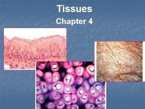
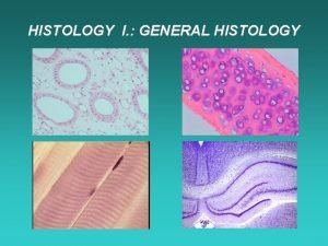
Chapter 4 Histology The Study of Tissues TISSUE


- Slides: 69

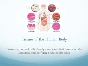
Chapter 4 Histology: The Study of Tissues TISSUE LEVEL OF ORGANIZATION: Tissues-groups of similar cells and substances surrounding them; with similar functions Classification of tissue type: based on cellular structure, function, composition of extracellular matrix Extracellular matrix: noncellular substances surrounding cells 1

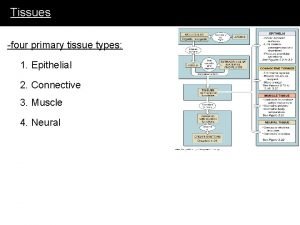
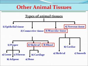
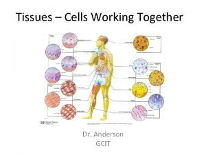
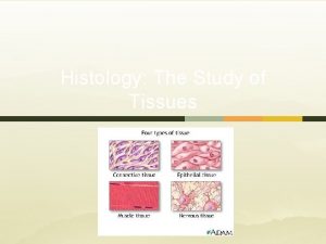
Tissues and Histology • 4 Primary Tissue Types – Epithelial – Connective – Muscle – Nervous • Histology: Microscopic Study of Tissues 2

Embryonic Tissue • Germ layers: the beginning of all adult structures can be traced back to one of them – Endoderm • Inner layer • Forms lining of digestive tract and its derivatives – Mesoderm • Middle layer • Forms tissues such as muscle, bone, blood vessels – Ectoderm • Outer layer • Forms skin and neuroectoderm 3

Characteristics of Epithelium • Consists almost entirely of cells that are attached to a basement membrane • Covers body surfaces and forms glands • Has free and basal surface • Specialized cell contacts • Avascular • Undergoes mitosis 4

Functions of Epithelia • • • Protecting underlying structures Acting as barriers Permitting the passage of substances Secreting substances Absorbing substances 5

6

Classification of Epithelium • Classified according to: – Number of cell layers, shape of the superficial cells • Simple (single layer) – Squamous, cuboidal, columnar • Stratified (more than one layer- top layer determines classification) – Squamous, cuboidal, columnar • Pseudostratified (appears to be more than one layer- each cell in contact with basement membrane) – Columnar • Transitional (a unique type of stratified) – Cuboidal or columnar when not stretched and squamous when stretched 7

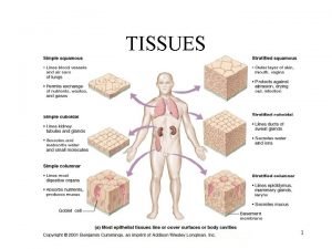
Simple Squamous Epithelium • Single layer of flat, hexagonal cells • Found in the lining of blood and lymph vessels, lining of small ducts, alveoli of lungs, loop of Henle in kidney tubules, lining of serous membranes (mesothelium), inner surface of eardrum • Function: Diffusion, filtration 8

Types of Epithelium 9

Simple Cuboidal Epithelium • Single layer of cube-shaped cells; some have cilia or microvilli • Found in: Kidney tubules, glands, and their ducts, lining of terminal bronchioles of lungs, surface of ovaries • Function: Secretion and absorption 10

Types of Epithelium 11

Simple Columnar Epithelium • Single layer of tall, narrow cells; some have cilia or microvilli • Lining of stomach, small and large intestines, gallbladder, bile ducts, bronchioles of lungs, ventricles of brains • Microvilli: projections that increase surface area; functions in secretion; absorption (found in intestines) • Cilia: functions in movement; bronchioles of lungs, auditory tubes, uterus, uterine tubes 12

Types of Epithelium 13

Stratified Squamous Epithelium • Multiple layers that are cuboidal in the basal layer and progressively flattened toward the surface • Condition of outermost layer • Moist (non-keratinized)- lining of structures • Consists of living cells in the deepest and outermost areas • Layer of fluid covers outermost cells • Mouth, throat, esophagus, larynx, anus, vagina, cornea • Keratinized-skin • Inner layers – livings cells • Outer layers – dead cells containing the protein, keratin • Durable, moisture- resistant and dry • Function: Protection against abrasion and infection 14

Types of Epithelium 15

Stratified Cuboidal Epithelium • Multiple layers of somewhat cube-shaped cells • Salivary gland ducts, sweat gland ducts, ovarian follicular cells • Function: Secretion, absorption, protection against infection 16

Types of Epithelium 17

Stratified Columnar Epithelium • Multiple layers of cells, with tall, thin cells resting on layers of more cuboidal cells • Larynx (ciliated), mammary gland duct, a portion of male urethra • Function: Protection and secretion 18

Types of Epithelium 19

Pseudostratified Columnar Epithelium • Single layer of tall, thin, cells; some reach free surface, some do not; the cells are almost always ciliated and have goblet cells that secrete mucus • Lining of nasal cavity, nasal sinuses, pharynx, trachea, bronchi of lungs, auditory tubes • Function: Synthesize and secrete mucus onto free surface; move mucus with foreign particles out of passages 20

Types of Epithelium 21

Transitional Epithelium • Stratified cells that appear cuboidal when organ is not stretched and squamous when organ stretched by fluid • Lining of urinary bladder, ureters, superior urethra • Function: Accommodates fluctuations in fluid volume; protection against the caustic effects of urine 22

Types of Epithelium ( 23

24

25

Functional Characteristics • Cell layers and shapes – Diffusion, Filtration, Secretion, Absorption, Protection • Cell surfaces – Microvilli: Increase surface area absorption or secretion – Cilia: Move materials across cell surface • Cell connections – Desmosomes, hemidesmosomes, tight, gap junctions • Glands – Secretory organs composed of epithelium – Exocrine: Have ducts – Endocrine: Have no ducts 26

Cell Connections • Functions – Bind cells together – Form permeability layer – Intercellular communication • Types – – Desmosomes Hemidesmosomes Tight Junctions Gap Junctions 27

Exocrine Glands • Most are multicellular – Can be classified according to structure of their ducts • Unicellular – Goblet cells of respiratory system 28

Multicellular Exocrine Glands 29

Exocrine Glands and Secretion Types Functional classification: How products leave the cell • Merocrine – Sweat glands – Most glands in the body – No loss of cell material • Apocrine – Mammary glands – Discharge fragments • Holocrine – Sebaceous glands – Shed entire cells 30

Connective Tissue • Abundant • Consists of cells separated by extracellular matrix • Diverse • Performs a variety of important functions 31

32

Functions of Connective Tissue • Enclosing and separating (sheets form capsules around organs) • Connecting tissues to one another: tendons and ligaments • Supporting and moving: bones • Storing: Adipose tissue (fat) • Cushioning and insulating: fat • Transporting: blood • Protecting: cells of the immune system 33

Connective Tissue Cells • Specialized cells that produce the extracellular matrix – Suffixes • -blasts: create the matrix • -cytes: maintain the matrix • -clasts: break the matrix down for remodeling • Adipose or fat cells • Mast cells that contain heparin and histamine • White blood cells that respond to injury or infection • Macrophages that phagocytize or provide protection • Stem cells 34

Extracellular Matrix • 3 Major Components: – Protein fibers • Collagen which is most common protein in body • Reticular fills spaces between tissues and organs • Elastic returns to its original shape after distension or compression – Ground substance • • Shapeless background hyaluronic acid-slippery; most abundant proteoglycans-branched-change shape adhesive molecules-hold proteoglycan aggregates together; chondronectin-cartilage; osteonectin-bone; fibronectin-fibrous connective tissue – Fluid: Plasma 35

Categories of Connective Tissue • Embryonic Connective Tissue – Mesenchyme: the embryonic tissue from which connective tissues, as well as other tissues, arise – Mucous connective tissue or Wharton’s jelly: embryonic tissue remaining in newborn umbilical cord • Adult Connective Tissue: 6 types – – – Loose Dense Connective tissue with special properties Cartilage Bone Blood 36

37

38

Loose Connective Tissue • • Also known as areolar tissue Loose packing material of most organs and tissues Attaches skin to underlying tissues Contains a variety of cells (fibroblasts, macrophages, lymphocytes) in a fine network of mostly collagen fibers 39

Dense Connective Tissue • Dense regular collagenous – Has abundant collagen fibers • Tendons: Connect muscle to bone • Ligaments: Connect bone to bone – Great tensile strength, stretch resistance • Dense regular elastic – Ligaments between vertebrae, in vocal cords, nuchal ligament in neck – Capable of stretching and recoiling due to elastin fibers • Dense irregular collagenous – Most of dermis of skin, sheaths, organ capsules, scars • Dense irregular elastic – Walls of elastic arteries 40

Dense Regular Connective Tissue 41

42

Dense Irregular Connective Tissue 43

Connective Tissue with Special Properties • Adipose tissue – Consists of adipocytes (fat cells) – Types • Yellow (white at birth) – most abundant, yellows with age • Brown – found only in specific areas of body as axillae (armpits), neck and near kidneys • Reticular tissue – Forms framework of lymphatic tissue, such as in spleen and lymph nodes, bone marrow, liver – Provides a superstructure for lymphatic and hematopoietic tissues 44

Adipose Tissue 45

Reticular Tissue 46

Cartilage • Composed of cartilage cells or chondrocytes located in spaces called lacunae • Next to bone, firmest structure in body • Cartilage matrix: collagen fibers, proteoglycan ground substance, fluid – Proteoglycan aggregates trap large amounts of water – Trapped water lets cartilage spring back after being compressed • Perichondrium: dense irregular connective tissue that covers the surface of all cartilage – Gives rise to chondrocytes and cartilage matrix • Cartilage has no blood vessels or nerves, except those in perichondrium; heals very slowly 47

Cartilage (cont’d) • 3 Types of cartilage: • Hyaline cartilage: most abundant • Appears to have a glassy, translucent matrix • Fibrocartilage • Elastic cartilage 48

Hyaline Cartilage • Found in areas for strong support and some flexibility – Rib cage, nasal, and cartilage in trachea and bronchi • Forms most of skeleton before replaced by bone in embryo • Involved in growth that increases bone length • Covers bone surfaces that move smoothly against each other 49

Fibrocartilage • Slightly compressible and very tough • Found in areas where a great deal of pressure is applied to joints – Intervertebral discs, knee, jaw 50

Elastic Cartilage • Rigid but elastic properties – External ears, epiglottis, auditory tubes 51

Bone • Hard connective tissue that consists of living cells (osteocytes) and mineralized matrix – Organic- protein fibers (collagen) – Inorganic-hydroxyapatite mineral (contains calcium and phosphate) – Bone, unlike cartilage, has a rich blood supply (easier repair) • 2 Types: – Cancellous or spongy bone- spaces between trabeculae or plates of bone; resembles a sponge • Interior of skull bones, vertebrae, sternum, pelvis, ends of long bones – Compact bone- more solid, almost no spaces between lamellae (thin layers) • Outer portion of all bones, shafts of long bones • Osteocytes located in holes in matrix, called lacunae; osteons or Haversian systems surrounding central canals 52

Bone 53

Blood and Bone Marrow • Blood: unusual with its fluid matrix • Formed by hemopoietic tissue • Found in bone marrow: soft connective tissue in cavities of bones – Yellow Marrow • Adipose tissue in shafts of long bones – Red Marrow • Hemopoietic tissue in ends of long bones and in short, flat, irregular bones 54

Bone Marrow 55

Muscle Tissue • Characteristics – Contracts or shortens with force – Moves entire body; pumps blood through heart, blood vessels • 3 Types: – Skeletal • Striated and voluntary • Attached to bones – Cardiac • Striated and involuntary • Found in heart – Smooth • Nonstriated and involuntary • Walls of hollow organs (stomach, intestines), blood vessels, eyes, glands, goose bumps in skin 56

Skeletal Muscle 57

Cardiac Muscle 58

Smooth Muscle 59

60

Nervous Tissue • Found in brain, spinal cord and nerves • Ability to produce electric signals called action potentials • Nerve Cells – Called neurons • Consist of dendrites, cell body, axons • Different types: multipolar, bipolar, unipolar – Neuroglia are support cells surrounding brain, spinal cord, peripheral neurons 61

Neurons 62

Neuroglia 63

Membranes • • • Mucous – Line cavities that open to the outside of body – Secrete mucus – Mesothelium+connective tissue Serous – Line cavities not open to exterior • Pericardial, pleural, peritoneal Mesothelium+connective tissue Synovial – Line freely movable joints – Produce fluid rich in hyaluronic acid; makes joint fluid very slippery – Connective tissue only; lack mesothelium 64

Inflammation • Response when tissues damaged or in an immune response • Manifestations – Redness, heat, swelling, pain, disturbance of function • Mediators – Include histamine, kinins, prostaglandins, leukotrienes – Stimulate pain receptors and increase blood vessel permeability – Causes edema 65

Tissue Repair • Substitution of viable cells for dead cells • Skin repair – Primary union: Edges of wound close together • • • Wound fills with blood Clot of fibrin forms Scab Pus Granulation tissue Scar – Secondary union: Edges of wound not close • Clot may not close gap • Inflammatory response greater • Wound contraction occurs leading to greater scarring 66

Tissue Repair 67

Tissues and Aging • Cells divide more slowly in older than younger people • Tendons and ligaments become less flexible and more fragile (due to irregular structure of collagen) • Arterial walls become less elastic • Rate of blood cell synthesis declines in elderly • Injuries heal more slowly in elderly 68

Cancer Tissue Cancermalignant spreading tumor and the illness that results from such a tumor Neoplasmabnormal tissue growth benign- does not spread from primary site malignant- spreads from primary site; by metastasis Carcinomamalignant neoplasm derived from epithelial tissue Sarcomamalignant neoplasm derived from connective tissue Causation- viruses, environmental toxins, other causes? Treatment. Surgery, Chemotherapy, Radiotherapy; Combined modality Early Detection-best prognosis for treatment 69
 Body tissue
 Body tissue
 Body tissues chapter 3 cells and tissues
 Body tissues chapter 3 cells and tissues
 Eisonophil
 Cns histology ppt
 Exercise 17 gross anatomy of the brain and cranial nerves
 Connective tissue histology quiz
 Stratified squamous epithelium characteristics
 Epithelial tissue histology pogil answer key
 Pogil epithelial tissue histology
 Tissue processing
 Parathyroid
 Tonsils
 Perforation plates
 Chapter 6 bones and skeletal tissues
 Chapter 3 cells and tissues
 Chapter 3 cells and tissues figure 3-1
 Four principal types of tissue
 Chapter 3 cells and tissues figure 3-1
 Simple squamos
 The microscopic study of diseased tissue.
 Chapter 8 oral embryology and histology
 A specialized connective tissue
 What are the 4 types of tissues
 Vascular bundles
 3 tissues of a plant
 3 tissues of a plant
 Jane campion tissues
 Upper epidermis function in photosynthesis
 Tissues causes of civil war
 Location
 Characteristics of connective tissues
 Tissues are groups of similar cells working together to
 Genetic effects on gene expression across human tissues
 Dense connective tissue description
 Types of tissues
 Cutaneous membrane
 Tissues are groups of similar cells working together to
 Tissues poem
 Types of tissues
 What macromolecule is a prominent part of animal tissues
 Division of anatomy
 Supportive tissues
 What is this tissue
 Predominant cell type
 Types of tissues
 Pearson endocrine system
 Four major tissue types
 Tissues working together
 Permanent tissue
 Tissues in the circulatory system
 Simple columnar epithelium tissue function
 Plastid in plant cell
 Goblet cells cilia and microvilli
 Periradicular disease
 What do all connective tissues have in common
 Gas exchange between tissues and capillaries
 Analogy of tissues
 Plant tissues
 A tissues
 What are tissues
 A group of cells similar in structure and function
 Meristematic tissue
 Sclerenchyma
 Periradicular tissues are
 Tissues in plants
 Table of tissues
 Fetal station chart
 Four basic tissues

