Chapter 4 Cellular Oncogenes 4 2 4 6


 The DNA from chemical-transformed tumor")


 Figure 4. 4 (part 2) The Biology of Cancer")
 The Biology of Cancer (© Garland Science 2007)")


")


 Malarial infection Figure 4. 12 The Biology of")


")

 of chronic myelogenous leukemia (CML) has t(9; 22)")
 (q 34; q 11) translocation (Abelson")

")
 Human gene protein")
- Slides: 33
Chapter 4 Cellular Oncogenes - 4. 2 ~ 4. 6 - Mar 22, 2007
Many retroviruses carrying oncogenes have been found in chickens and mice
However, attempts undertaken during the 1970 s to isolate viruses from most types of human tumors were unsuccessful. Even reverse transcriptase-containing virus particles are difficult to find in human tumor samples.
4. 2 Transfection of DNA provides a strategy for detecting nonviral oncogenes (NIH 3 T 3 cells) transformed by 3 -methycholanthrene Sidebar 4. 2 Figure 4. 2 (part 1 of 2) The Biology of Cancer (© Garland Science 2007)
DNA from tumor cells (NIH 3 T 3 cells) The DNA from chemical-transformed tumor cells was able to convert non-tumorigenic NIH 3 T 3 fibroblasts into tumorigenic cells. Figure 4. 2 (part 2 of 2) The Biology of Cancer (© Garland Science 2007)
DNA from human cancers also cause focus formation cells in the focus surrounding untransformed monolayer cells Transfection of DNA from T 24 human bladder carcinoma cell line into NIH 3 T 3 cells Figure 4. 3 The Biology of Cancer (© Garland Science 2007)
4. 3 Oncogenes discovered in human tumor cell lines are related to those carried by transforming retroviruses Southern blotting (DNA) Northern blotting (RNA)
nitrocellulose paper ( 32 P-DNA) Figure 4. 4 (part 2) The Biology of Cancer (© Garland Science 2007)
Figure 4. 4 (part 3) The Biology of Cancer (© Garland Science 2007)
Homology between transfected oncogenes and retroviral oncogenes probe used: H-ras oncogene present in Harvey rat sarcoma virus _______________ 11 (a ~ k) NIH 3 T 3 cell lines transfected with DNA extracted from a human bladder carcinoma cell line Figure 4. 5 The Biology of Cancer (© Garland Science 2007) untransfected NIH 3 T 3
TK : tyrosine kinase Table 4. 1 The Biology of Cancer (© Garland Science 2007)
Amplification of the erb. B 2/HER 2/neu oncogene in breast cancers Kaplan-Meier plot Figure 4. 6 a The Biology of Cancer (© Garland Science 2007)
erb. B 2/HER 2/neu oncogene can be amplified or overexpressed in human breast carcinoma cells erb. B 2/neu oncogene is amplified erb. B 2/neu m. RNA is overexpressed increased level of erb. B 2/neu -encoded protein Figure 4. 6 c The Biology of Cancer (© Garland Science 2007)
Cloning of transfected human oncogenes Alu sequence present in about 106 copies scattered throughout human genome Figure 4. 8 The Biology of Cancer (© Garland Science 2007)
Localization of the mutation responsible for oncogene activity cloned DNA of a human bladder carcinoma oncogene Figure 4. 9 The Biology of Cancer (© Garland Science 2007)
A point mutation is responsible for H-ras oncogene activation Figure 4. 10 The Biology of Cancer (© Garland Science 2007)
4. 4 Proto-oncogenes can be activated by genetic changes affecting either protein expression or structure
4. 5 The myc oncogene can arise via at least three distinct mechanisms homogeneous staining regions (HSR) The N-myc gene amplification is found in 30% of human childhood neuroblastoma. Astrocytoma, retinoblastoma and small-cell lung carcinomas (neuroendocrinal traits) also often exhibit amplified N-myc genes. * N-myc is a close relative of c-myc. Figure 4. 11 a The Biology of Cancer (© Garland Science 2007)
event - free survival Kaplan-Meier plot of childhood neuroblastoma
Table 4. 3 The Biology of Cancer (© Garland Science 2007)
Activation of the myc protooncogenes 1. Gene amplification 10 ~ 30 copies or 100 ~ 150 copies shown as homogeneously staining regions (HSR) or double minutes (DM) 2. Provirus integration - insertional mutagenesis constitutive expression by insertion of retroviruses 3. Chromosomal translocation
Insertional mutagenesis transcription of myc gene is controlled by viral promoters excessive myc protein Figure 3. 23 b The Biology of Cancer (© Garland Science 2007)
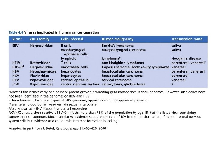
Burkitt’s lymphoma in Africa (Aedes simpsoni) Malarial infection Figure 4. 12 The Biology of Cancer (© Garland Science 2007) Epstein-Barr virus (EBV) genome in Burkitt’s lymphoma cells
Chromosome translocations in Burkitt’s lymphoma The expression of c-myc gene is placed under control of the trancriptioncontrolling enhancer sequences of an immunoglobulin heavy chain (Ig. H) gene. Figure 4. 13 a The Biology of Cancer (© Garland Science 2007)
Genetic map of the translocation event of c-myc gene The c-myc gene is translocated into chromosome 8, under the control of the immunoglobulin heavy-chain (Ig. H) sequences present on human chromosome 14 Figure 4. 13 b The Biology of Cancer (© Garland Science 2007)
Table 4. 4 The Biology of Cancer (© Garland Science 2007)
4. 6 A diverse array of structural changes in proteins can also lead to oncogene activation (GF) Figure 4. 14 The Biology of Cancer (© Garland Science 2007)
The great majority (> 95 %) of chronic myelogenous leukemia (CML) has t(9; 22) (q 34; q 11) translocation Philadelphia chromosome (Ph 1) Figure 2. 23 a The Biology of Cancer (© Garland Science 2007)
Formation of the bcr-abl oncogene after t(9; 22) (q 34; q 11) translocation (Abelson murine leukemia virus) (breakpoint cluster region) Figure 4. 15 a The Biology of Cancer (© Garland Science 2007)
Different breakpoints in bcr results in different types of human leukemia ↓ ↓a ↓b ↓c ↓ a. acute lymphocytic leukemia b. c. Bcr ↑ Abl Bcr-Abl fusion protein Figure 4. 15 b The Biology of Cancer (© Garland Science 2007) chronic myelogenous leukemia chronic neutrophilic leukemia
Table 4. 5 The Biology of Cancer (© Garland Science 2007)
Notations used for proto-oncogenes and oncogenes Non-human (chicken, mouse, etc. ) Human gene protein src, myc Src, Myc SRC, MYC