CHAPTER 3 The Neuron Synaptic Transmission Neurotransmitters and


 – After action in postsynaptic cleft,")


- Slides: 101
CHAPTER 3 The Neuron, Synaptic Transmission, Neurotransmitters and the CNS
How do neurons communicate?
a b c
How do neurons communicate? Need to think about this question 2 ways
How do neurons communicate? 1. within neurons – 2. between neurons-
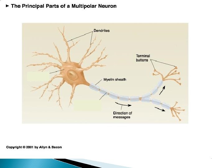
Neuron receiving info Information traveling down neuron
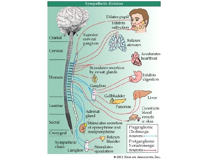
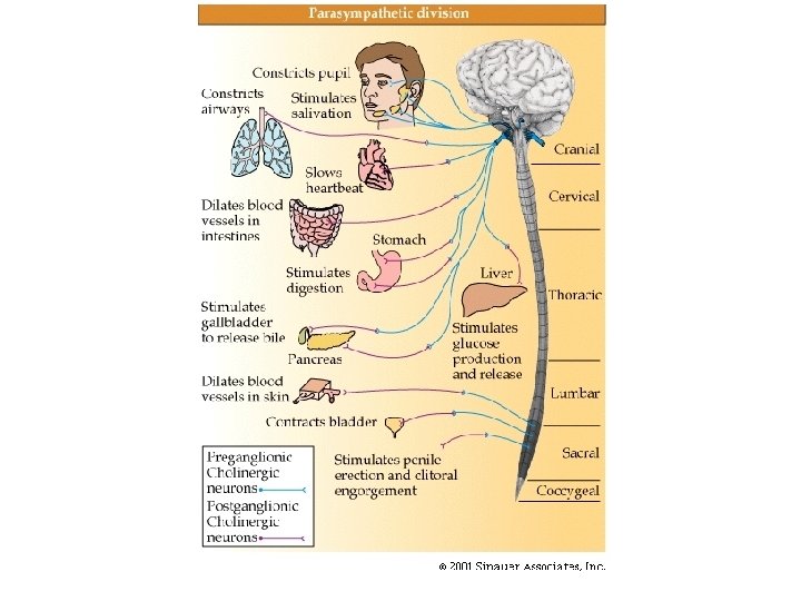
How do neurons communicate � within neurons – electrically � between neurons – chemically ◦ Synapse – space between neurons
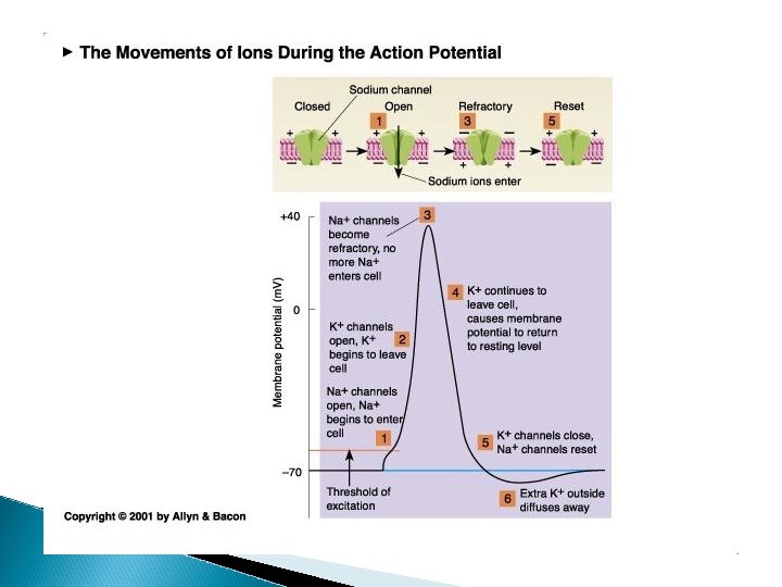
Neurons can exist in one of 3 states � the “resting” state � the “active” state � the “refractory” state ◦ neuron is firing ◦ action potential
At rest: � inside of the axon has a slightly negative charge relative to outside the axon ◦ called the membrane potential ◦ usually around -70 m. V
At rest: � inside of the axon has a slightly negative charge relative to outside the axon ◦ called the membrane potential � why?
action potential or spike
Neuron stimulated (either electrically or by receiving a “message” � see depolarization (change from negative inside neuron to more positive)
action potential or spike
Neuron stimulated (either electrically or by receiving a “message” � see depolarization (change from negative inside neuron to more positive) ◦ “threshold” – if a great enough depolarization occurs, an action potential will occur ◦ action potential – very quick – milliseconds �Other terms – spike, firing, generating an AP
action potential or spike
� Hyperpolarization �return to negative �this is the refractory or recovery period
action potential or spike
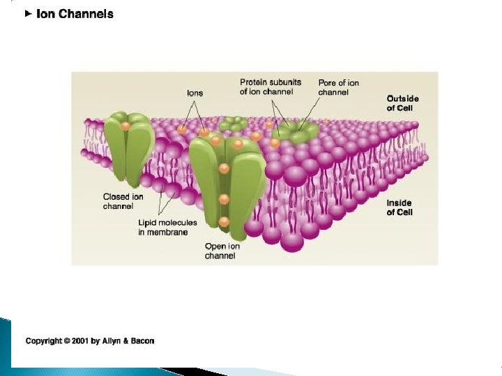
What causes these changes in electrical potential and the action potential? � All axons and cells have a membrane �thin lipid (fat) bilayer � The membranes have channels (to allow ions in or out) � Ions – molecules with a charge � These channels can be open or shut
What causes these changes in electrical potential? � Ions flowing across the membrane causes the changes in the potential � Ions are molecules that contain a positive or negative charge �anion – negative charge �cation – positive charge
Some important ions for neuronal communication �Na+ sodium ◦ HIGHER CONCENTRATION OUTSIDE THE AXON �Cl- chloride �K+ potassium ◦ HIGHER CONCENTRATION OUTSIDE AXON ◦ higher concentration inside the axon �A- anions -large (-) molecules with a negative charge (stuck inside the axon)
Some forces that play a role in maintaining membrane potential � concentration gradient – ◦ ions diffuse from higher concentration to lower concentration
example of concentration forces
Concentration Gradient Na+ would enter axon K+ K+ would leave axon Cl- would enter axon
Some forces that play a role in maintaining membrane potential � concentration gradient – ◦ ions diffuse from higher concentration to lower concentration � electrical gradient - ◦ opposite charges attract so ions are attracted to an environment that has a charge that is opposite of the charge they carry!
example of electrostatic forces
Electrical Gradient Na+ go in K+ stay in Cl- stay out
Concentration Gradient Electrical Gradient Na+ go in K+ go out stay in Cl- go in stay out
What drives the action potential? � opening ions of Na+ channels and influx of Na+
What happens if sodium channels are blocked? lidocaine, novocaine, cocaine TTX – tetrototoxin Sagitoxin◦ red tides
Concentration Gradient Na+ go in K+ go out Cl- go in Electrical Gradient as cell is depolarized (+ intracellular)
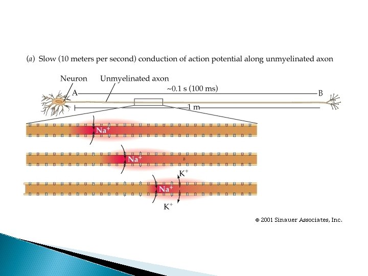
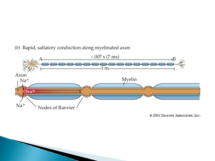
nodes of ranvier
nodes of ranvier
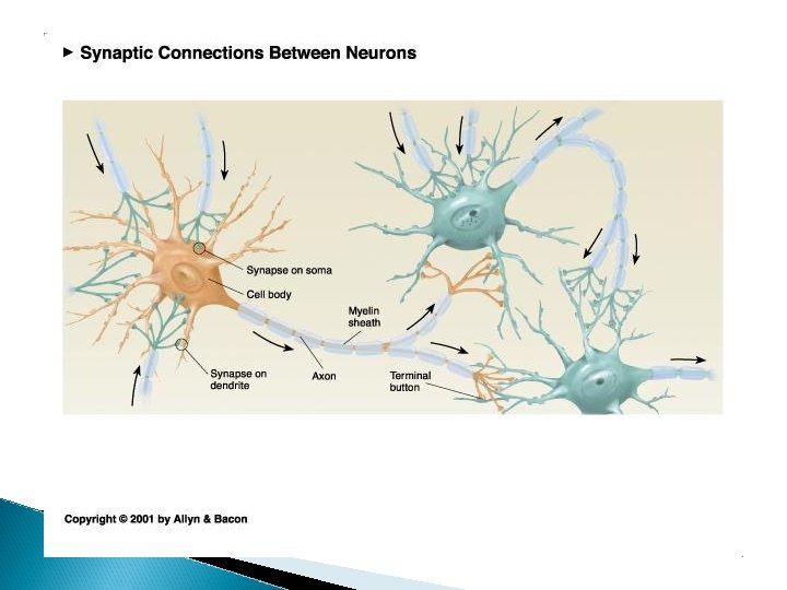
What about communication between neurons?
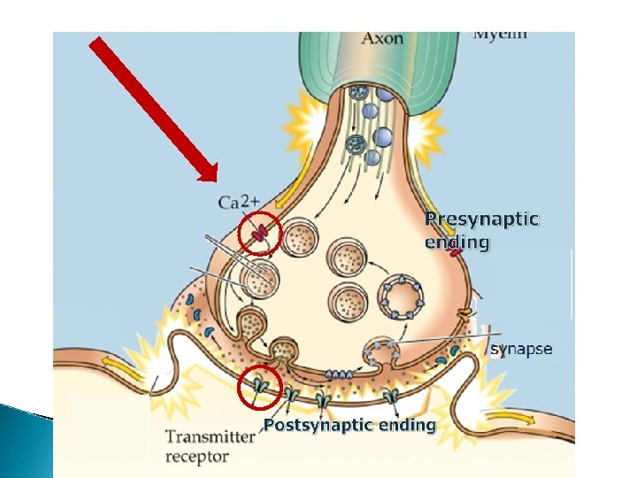
Some terms……. � presynaptic ending – ◦ portion of the axon conveying information to the next neuron
Some terms……. � presynaptic ending – ◦ the portion of the axon that is conveying information to the next neuron � synapse or synaptic cleft ◦ the space between neurons where communication occurs
Some terms……. � presynaptic ending – ◦ the portion of the axon that is conveying information to the next neuron � synapse or synaptic cleft ◦ the space between neurons where communication occurs � postsynaptic membrane ◦ the portion of the neuron (usually dendrite) that receives information
Some terms……. � � presynaptic ending – ◦ the portion of the axon that is conveying information to the next neuron synapse or synaptic cleft ◦ the space between neurons where communication occurs postsynaptic membrane ◦ the portion of the neuron (usually dendrite) that receives information pre and postsynaptic receptors ◦ proteins in both the presynaptic and postsynaptic ending that allow for information to be transferred
� synaptic vesicles --small enclosed membranes that contain neurotransmitter found in presynaptic ending � neurotransmitter – substance in vesicles that are released in synapse and convey info to the next neuron
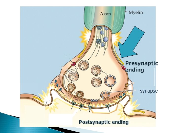
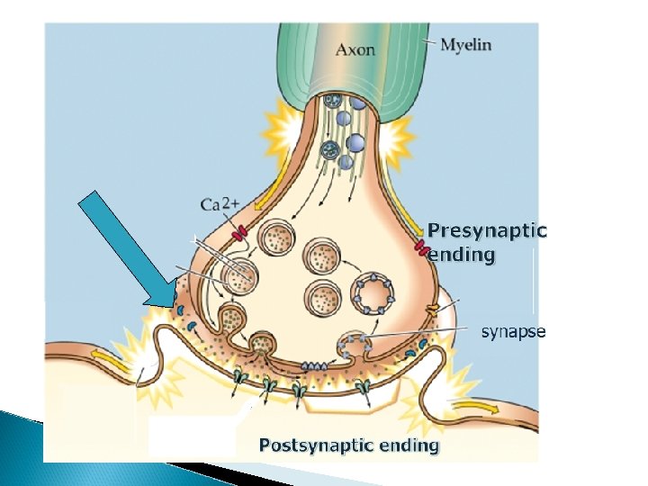
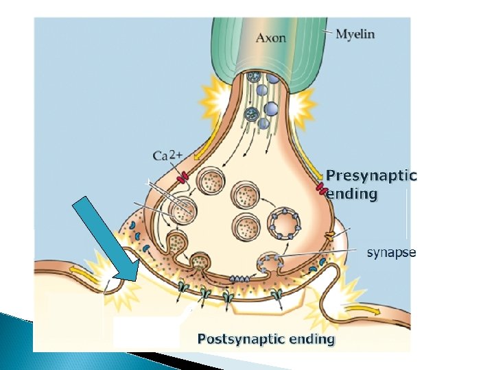
Presynaptic ending synapse Postsynaptic ending
What happens at level of synapse? � AP reaches presynaptic ending- � Ca+2 channels in presynaptic ending open and Ca+2 enters
Why are Ca+2 ions important? Ca+2 entry into the presynaptic ending critical for neurotransmitter release
Figure 3. 5 A. Photomicrograph of a synapse in action, taken with the electron microscope. B. Schematic of the process Julien: A Primer of Drug Action, Eleventh Edition Copyright © 2008 by Worth Publishers
postsynaptic receptors � protein embedded in membrane � mechanism for neurotransmitter to influence postsynaptic activity by binding to receptor
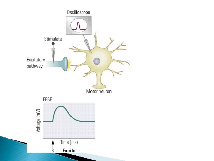
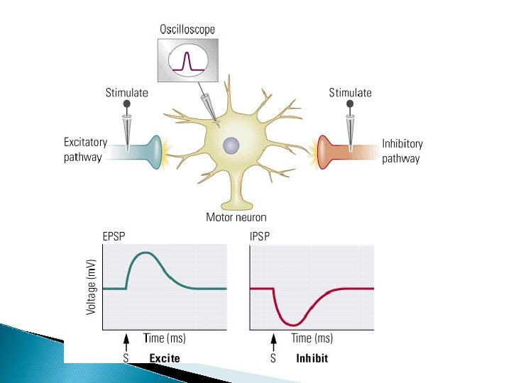
Summary � NT binds to postsynaptic receptors and causes small local changes in electrical potential (depolarizations or hyperpolarizations)◦ Called graded potentials
What happens to convey info from one neuron to the next � ◦ Graded Potentials- increase or decrease the likelihood of the neuron receiving info to generate an action potential
Graded potentials � graded potentials that increase the likelihood of an action potential are called EPSPs (excitatory postsynaptic potentials)
Graded potentials � graded potentials that increase the likelihood of an action potential are called EPSPs (excitatory postsynaptic potentials) � graded potentials that decrease the likelihood of an action potential are called IPSPs (inhibitory postsynaptic potentials)
How does the neurotransmitter cause EPSPs and IPSPs? � NT binding to postsynaptic receptors cause local ion channels to open
How does the neurotransmitter cause EPSPs and IPSPs? �– chemically dependent ion channels � in contrast with electrically dependent ion channels
How does the neurotransmitter cause EPSPs and IPSPs?
How does the neurotransmitter cause EPSPs and IPSPs? � postsynaptic receptors open ion channels – ◦ ion channels in postsynaptic membrane (that we need to worry about) include Na+, Cl- and K+
Two kinds of Graded Potentials � EPSPs – excitatory postsynaptic potentials �- increase the likelihood of an AP �- opening of
�EPSPs – excitatory postsynaptic potentials �opening of local Na+ channels �IPSPs – inhibitory postsynaptic potentials
◦ IPSPs – inhibitory postsynaptic potentials • decreases the liklihood of an action potential �opening of
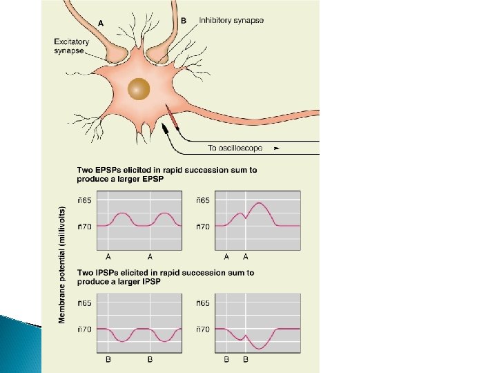
What happens to convey info from one neuron to the next? ◦ graded potentials are summed at axon hillock
Axon hillock
What happens to convey info from one neuron to the next? ◦ EPSPs and IPSPs are summed a axon hillock……. . AND
What happens to convey info from one neuron to the next Graded potentials are localized – has impact in limited region; AP travels down the axon
Neurotransmitters and Receptors General Principles • Synthesis 1. Formation of transmitters 2. Precursors are the main ingredient. • Brought to the neuron by the bloodstream. • Taken up by cell body and/or terminal. • Often come from substances in the diet. 3. Enzymes put the ingredients together.
Neurotransmitters and Receptors Transmitters Stored in Vesicles 1. Concentration 2. Protection
Neurotransmitters and Receptors Release = exocytosis – Vesicles fuse with presynaptic membrane and release transmitters into the synapse. Binding = attachment of transmitter to receptor
Neurotransmitters and Receptors There are different varieties of receptors. – Some respond fast – Called Ionotropic – Direct reaction to the transmitter
Neurotransmitters and Receptors Different varieties of receptors: – Other types of receptors respond more slowly. – Indirectly – Called Metabotropic, or G protein-coupled – Initiates a second signal (messenger) inside the neuron.
Neurotransmitters and Receptors Inactivation: Termination of Synaptic Transmission 1. Metabolism 2. Re‑uptake 3. Re-uptake by glial cell (glutamate only)
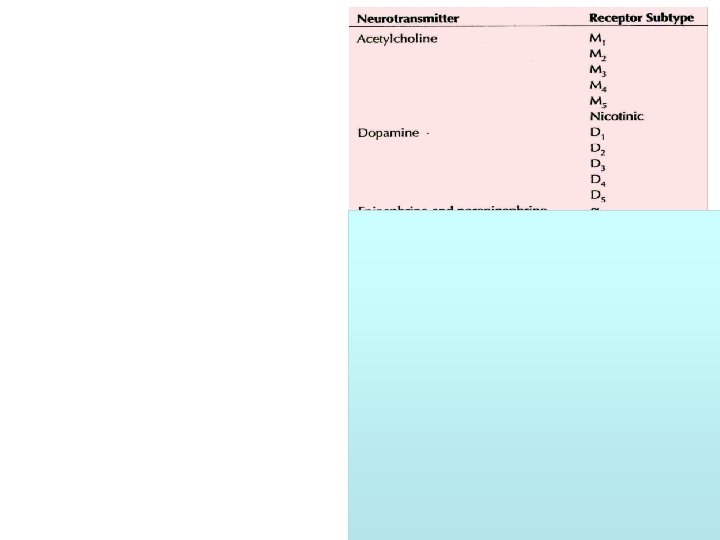
Neurotransmitters • Acetylcholine • Catecholamines – norepinephrine – dopamine • Indoleamines – serotonin • amino acids – gaba – glutamate • peptides – opiates • biogenic amines – histamine
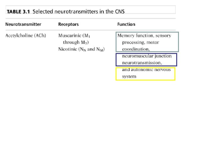
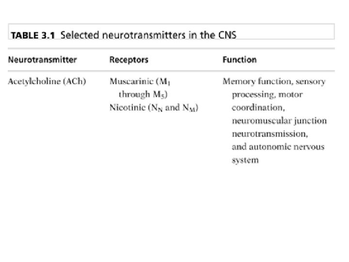
Neurotransmitters and Receptors Acetylcholine—first to be recognized, because of peripheral actions • Synthesis – Acetyl-Co. A (in mitochondria) + choline (from diet)
Published in 1939
Neurotransmitters and Receptors Inactivation: – Acetylcholinesterase (ACh. E) – After action in postsynaptic cleft, ACh. E degrades ACh to choline and acetate, which are taken back up into the neuron.
Neurotransmitters and Receptors Where is ACh produced? • Septal nucleus and nucleus basalis – Projects to forebrain. • Midbrain – Projects to reticular formation, pons, cerebellum, and cranial nerve nuclei. Ach NE Ach
Cholinergic system
Neurotransmitters and Receptors • Receptors – Nicotinic – Muscarinic • ACh. E Inhibitors – Irreversible • Often toxic • Include pesticides and nerves gases – Reversible • Cognitive enhancers • Treating Alzheimer’s
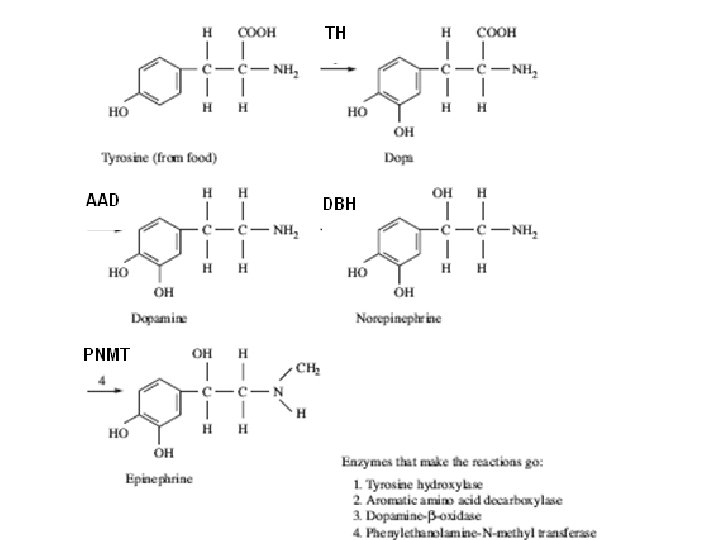
Neurotransmitters and Receptors Catecholamines • Synthesis – Tyrosine • Dopamine – Norepinephrine • Termination – Re-uptake – Monoamine oxidase (MAO)
Neurotransmitters and Receptors • DA Pathways – 3 classic circuits • Hypothalamus to pituitary gland – tuberofundibular; hormonal • Substantia nigra to basal ganglia – nigrostriatal pathway - movement • VTA to cortex and limbic system – mesolimbic – mesocortical – mesolimbicortical
DA Pathways
Neurotransmitters and Receptors: DA
Neurotransmitters and Receptors • Receptors – Dopamine • Two families: D 1 and D 2 • D 1 – D 5