Chapter 3 Macromolecules General Biology I BSC 2010
Pixabay, Public domain")
Chapter 3 Macromolecules General Biology I BSC 2010 Caption: DNA Structure (c)Pixabay, Public domain

Types of Macromolecules • Monomer – small, similar chemical subunits • Polymer – built by linking monomers Monomers Polymers

Types of Macromolecules: large biological molecules 1. Proteins: polymers of amino acids monomers 1. Carbohydrates: polymers of monosaccharides 3. Nucleic acids: polymers of nucleotides 4. Lipids: aren’t built of monomers but form large complexes considered macromolecules

Building Macromolecules Dehydration Synthesis • Formation of large molecules by the removal of water • Monomers are joined to form polymers Monomers Caption: Dehydration Synthesis and Hydrolysis (c) Open. Stax College, Public domain Polymer

Breaking Macromolecules Hydrolysis • Breakdown of large molecules by the addition of water (hydro=water, lysis=break) • Polymers are broken down to monomers Polymer Caption: Dehydration Synthesis and Hydrolysis (c) Open. Stax College, Public domain Monomers

Carbohydrates • Molecules with a 1: 2: 1 ratio of carbon, hydrogen, oxygen • Empirical formula (CH 2 O)n • C-H covalent bonds hold a lot of energy and are quickly broken down – Carbohydrates are good energy storage molecules – Examples: starch, glycogen, glucose
 Monosaccharaides 2) Disaccharides 3) Polysaccharides Know definition, example, functions,")
Carbohydrates Three Major Groups: 1) Monosaccharaides 2) Disaccharides 3) Polysaccharides Know definition, example, functions, and how they form for each. Example: Starch is an energy storage molecule that is formed of glucose molecules linked by covalent bonds through dehydration synthesis.

Monosaccharides • Simplest carbohydrate • 6 carbon sugars play important roles • Glucose C 6 H 12 O 6 Glucose (C 6 H 12 O 6) Caption: Alpha–D-glucose (c)Ben Mills, Public domain

Monosaccharides Examples of Monosaccharides: • Glucose • Fructose is a structural isomer of glucose • Galactose is a stereoisomer of glucose – used for structural needs • Ribose is a chemically different sugar - used for nucleic acids • Enzymes that act on different sugars can distinguish structural and stereoisomers of this basic six-carbon skeleton

• Monosaccharides are classified based on the position of their carbonyl group and the number of carbons in the backbone. • Trioses, pentoses, and hexoses have three, five, and six carbon backbones, respectively. Download for free at http: //cnx. org/contents/185 cbf 87 -c 72 e-48 f 5 -b 5 1 e-f 14 f 21 b 5 eabd@10. 61
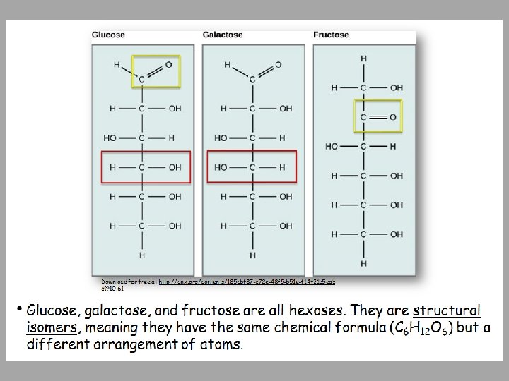

• Five and six carbon monosaccharides exist in equilibrium between linear and ring forms. When the ring forms, the side chain it closes on is locked into an α or β position. • Fructose and ribose also form rings, although they form five-membered rings as opposed to the sixmembered ring of glucose. Download for free at http: //cnx. org/contents/185 cbf 87 -c 72 e-48 f 5 -b 51 e-f 14 f 21 b 5 eabd@10. 61

Disaccharides • 2 monosaccharides linked together by dehydration synthesis • Used for sugar transport or energy storage • Examples: sucrose, lactose, maltose

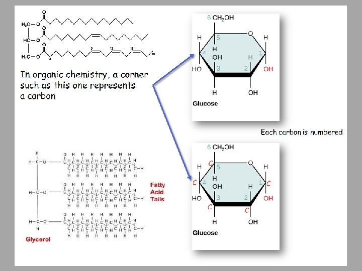
• Sucrose is formed when a monomer of glucose and a monomer of fructose are joined in a dehydration reaction to form a glycosidic bond. In the process, a water molecule is lost. By convention, the carbon atoms in a monosaccharide are numbered from the terminal carbon closest to the carbonyl group. In sucrose, a glycosidic linkage is formed between carbon 1 in glucose and carbon 2 in fructose. Download for free at http: //cnx. org/contents/185 cbf 87 -c 72 e-48 f 5 -b 51 e-f 14 f 21 b 5 eabd@10. 61

Polysaccharides • Long chains of monosaccharides – Linked through dehydration synthesis Four major classes: 1) Starch: energy storage in plants (rice, pasta, potatoes) 2) Glycogen: short term energy storage in animals 3) Cellulose: structural molecule of plants (dietary fiber: this is why fiber is listed under the carbohydrate section of food label). 4) Chitin: structural support in insects, arthropods (exoskeleton), fungal cell walls

Note the parallel array of monomeric sugars note the anti parallel array of monomeric sugars Download for free at http: //cnx. org/contents/185 cbf 87 -c 7 2 e-48 f 5 -b 51 e-f 14 f 21 b 5 eabd@10. 61

Download for free at http: //cnx. org/contents/185 cbf 87 -c 72 e-48 f 5 -b 51 e-f 14 f 21 b 5 eabd@10. 61 Credit: Louise Docker • Insects have a hard outer exoskeleton made of chitin, a type of polysaccharide (like cellulose, but made of glucosamine instead of glucose).

• Carbohydrates: • Monomer – monosaccharides • Covalent bond name – glycosidic linkage

Proteins Protein functions include: 1. Enzyme catalysis 2. Defense 3. Transport 4. Support 5. Motion 6. Regulation 7. Storage
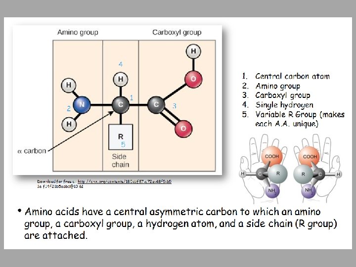

• There are 20 common amino acids commonly found in proteins, each with a different R group (blue box) that determines its chemical nature. • You do not have to memorize all 20 individual amino acids • You need to know that some are: Download for free at http: //cnx. org/contents/185 cbf 87 -c 72 e-48 f 5 -b 51 e-f 14 f 21 b 5 eab d@10. 61 • Nonpolar • Polar uncharged • Polar charged

Download for free at http: //cnx. org/contents/185 cbf 87 -c 72 e-48 f 5 -b 51 e-f 14 f 2 1 b 5 eabd@10. 61 • Peptide bond formation is a dehydration synthesis reaction. The carboxyl group of one amino acid is linked to the amino group of the incoming amino acid. In the process, a molecule of water is released.

Proteins • Proteins are polymers made of Amino Acid monomers • There are 20 amino acids • Combinations of these 20 AA make up long chains from 2 to over 5, 000 or more AA in length • Each chain is a polypeptide • AA link together by a peptide bond to form the long chain polymers of proteins

4 Levels of Protein Structure Primary structure: sequence of amino acids § Amino acids linked by peptide bonds § Millions of different proteins made from the 20 A. A. § A. A. sequence: amino acids given 3 or 1 letter abbreviation Caption: Primary Protein Structure (c) National Human Genome Research Institute, Public domain

Secondary Protein Structure Download for free at http: //cnx. org/contents/185 cbf 87 -c 72 e-48 f 5 -b 51 e-f 14 f 2 1 b 5 eabd@10. 61 • The α-helix and β-pleated sheet are secondary structures of proteins that form because of hydrogen bonding between carbonyl and amino groups in the peptide backbone. Certain amino acids have a propensity to form an α-helix, while others have a propensity to form a β-pleated sheet.

Download for free at http: //cnx. org/contents/185 cbf 87 -c 72 e-48 f 5 -b 51 e-f 14 f 21 b 5 eabd@10. 61 • The tertiary structure of proteins is determined by a variety of chemical interactions. These include hydrophobic interactions, ionic bonding, hydrogen bonding and disulfide linkages. The final level of structure for proteins that consists of only a single polypeptide chain.
 in a protein with 2 or more")
Quaternary structure: arrangement of individual chains (subunits) in a protein with 2 or more polypeptide chains. Caption: Main Protein Structure Levels (c)Ladyof. Hats, Public domain

• The four levels of protein structure can be observed in these illustrations. (credit: modification of work by National Human Genome Research Institute) • Know the names of all four levels Download for free at http: //cnx. org/contents/185 cbf 87 -c 72 e-48 f 5 -b 5 1 e-f 14 f 21 b 5 eabd@10. 61

Additional Structural Characteristics • Domains – Functional units within a larger structure – Often each tertiary structure has its own single domain – Most proteins made of multiple domains that perform different parts of the protein’s function –Example: enzyme’s active site –Example: bind substrate –Example: anchor to membrane, actin, DNA

Chaperones • Chaperone proteins: help proteins fold correctly or keep folded properly • Deficiencies in chaperone proteins implicated in certain diseases – Cystic fibrosis is a hereditary disorder • In some individuals, protein appears to have correct amino acid sequence but fails to fold

Denaturation • Denature: Protein loses structure and function • Due to environmental conditions – p. H – Temperature – Ionic concentration of solution • Sometimes reversible; usually not • Think fried eggs, yogurt

Dissociation • Subunits may be dissociated without losing their individual tertiary structure Hemoglobin Caption: 1904 Hemoglobin (c)Open. Stax College, Public domain
Monomer – amino acids b)Covalent bond – peptide bond")
*Proteins a)Monomer – amino acids b)Covalent bond – peptide bond

Nucleic Acids Polymer: nucleic acids Monomers: nucleotides • Nucleotide: sugar + phosphate + nitrogenous base • Sugar is deoxyribose in DNA or ribose in RNA Caption: DNA Nucleotides (c) Open. Stax College, Public domain

Nitrogeneous bases in Nucleotides • A nucleotide is made up of three components: 1. A nitrogenous base (A, T, G, C, U) 2. A pentose sugar 3. One or more phosphate groups. • Carbon residues in the pentose are numbered 1′ through 5′ (the prime distinguishes these residues from those in the base, which are numbered without using a prime notation). • Bases can be divided into two categories: 1. Purines have a double ring structure 2. Pyrimidines have a single ring.

• Sugars • in Nucleotides Two types of pentose are found in nucleotides, deoxyribose (found in DNA) and ribose (found in RNA). Deoxyribose is similar in structure to ribose, but it has an H instead of an OH at the 2′ position. RNA!!!!!
 Mirmillon, Public domain")
Caption: Nucleotide (c) Mirmillon, Public domain

Download for free at http: //cnx. org/contents/185 cbf 87 -c 72 e-48 f 5 -b 51 e-f 14 f 21 b 5 eabd@10. 61 • In a double stranded DNA molecule, the two strands run antiparallel to one another so that one strand runs 5′ to 3′ and the other 3′ to 5′. • The phosphate backbone is located on the outside, and the bases are in the middle, aligned like stairs stacked on top of one another along a spiral staircase. • Adenine forms hydrogen bonds (or base pairs) with thymine, and guanine base pairs with cytosine.
 • Genes: Encodes information for amino acid sequence of proteins –")
Deoxyribonucleic Acid (DNA) • Genes: Encodes information for amino acid sequence of proteins – Sequence of bases (A, T, C, G) • Double helix • Base-pairing rules • A with T (or U in RNA) • C with G DNA Strand 1: T T A C G G A T C DNA Strand 2: _ _ _ _ _

C T A G T C Caption: The Chemical Structure of a Four Base Pair Fragment of a DNA Double Helix (c)Thomas Shafee, Public domain Nucleotides: sugar + phosphate + nitrogenous base Nitrogenous bases include Purines: adenine and guanine Pyrimidines: thymine, cytosine, uracil (RNA only)
 • RNA similar to DNA except – Contains ribose instead of")
Ribonucleic Acid (RNA) • RNA similar to DNA except – Contains ribose instead of deoxyribose – Contains uracil instead of thymine • Single stranded • RNA plays many roles in the cell • Messenger RNA (m. RNA): DNA encodes the information to make proteins, m. RNA relays this info to the structures that make proteins • DNA is protected from the protein production process • Ribosomal RNA (r. RNA) and Transfer RNA (t. RNA) also needed for protein production

Download for free at http: //cnx. org/contents/185 cbf 87 -c 72 e-48 f 5 -b 51 e-f 14 f 21 b 5 eabd@10. 61 • A ribosome has two parts: a large subunit and a small subunit. • The m. RNA sits in between the two subunits, anchored and oriented by the small subunit. • A t. RNA molecule recognizes a codon on the m. RNA, binds to it by complementary base pairing, and adds the correct amino acid to the growing peptide chain.
Sponk, Public domain")
Caption: Difference in DNA and RNA (c)Sponk, Public domain

Other nucleotides • ATP Adenosine Tri. Phosphate – Primary energy currency of the cell Caption: Figure 06 04 01 – Adenosine Triphosphate (c)CNX Openstax, Public domain adenosine

Other nucleotides • GTP Guanosine Tri. Phosphate – Signal transduction activating molecule • NAD Nicotine Adenine Dinucleotide – Cofactor for oxidation/reduction reactions • FAD Flavin Adenine Dinucleotide – Cofactor for oxidation/reduction reactions

*Nucleic Acids • Monomer – nucleotides (made up of phosphate group, sugar and nitrogeneous base) • Covalent bond linking one nucleotide to another – phosphodiester bond • Remember that hydrogen bonds hold the 2 strands of DNA together

Lipids • Group of nonpolar compounds • hydrophobic • insoluble in water • Not polymers • Examples: Fats, oils, waxes, cholesterol, hormones, some vitamins • Excellent for energy storage • Due to high number of C-H bonds
 Premier, Public domain • Hydrophobic lipids in the blubber of")
Caption: Premier Beluga (c) Premier, Public domain • Hydrophobic lipids in the blubber of aquatic mammals, such as this whale, protect them from the elements.

Lipids: Fats Fatty acids • Saturated: no double bonds between carbon atoms • Higher melting point, usually animal origin • Solid at room temperature • Unsaturated: 1 or more double bonds • Low melting point, usually plant origin • Liquid at room temperature • Trans fats: produced industrially
 Keith Weller, Public domain Caption: Saturated Fatty Acid")
Caption: Cow Female Black White (c) Keith Weller, Public domain Caption: Saturated Fatty Acid (c)Bruce. Blaus, Public domain Saturated: • No double bonds • Solid at room temperature • Animal origin
USDA, Public domain Caption: Alpha-Linolenic-acid-3 D-spacefill (c) Jynto, Public")
Caption: Extra Virgin Olive Oil (c)USDA, Public domain Caption: Alpha-Linolenic-acid-3 D-spacefill (c) Jynto, Public domain Unsaturated: • 1 or more double bonds • Liquid at room temperature due to “kinks” • Plant origin

• Stearic acid is a common saturated fatty acid. (No double bonds between carbon atoms) Download for free at http: //cnx. org/contents/185 cbf 87 -c 72 e-48 f 5 -b 51 e-f 14 f 21 b 5 eabd@10. 61

• Oleic acid is a common unsaturated fatty acid. (One or more double bonds between carbon atoms) Download for free at http: //cnx. org/contents/185 cbf 87 -c 72 e-48 f 5 -b 51 e-f 14 f 21 b 5 eabd@10. 61

Download for free at http: //cnx. org/contents/185 cbf 87 -c 72 e-48 f 5 -b 51 ef 14 f 21 b 5 eabd@10. 61 Credit: Roger Griffith • Waxy coverings on some leaves are made of lipids.

Lipids: Fats • Triglycerides Composed of 1 glycerol and 3 fatty acids Caption: Triglycerides (c)Open. Stax College, Public domain Caption: Trimyristin 3 D (c)Wikimedia Commons, Public domain

• Triacylglycerol is formed by the joining of three fatty acids to a glycerol backbone in a dehydration reaction. Three molecules of water are released in the process. Download for free at http: //cnx. org/contents/185 cbf 87 -c 72 e-48 f 5 -b 51 e-f 14 f 21 b 5 eabd@10. 61

• A phospholipid is a molecule with two fatty acids and a modified phosphate group attached to a glycerol backbone. • The phosphate may be modified by the addition of charged or polar chemical groups (The R group). • Four chemical groups that may modify the phosphate (choline, ethanolamine, serine, and inositol), attach to the phosphate group at the position labeled R via the hydroxyl group indicated in green. Download for free at http: //cnx. org/contents/185 cbf 87 -c 72 e-48 f 5 -b 51 e -f 14 f 21 b 5 eabd@10. 61

Download for free at http: //cnx. org/contents/185 cbf 87 -c 72 e-48 f 5 -b 51 e-f 14 f 21 b 5 eabd@10. 61 • The phospholipid bilayer is the major component of all cellular membranes. The hydrophilic head groups of the phospholipids face the aqueous solution. The hydrophobic tails are sequestered in the middle of the bilayer. This provides a continuous molecular interface separating the two aqueous regions so any molecule can contact the membrane (necessary to transport).

• Steroids such as cholesterol and cortisol are composed of four fused hydrocarbon rings. *Cholesterol is mainly synthesized in the liver and is the precursor to many steroid hormones such as testosterone, cortisol, and estradiol *Cholesterol is also the precursor for digestive detergents called bile salts *Cholesterol is precursor to vitamin D and important in plasma membranes Download for free at http: //cnx. org/contents/185 cbf 87 -c 72 e-48 f 5 -b 51 e-f 14 f 21 b 5 eabd@10. 61
- Slides: 60