Chapter 3 Lecture Outline Cells Copyright The Mc


 Stacks of flattened discs or")


")


- Slides: 53
Chapter 3 Lecture Outline Cells Copyright © The Mc. Graw-Hill Companies, Inc. Permission required for reproduction or display.
Outline v History of Cell Theory v Modern Microscopes v Eukaryotic and Prokaryotic Cells v Cell Structure and Communication v Cell Components v Cellular Reproduction v Higher Plant Cells Versus Animal Cells
History of Cell Theory v Cell Theory - living organisms are composed of cells - cells form a unifying structural basis of organization. - all cells come from previously existing cells v v 1665 - Cells discovered by Robert Hooke. 1838 - Matthias Schleiden and Theodor Schwann – developed cell theory 1858 - Rudolf Virchow argued there is no spontaneous generation of cells. Louis Pasteur experimentally disproved spontaneous generation in 1862.
Modern Microscopes v Light Microscopes - Increase magnification as light passes through a series of transparent lenses made of glass or calcium fluoride crystals • Compound Microscopes – Light passes through thinly sliced material – In general can distinguish organelles 2 micrometers or larger in diameter – Can magnify up to 1500 x • Dissecting Microscopes (Stereomicroscopes) – Allow three-dimensional viewing of opaque objects – Can magnify up to 30 x
Modern Microscopes v Electron Microscopes • Use a beam of electrons produced when high- voltage electricity is passed through a wire • Transmission Electron Microscopes – Up to 200, 000 x magnification, but material must be sliced extremely thin • Scanning Electron Microscope – Up to 10, 000 x magnification – Surface detail can be observed on thick objects.
Modern Microscopes • Scanning Tunneling Microscope – Uses a probe that tunnels electrons upon a sample – Produces a map of sample surface – Even atoms can become discernible – First picture of DNA segment showing helical structure
Eukaryotic versus Prokaryotic Cells v Prokaryotic - Cells lack a nucleus. • Bacteria v Eukaryotic - Cells contain a nucleus. • Plants and animals • Cell walls - Rigid boundary of cells • Organelles - Membrane-bound bodies found within eukaryotic cells
Cell Structure and Communication v v Cell Wall surrounds protoplasm. Protoplasm consists of all living cell components. • Bound by plasma membrane – Cytoplasm consists of all cellular components between the plasma membrane and the nucleus. o Cytosol - Fluid within cytoplasm containing organelles o Organelles - Persistent structures of various shapes and sizes with specialized functions « Most, but not all, are bound by membranes.
Cell Structure and Communication
Cell Structure and Communication Cell Size v Cells of higher plants generally vary in length between 10 and 100 micrometers. v Plasma membrane surface area – squared increase v Interior Volume – cubed increase • Smaller cells have relatively large surface to volume ratios enabling faster and more efficient cellular communication.
Cell Structure and Communication Cell Wall v Main structural component of cell walls is cellulose (long chains of glucose monomers). • Also contain matrix of: v – Hemicellulose - Holds cellulose fibrils together – Pectin - Gives stiffness (like in fruit jellies) – Glycoproteins - Proteins with associated sugars Middle lamella first produced when new cell walls are formed. • Shared by two adjacent cells
Cell Structure and Communication Cell Wall v v Flexible primary walls laid down on either side of middle lamella. Secondary walls produced inside primary walls. • Derived from primary walls by thickening and inclusion of lignin • Cellulose microfibrils embedded in lignin for strength. Secondary cell wall structure
Cell Structure and Communication Between Cells v Fluids and dissolved substances can pass through primary walls of adjacent cells via plasmodesmata. • Plasmodesmata are cytoplasmic strands that extend between cells through minute openings. Two adjacent cells connected by plasmodesmata
Cell Components Plasma Membrane v Plasma Membrane is the semipermeable outer boundary of the living part of the cell. • Regulates movement of substances into and out of cell • Composed of phospholipids arranged in two layers, with proteins interspersed throughout – Fluid mosaic model - Plasma membrane is dynamic structure
Cell Components Nucleus v Nucleus is control center of cell and contains DNA. • Sends coded messages from DNA to be used in other parts of the cell • Bound by two membranes, which together constitute the nuclear envelope • Structurally complex pores occupy up to one-third of the total surface area. – Permit only certain kinds of molecules to pass between nucleus and cytoplasm
Cell Components Nucleus v Contains fluid nucleoplasm in which are: • Nucleoli - Composed primarily of RNA • Chromatin Strands – Composed of DNA and proteins – Coil and become chromosomes
Cell Components Endoplasmic Reticulum v Endoplasmic reticulum is enclosed space consisting of a network of flattened sacs and tubes forming channels throughout the cytoplasm. • Facilitates cellular communication and channeling of materials • Synthesizes membranes for other organelles and modifies proteins
Cell Components Endoplasmic Reticulum v Rough ER - Ribosomes distributed on outer surface of ER. • Associated with protein synthesis and storage v Smooth ER - Devoid of ribosomes and associated with lipid secretion
Cell Components Ribosomes v Ribosomes - consist of two subunits that are composed of RNA and proteins • Link amino acids to construct complex proteins • Subunits assembled in nucleolus. • May occur on outside of rough ER, or in cytoplasm, chloroplasts or other organelles • No bounding membranes
Cell Components Dictyosomes v Dictyosomes (Golgi bodies in animals) Stacks of flattened discs or vesicles Dictyosome
Cell Components Dictyosomes v Dictyosomes function: • To modify carbohydrates attached to proteins that are synthesized and packaged in the ER. • To assemble polysaccharides and collect them in small vesicles. – Vesicles pinched off from margins of dictyosomes. – Vesicles migrate to plasma membrane, fuse with it, and secrete contents to the outside of cell. o Contents may include cell wall polysaccharides, floral nectars, and essential oils in herbs.
Cell Components Plastids v Chloroplasts are the most conspicuous plastids. • Bound by double membrane and contain: – Grana made up of thylakoids o Thylakoid membranes contain chlorophyll. o First steps of photosynthesis occur in thylakoid membranes. – Stroma - Matrix of enzymes involved in photosynthesis – Small circular DNA molecule o Encodes for production of certain proteins for photosynthesis
Cell Components Plastids
Cell Components v Other types of plastids include: Plastids • Chromoplasts – Synthesize and accumulate carotenoids (yellow, orange, red) which color fruits – oilsoluble • Leucoplasts – Colorless – May synthesize starches (amyloplasts) – Or oils (elaioplasts) Chromoplasts in red pepper cells
Cell Components Mitochondria v Mitochondria release energy produced from cellular respiration. • Bound by two membranes – Inward membrane forms numerous folds = cristae. o Increase surface area available to enzymes in matrix • Matrix also includes DNA and RNA.
Cell Components Microbodies v Microbodies are small, spherical bodies distributed throughout the cytoplasm that contain specialized enzymes. • Bound by a single membrane • Peroxisomes - Serve in photorespiration • Glyoxisomes - Aid in conversion of fat to carbohydrates
Cell Components Vacuoles v In mature cells, 90% of volume may be taken up by central vacuoles. • Bounded by vacuolar membranes, tonoplasts • Filled with watery fluid called cell- sap – Contains dissolved substances, such as salts, sugars, organic acids and small proteins – Also frequently contains water-soluble pigments called anthocyanins (red, blue, purple) – flower coloration – Functions: maintenance of cell pressure and p. H, storage of numerous cell metabolites and waste products. Inside the vacuole is a watery fluid called cell sap, which is slightly to moderately acidic.
Cell Components Cytoskeleton v Cytoskeleton - Involved in movement within cell and in cell’s architecture • Network of microtubules and microfilaments • Microtubules: – Control addition of cellulose to cell wall – Involved in movement of flagella and cilia – Found in fibers of spindles and phragmoplasts in dividing cells – Are thin, hollow, tubelike and composed of tubulins (proteins) • Microfilaments - Role in cytoplasmic streaming
Cellular Reproduction Cell Cycle v Cell cycle - Orderly series of events when cells divide • Divided into interphase and mitosis v Interphase • Occupies up to 90% of cell cycle • Period when cells are not dividing – G 1 - Cell increases in size. – S - DNA replication takes place. – G 2 - Mitochondria and other organelles divide, and microtubules are produced.
Interphase
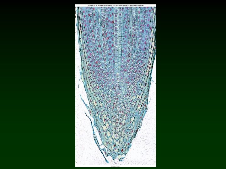
Cellular Reproduction Mitosis v Mitosis refers to the process of cellular division. • Produces two daughter cells with equal amounts of DNA and other substances duplicated during interphase • Each daughter cell is an exact copy of the parent cell. • Mitosis occurs in meristems. • Although mitosis is a continuous process, it is divided into 4 phases: prophase, metaphase, anaphase and telophase.
Cellular Reproduction Mitosis v Prophase • Chromosomes condense by coiling and tightening to become shorter and thicker. – Chromosomes made of two identical chromatids held together by centromeres. o Kinetochore (protein complex) is located on the outer surface of each centromere. o Spindle fibers (microtubules) become attached to the kinetochore and anchored to two poles of the cell. • Nuclear envelope dissociates and nucleolus disintegrates
Cellular Reproduction Mitosis v Metaphase Prophase • Chromosomes align between the poles around the circumference of the spindle at the cell’s equator. – Spindle fibers collectively referred to as the spindle. – At the end of metaphase, centromeres holding each sister chromatid separate lengthwise. Metaphase
Cellular Reproduction Mitosis v Anaphase • Sister chromatids separate in unison and are pulled to opposite poles, with centromeres leading the way. – – Spindle fibers gradually shorten as material is continuously removed from the polar ends. Chromatids after separation are called daughter chromosomes. Anaphase
Cellular Reproduction Mitosis v Telophase • Each group of daughter chromosomes become surrounded by a nuclear envelope. • Daughter chromosomes become longer and thinner and eventually, indistinguishable. Telophase • Nucleoli reappear. • Spindle fibers disintegrate. • Phragmoplast and cell plate form at equator.
Cellular Reproduction Mitosis v Cell Plate Formation: • Phragmoplast develops between daughter cell nuclei. – Phragmoplast is a complex of microtubules and ER. • Microtubules trap dictyosome- derived vesicles. • Vesicles fuse to form cell plate. – Cell plate grows outward toward mother cell walls. Cell plate formation • Portions of ER are trapped between vesicles, forming plasmodesmata.
Copyright © The Mc. Graw-Hill Companies, Inc. Permission required for reproduction or display. Prophase 6. 2 µm Figure 5. 5 cell wall Chromosomes © R. Calentine/Visuals Unlimited
Copyright © The Mc. Graw-Hill Companies, Inc. Permission required for reproduction or display. Metaphase Figure 5. 5 spindle fibers © R. Calentine/Visuals Unlimited 6. 2 µm
Copyright © The Mc. Graw-Hill Companies, Inc. Permission required for reproduction or display. Anaphase 6. 2 µm Figure 5. 5 © R. Calentine/Visuals Unlimited
Copyright © The Mc. Graw-Hill Companies, Inc. Permission required for reproduction or display. Telophase cell plate Figure 5. 5 © Jack M. Bostrack/Visuals Unlimited 25 µm
Copyright © The Mc. Graw-Hill Companies, Inc. Permission required for reproduction or display. MITOSIS Prophase Metaphase 6. 2 µm cell wall Chromosomes 6. 2 µm spindle fibers Anaphase Telophase 6. 2 µm 25 µm cell plate (prophase, metaphase, anaphase): © R. Calentine/Visuals Unlimited; (telophase): © Jack M. Bostrack/Visuals Unlimited Figure 5. 5
Prophase
Metaphase
Anaphase
Telophase
Prophase
Metaphase
Anaphase
Telophase
Daughter Cells (interphase)
Higher Plant Cells Versus Animal Cells v Plants: • Cell walls • Cell plate and plasmodesmata • Plastids and vacuoles v Animals: • Internal or external skeletons; no cell walls • Plasma membrane called cell membrane. • Divide by pinching in two; no cell plate nor plasmodesmata • Centrioles present during cell division. • No plastids nor vacuoles
Review v History of Cell Theory v Modern Microscopes v Eukaryotic and Prokaryotic Cells v Cell Structure and Communication v Cell Components v Cellular Reproduction v Higher Plant Cells Versus Animal Cells