Chapter 3 Connective Tissue Proper Department of Histology


 Function Fibril-forming")


 Proteoglycans")
 F. B.")

 in the electron microscope E=elastin; C, collagen fibrils M/L=microfibrils")

 EM: fibrils w/ periodic cross-bandings at intervals of")

 and collagen (right) fibers. Note")

 1. 2. 3. Glycosaminoglycans (GAG) • linear")


: produced by epithelial cells and fibroblasts play a role in events")
 2. 3. 4. 5. Free (transient")

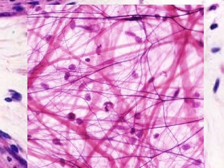

 shows several fibroblasts (F), which are elongated cells. H&E")


. L N")


 connective tissue – delicate, vascularized, cellular; supports")


 consists of a small population of fibroblasts and lots of")
 Wall of large artery: ①Elastic membrane")
 HE: /")
 Adipose Tissue Present in newborns, e. g. hips, back, (and hibernating 冬眠")


- Slides: 86
Chapter 3 Connective Tissue Proper 霍冠华 滨州医学院组织胚胎学教研室 Department of Histology & Embryology Binzhou Medical University September, 2013
Connective Tissue v v The different types of connective tissues are responsible for providing and maintaining form in the body. Functioning mechanically, they provide a matrix that connects and binds the cells and organs and ultimately gives support to the body. Structurally, connective tissue is formed by three classes of components: cells, fibers, and ground substance. The major constituent of connective tissue is the extracellular matrix. Extracellular matrices consist of different combinations of protein fibers (collagen, reticular, and elastic) and ground substance. Connective tissue includes connective tissue proper and specialized connective tissue.
CONNECTIVE TISSUE PROPER
一、CHARACTERISTICS: v v v Fewer cells Large amount of intercellular matrix Cell: separated and no polarity Intercellular matrix: fibers + ground substance + tissue fluid Filled with blood vessels, Lymphatic vessels and nerves Function: support, connect, nourish, defence, and repair etc. Architectural framework of the body Bind together and provide mechanical support for other tissue (metabolic, defense, transport, storage) Wound repair / inflammatory response
AF - Active Fibroblast IF - Inactive Fibroblast C - Collagen EF - Elastic Fibers
二、CLASSIFICATION v Connective tissue proper: v v Loose C. T. Dense C. T. Adipose tissue Reticular tissue Cartilage and bone Blood and Lymph Broadly speaking
Connective Tissue Epithelium Connective Tissue papilla ridge Connective Tisue Muscle
Connective Tissue v Extracellular Matrix Fibers – collagen, elastic, reticular v Ground substance Cells
Fibers in Connective Tissue v v v Collagen Fibers most abundant protein in human body (up to 30% dry weight) multiple types: fibril-forming or fibril-associated (in skin, tendon, cartilage, bone, dentin, blood vessels); cross-linked networks (in all basement membranes) Reticular Fibers – specialized type of collagen (Type III; reticulin) associated with smooth muscle in organs subjected to changes in volume, forms the stroma in lymphatic and hematopoietic organs Elastic Fibers –thin fibers or fenestrated sheets composed of various glycoproteins, including the protein elastin, providing elastic properties to tissues that experience repeated deformation (in skin, blood vessels, lung, bladder)
Major Collagen Fiber Types (out of Collagen Type Tissues at least 20) Function Fibril-forming collagens (these are visible) I (most abundant) Skin, tendon, bone, dentin Resistance to tension II Cartilage, vitreous of eye Resistance to pressure III (reticulin) Skin, muscle, blood vessels, liver, etc. Structural framework and stability All basement membranes Support and filtration Network-forming collagens IV Fibril-associated collagens with interrupted triple helices (FACIT) VI, IX Assoc. w/ type I and II fibrils Fibril-fibril / fibril-ECM binding Epithelia Epidermis to basal lamina Anchoring filament collagens VII
Collagen fibers viewed by light microscopy H&E Trichrome
Collagen Fibers vs. Fibrils H&E fibers fibrils
Assembly of collagen fiber bundles
① Fibroblast Synthesize: In r. ER / GL: → Polypeptide αchains ① → hydroxylated αchains ② → Procollagen ③ In Extracellular space: → Tropocollagen ④ → fibrils ⑤ → collagenous f. ⑥ ② ③ ④ 280 x 1. 4 nm ⑤ ⑥
Clinical disorders resulting from defects in collagen synthesis Type Disease Symptoms I Osteogenesis imperfecta Spontaneous fractures, progressive hearing loss, cardiac insufficiency III Ehlers-Danlos (type IV) Hypermobility of digits, early morbidity/mortality from rupture of aorta or intestine multiple Scurvy (lack of vit. C, Ulceration of gums, hemorrhages a cofactor for prolyl and lysyl hydroxylase)
Ehlers-Danlos Syndromes • A series of genetic diseases with faulty assembly of collagens (lysyl hydroxylase deficiency). • Hyperextensible skin and hypermobile joints • In some forms (e. g. , type IV), weakness in blood vessels or intestines are life threatening.
Noncollagen Components of the Extracellular Matrix v Elastin v “Ground substance” Glycosaminoglycans (GAG’s) Proteoglycans Multiadhesive matrix proteins vlaminin vfibronectin
2、Elastic fibers Structure: LM: EM: elastin + microfibrils (no bandings) F. B.
Elastic Fibers LM: Visualized by selectively staining with Weigert’s, resorcin-fuchsin, or aldehyde-fuchsin EM: Consist of amorphous core of elastin surrounded by microfibrillar glycoprotein, fibrillin (8 -10 nm). Elastin: is rich in glycine and proline, but it contains little or no hydroxyproline and hydroxylysine. uniquely contains desmosine and isodesmosine, which are thought to cross-link the molecules into a network of randomly coiled chains. This cross-linking is responsible for its rubber-like properties. Confers elasticity: present in large amounts in ligaments, lung, skin, bladder, and walls of blood vessels. Marfan Syndrome: defect in elastic fiber synthesis; reduced elasticity in skin and lungs, skeletal defects (bones are longer and thinner than usual), cardiovascular complications (aneurism, valve prolapse)
Elastin appears amorphous (not fibrillar) in the electron microscope E=elastin; C, collagen fibrils M/L=microfibrils of fibrillin, a scaffolding glycoprotein involved elastin deposition Marfan Syndrome: defect in fibrillin gene, results in weakened elastic fibers
Elastin molecules are joined by covalent bonds to generate an extensive cross-linked network. Because each elastin molecule in the network can expand contract like a random coil, the entire network can stretch and recoil like a rubber band.
3、Reticular fibers Sructure: LM: argyrophilia, PAS(+) EM: fibrils w/ periodic cross-bandings at intervals of 64 nm, type. III collagen covered carbohydrate
Reticular fibers in liver Reticular fibers in lymph node
v Electron micrograph of cross sections of reticular (left) and collagen (right) fibers. Note that each fiber type is composed of numerous smaller collagen fibrils. Reticular fibrils (R) are significantly narrower in diameter than collagen fibrils of collagen fibers (C; see histogram inset); in addition, the constituent fibrils of the reticular fibers reveal an abundant surfaceassociated granularity not present on regular collagen fibrils (right). x 70, 000.
Ground Substance
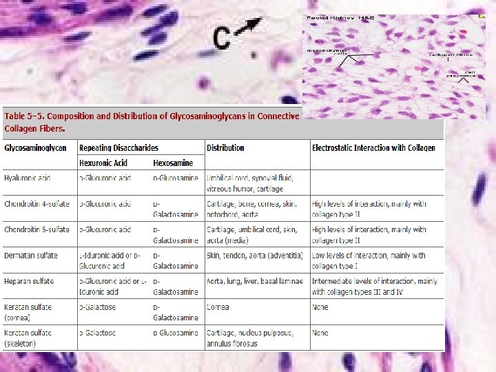
Ground Substance of the Extracellular Matrix (ECM) 1. 2. 3. Glycosaminoglycans (GAG) • linear (unbranched) polysaccharides, e. g. heparan sulfate, condroitin sulfate, keratan sulfate, hyaluronic acid • very hydrophilic due to abundant negative charges (e. g. SO 4 groups). • except for hyaluronic acid, are usually bound covalently to protein core as part of a proteoglycan Proteoglycans • core protein + GAG side chains (like a bottle brush) • bind cells, other proteins, and/or ECM components Multiadhesive glycoproteins • small glycosylated proteins containing NUMEROUS binding sites to cells, signaling molecules, and other ECM components • e. g. fibronectin and laminin: important for adhesion of epithelial cells to the basal lamina via transmembrane integrin receptors.
Ground substance Ø Jelly-like & amorphous substance; Ø Proteoglycan: in molecular sieve hyaluronic acid GAG(glycosaminoglycan) chondroitin sulfate keratin sulfate etc. protein: core protein & link protein glycoprotein: fibronectin (FN) laminin (LN) chondronectin (Ch. N);
Side chain subnit hyaluronic acid Collagenous fiber Ground substance Intercellular material
hyaluronic acid molecular sieve: hyaluronic acid -- link pr. -- side chain subunit: core protein + chondroitin sulfate & keratin sulfate tissue fluid: flowing through the sieve pores side chain subunit Core pr. link pr. chondroitin sulfate Link pr. keratin sulfate core pr.
glycoprotein: fibronectin (FN): produced by epithelial cells and fibroblasts play a role in events of identification, adhesion, migration and proliferation v laminin (LN) : located in B. M. , produced by epi. cells, function: adhesion the epi. cells and B. M. v chondronectin (Ch. N): in cartilage tissue, fuction: a component of ground substances; adhesion chondrocyte. S and colagen type. II v
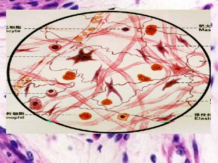
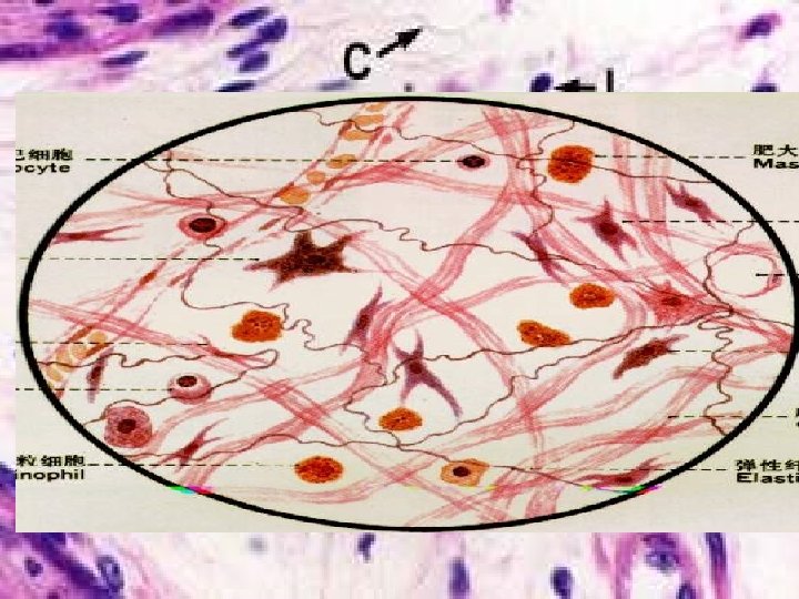
Cells in Connective Tissue 1. Fixed (permanent residents) 2. 3. 4. 5. Free (transient residents) • • • Fibroblasts Adipose (fat) cells Macrophages** Mast cells** undifferentiated mesenchymal cells Lymphocytes Plasma Cells (differentiated B-cells) ** Leukocytes** (specifically, neutrophils, eosinophils, & basophils) ** derived from hematopoietic stem cells and involved in immune function and inflammation
Connective Cell Lineages
1. Fibroblasts are the most common cells in connective tissue v Synthesize & secrete collagenous pro. & elastic pro. (to form collagenous f. , elastic f. & reticular f. ); and ground substance (proteoglycan & glycoprotein). v Active and quiescent stages (when quiescent sometimes called fibrocytes or mature fibroblasts). v Synthesize growth factors. v Rarely undergo cell division unless tissue is injured, which activates the quiescent cells. v Play a major role in the process of wound healing and respond to an injury by proliferating and enhanced fiber formation.
A connective tissue layer (dermis) shows several fibroblasts (F), which are elongated cells. H&E stain. v Structurally, fibroblasts are of 2 types, one of which resembles mesenchymal cells. This type is stellate, with long cytoplasmic processes and a large, ovoid, pale-staining nucleus. The cytoplasm contains abundant RER and Golgi complex, and this type is important in the production of collagen and other matrix components. Also, lysosomes and vacuoles of secretion product are prominent features of the cells. Fibroblast secrets collagen, fibronectin, glycoproteins, and proteoglycans. The cells have many actin-containing microfilaments because they are highly motile cells. They also contain numerous microtubules, which probably help maintain the cell morphology.
v Electron micrograph revealing portions of several flattened fibroblasts in dense connective tissue. Abundant mitochondria, rough endoplasmic reticulum, and vesicles distinguish these cells from the less active fibrocytes. Multiple strata of collagen fibrils (C) lie among the fibroblasts. x 30, 000.
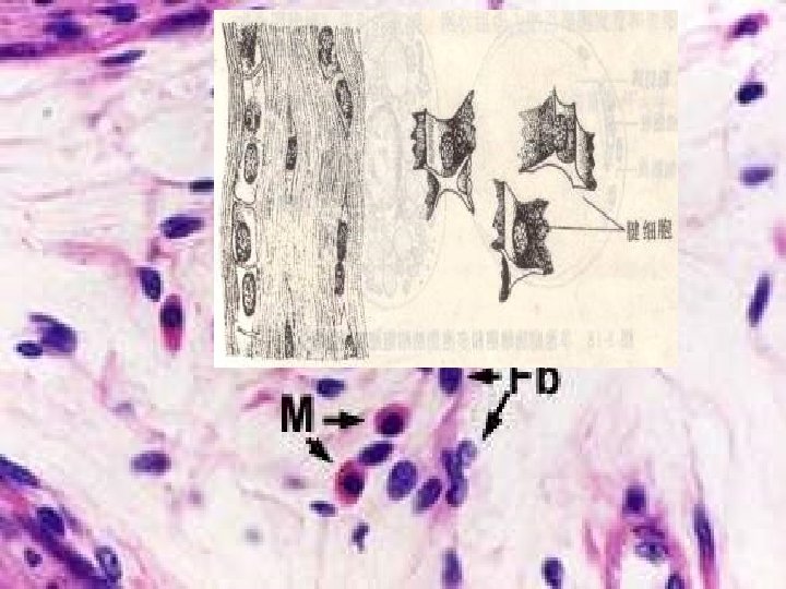
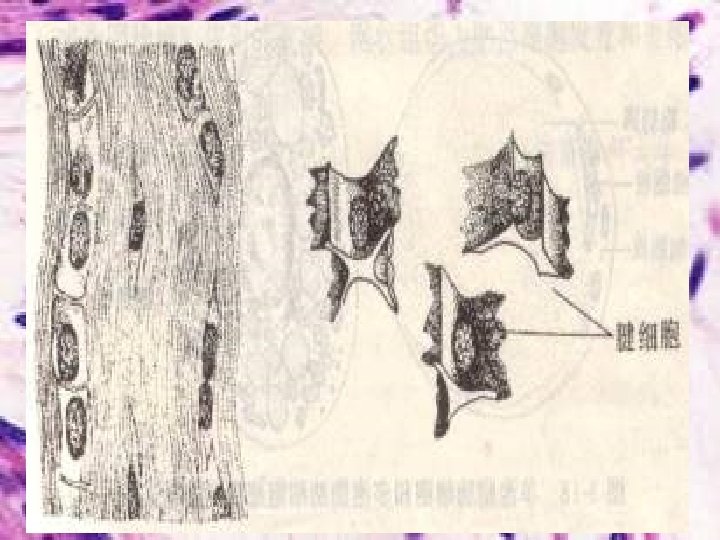
v Cells of the second type are less active and are sometimes termed fibrocytes, because they are believed to be more mature. They are smaller and spindleshaped, with a dark, elongate nucleus and fewer cytoplasmic organelles. They can revert to the fibroblast state and participate in tissue repair.
External morphologic characteristics and ultrastructure of each cell are shown. Fibroblasts that are actively engaged in synthesis are richer in mitochondria, lipid droplets, Golgi complex, and rough endoplasmic reticulum than are quiescent fibroblasts (fibrocytes). Active quiescent
Quiescent fibroblasts are elongated cells with thin cytoplasmic extensions and condensed chromatin. Pararosaniline-toluidine blue (PT) stain.
2. Macrophages LM: Large, stellate, cytoplasm is acidophilic Cytoplasmic granules Vacuoles
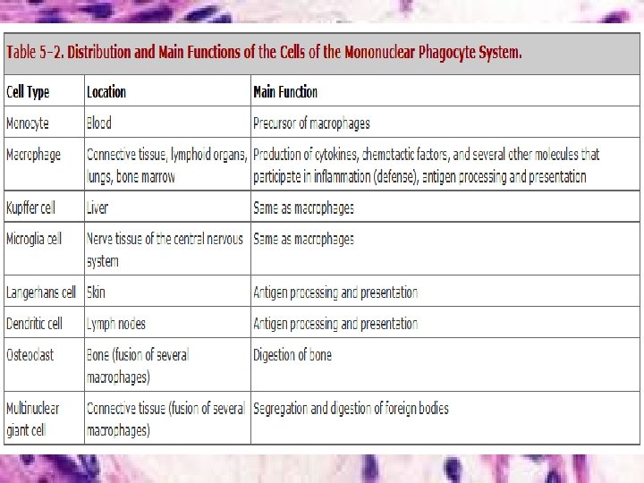
Macrophages v v v Derived from monocytes EM: a large number of Ly. , pinosome & phagosomes Well-developed Golgi complex bundles of microtubules and microfilaments Mononuclear phagocyte system: monocytes, macrophages, Kupffer cells (liver), osteoclasts (bone), dust cells (lung), Langerhans cells (skin), microglial cells (central nervous system)
Ultrastructural features of a Macrophage secondary lysosomes phagocytic vacuoles
Function: Mobility chemotaxis chemotactic factor: complement C 5 a, bacterial products etc. Ø Phagocytize specific: depend on identify factors: Ab, C, Fibronectin etc (their receptors are on surface of the macrophge). non-specific: independently Ø indigestible remnant adsorb phagosome fuse
v v v Functions: Their role is to phagocytose, or engulf and then digest, cellular debris and pathogens, either as stationary or as mobile cells. They are specialized phagocytic cells that attack foreign substances, infectious microbes and cancer cells through destruction and ingestion. They participate in the immune response by presenting phagocytosed antigens to lymphocytes, and also stimulate lymphocytes and other immune cells to respond to pathogens. Secret bioactivators (bioactive substances ): lysozymes kill bacteria, complement participating in process of inflammation Cytokines: IL-1, Interferon, hematopoietic cell colony stimulating factor, tumour necrosis factor (TNF) etc. Identified by specific expression of a number of proteins including CD 14, CD 40, CD 11 b, CD 64, F 4/80 (mice)/EMR 1 (human), lysozyme M, MAC-1/MAC-3 and CD 68
antigens ingest phagocytic vacuole Primary lysosomes secondary lysosome MHC-II moleculesantigen complex MHC: major Histo-compatibility complex molecules type II sets specially at surface of some immune cells, type I occurs in all types of cells. Macrophage functions as a antigen presenting cell
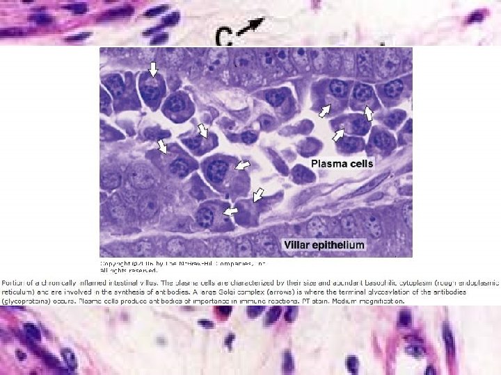
3. Plasma Cells, the mature B lymphocytes that constitutively secrete antibodies chromatin Two more plasma cells (P) are shown in this field. One shows the cartwheel nucleus well. The cytoplasm is basophilic. Also, note three macrophages which are detected by their content of ingested black deposits. Perhaps this came from a lymph node near the lung (of a smoker). The cell labeled is an Eosinophil.
v The plasma cells are characterized by their size and abundant basophilic cytoplasm (rough endoplasmic reticulum) and are involved in the synthesis of antibodies. A large Golgi complex (arrows) is where the terminal glycosylation of the antibodies (glycoproteins) occurs. Plasma cells produce antibodies of importance in immune reacions.
Plasma cells § Structure: Nu. : eccentrically,heterochromatin in wheel shape Cytoplasm: strong basophilic w/ lightlystained area near Nu.
Plasma Cells EM:rich in r. ER & ribosomes, developed Golgi complex § Source: B lmphocytes § Function: Ø synthesize antibody & cytokines Ø participate in humoral immunity
Golgi complex RER
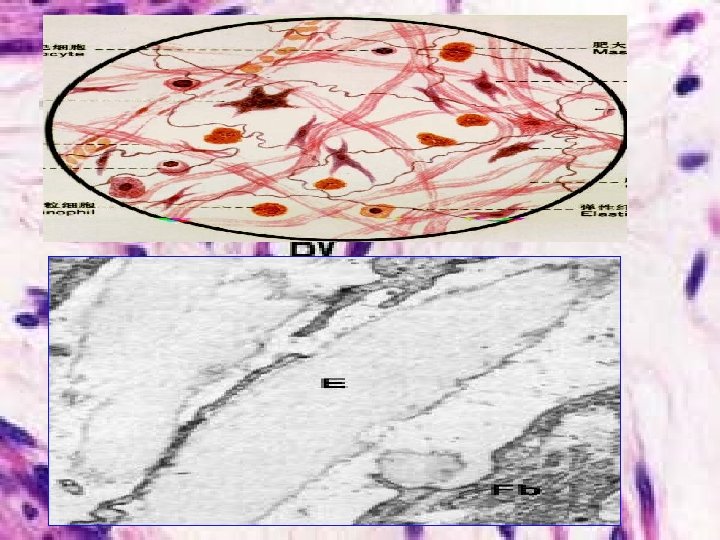
4. Mast cell v v The thin strands: Elastic fibers The thick strands: Collagen fibers The small dark purple structures: Nuclei of fibroblasts The large granular-looking cell: Mast cell
Basophilic granules Metachromasia – when stained with toluidine blue, the granules bind the dye and change its color to red.
EM: a great number of granules w/ crystals, containing: heparin, histamine, leukotriene, slow-reacting substance, eosinophil chemotactic factors (ECF-A)
degranulation
Functions v v v v Granules: degranulation Heparin: is an anticoagulant, anti coagulation Histamine: causes vasoconstriction and increased permeability of small venules eosinophil chemotactic factor: attract eosinophils migration to the allergic reaction site Cells secret Luekotriene: cytoplasm, likes histamine Principal function is storage in secretory granules and REGULATED release (degranulation) of histamine and other vasoactive mediators of inflammation. Responsible for the immediate hypersensitivity response characteristic of allergies, asthma[‘æzmə] and anaphylactic shock.
v Mast cells are filled with conspicuous large granules containing Heparin and Histamine, mast cells can "degranulate" - often in response to injury - and release Heparin, which slows blood clotting, and Histamine, which increases capillary permeability. In this manner, mast cells participate in the inflammatory response; they promote the flow of blood out of the bloodstream and into tissue spaces, where blood cells and antibodies can fight infection.
5. Adipocytes, predominate in adipose tissue Very active cells with many functions: • Triglyceride storage and glucose metabolism (insulin and glucagon receptors) • Secretion of many bioactive molecules: leptin (regulates satiety); angiotensinogen (blood pressure); steroids (glucocorticoids & sex hormones); growth factors (e. g. insulin-like growth factor, tumor necrosis factor ); cytokines (e. g. interleukin-6)
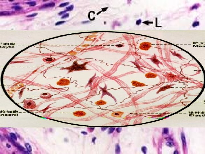
6. Leucocytes
Leucocytes v Neutrophils: Small cells with multi-lobed, heterochromatic nuclei (or “polymorphonuclear neutrophils”). L N
7. Undifferentiated mesenchyme cells
Tissue fluid dynamic balance
Function: Tissue fluid nourishes the cells & tissues; v Molecular sieve acts as a barrier: to prevent the spread of bacteria & other microorganisms *haemolytic streptococci produce hyaluronidase & promote the invasion § Glycoproteins (FN/LN/chondronectin): identification/ adherence/ migration/ proliferation etc. v
v Edema: swelling from excessive accumulation of serous fluid in tissue. Tends to appear first where tissue turgor is normally lowest. E. g. chronic nephritis, nephrotic syndrome, hepatitis, heart failure, cancer. Eyelid edema: common in nephritis, chronic liver disease, malnutrition, anemia, blood vessels, such as edema. Edema of lower limb. v v v Dehydration: to lose too much water from body; to make a person's body lose too much water. E. g. got diarrhoea(diarrhea ) and is suffering from dehydration; excessive sweating; burn The eyes are often retracted from dehydration. The major factors causing death are electrolyte imbalance and dehydration, leading to cardiac arrest.
Types of Connective Tissue Proper Loose (areolar) connective tissue – delicate, vascularized, cellular; supports the epithelia of the major organs and glands and fills the space between muscle tissue. - not very resistant to stress Dense connective tissue (many more fibers than cells) Dense irregular: meshwork of coarse fibers; dermis of skin, organ capsules, fascia - resists multi-directional forces Dense regular: vcollagenous: fibers aligned in defined pattern; tendons, ligaments, etc. - resists linear mechanical stresses velastic: elastin and microfibrils (fibrillin) - elasticity Adipose - fat storage, glucose regulation, satiety Reticular - argyrophilic fibers of type III collagen - forms stroma of highly cellular organs (e. g. liver, lymph nodes, spleen)
LOOSE CONNECTIVE TISSUE I. Characteristics: v v v A small amount of cells & many kinds of cells small number of fibers & great amount of ground substance The cells have no polarity Sponge-like structure (areolar tissue) Distributed widely between cells, tissues and organs. Function: support, connect, defence, and repair etc.
Loose connective tissue: delicate, vascularized, flexible; facilitates transport of cells and materials (secretion, absorption, immunity) a n i lam ia r p ro p small intestine lamina propria mammary gland intralobular connective tissue
Dense Irregular CT Densely packed collagen fibers, often in perpendicular bundles; resists tension in many directions and provides mechanical support. Collagen Fibroblast nucleus Skin dermis, H &E
Dense Irregular CT from human dermis contains thick bundles of collagen fibers, fibroblast nuclei (arrow) , and a few small blood vessels (bv). H&E stain.
Dermis of Skin has both Loose and Dense Irregular CT Loose CT sweat gland H&E
Dense Regular CT (collagenous) consists of a small population of fibroblasts and lots of collagenous fibers
Dense Regular CT (elastic) Wall of large artery: ①Elastic membrane
Adipose Tissue Structure: Loose C. T. + fat cells (in large aggregations) HE: / Osmic acid: Function: energy storage, shock absorber, insulating layer Unilocular adipose tissue.
Brown (Multilocular) Adipose Tissue Present in newborns, e. g. hips, back, (and hibernating 冬眠 mammals) and involved in thermoregulation Mitochondria of brown fat cells express uncoupling protein which “short circuits” the electron transport chain producing HEAT rather than ATP.
RETICULAR TISSUE Structure: § Reticular cells + reticular f. + ground substance v Reticular cell: stellate, pale nucleus, obvious nucleoli, processes (rich in RER) Function: architectural framework of lymphatic & hematopoietic tissues cells Lymph node fibers
macrophage reticular fiber reticular cell lymphocyte reticular cell: cytoplasmic processes, cross-linked networks; in rich in cytoplasm, more RER Function: produces reticular fibers and ground substances.