Chapter 3 Connective tissue proper Content n Characteristics


 comprises a diverse group of cells and specific extracellular matrix.")


and interferon(INF) C. antigen presenting function: *capture")


 ---structure: n n large, round or polygonal flattened ovoid nucleus")

 and monocyte")
 v Reticular fiber v Elastic fiber Elastin")
 Collagen fibers made of collagen type I are the most")

 → process")
 Collagen (type I, III) triple helix α chain")


 LM: l thinner and less, 0. 1 -1. 0")


 irregular DCT: • Fiber arranged in bundles, running in different directions • Fibroblast")

 Regular DCT: • parallel-arranged collagen fibers • tendon cells /special fibroblast Distribution: tendons,")


Unilocular (common or yellow) adipose tissue n n single droplet")

. Multilocular adipose tissue (brown fat) ♣ fat cells contain many small lipid droplets,")


- Slides: 69
Chapter 3 Connective tissue proper
Content n Characteristics and classification of connective tissue n Connective tissue proper Loose connective tissue (cells, fibers) Dense connective tissue Adipose tissue Reticular tissue
Introduction Connective tissue (CT) comprises a diverse group of cells and specific extracellular matrix.
All of CTs originate from mesenchyme embryonic CT mesenchymal cells
Components of CT Cell Connective tissue Ground substance Extracellular Fiber matrix Tissue fluid
Classification of CT A. Connective tissue proper Ø Ø Loose connective tissue Dense connective tissue Reticular tissue Adipose tissue B. Specialized connective tissue Ø Ø Blood Cartilage Bone Lymph
A variety of connective tissue
Characteristics of connective tissue n n n A. Have small number of cells but have much extracellular matrix. B. The cells in connective tissue have no polarity, and they are widely separated in the extracellular matrix. C. Have no basement membrane, but have blood vessel and nerve.
Function CT have functions of connecting, supporting, protecting, nutrition, defense and repairing.
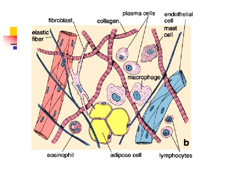
loose connective tissue 7 types of cells present in LCT Fibroblast, Macrophage, Plasma cell, Mast cell, Fat cell, undifferentiated mesenchymal cell , Leukocytes. 3 types fibers Collagen fibers, Elastic fibers and Reticular fibers
Loose connective tissue
Loose connective tissue Loosely arranged fibers and abundant cells
Loose connective tissue 3 6 1. fibroblast 2. macrophage 8 3. mast cell 4 4. fibrocyte 5. fat cell 6. collagenous fiber 7. elastic fiber 8. capillary 2 5 1
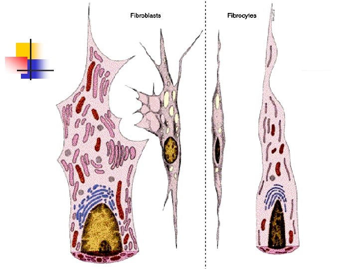
Seven types of cells present in loose connective tissue ① Fibroblast---active Fibrocyte----quiescent
① Fibroblast Structure LM: • Large, flattened cell stellate in shaped • Large ovoid pale nucleus-contain more fine chromatin, with clear one-two nucleoli • Weakly basophilic
① Fibroblast EM: n n Rich in RER, Golgi appatatus and free ribosome Fibroblast---active ---Function: synthesize fibers and ground substance
*Fibrocyte: still state or inactive fibroblast ---structure: LM: n spindle-shaped, small n Nucleus: small, dark stained n Acidophilic cytoplasma EM: less organelles ---Function: become into fibroblast for repairing
②Macrophage ---structure LM n n n round or ovoid-irregular in shape Small and dark nucleus Acidophilic cytoplasm
②Macrophage EM: rich in n a. lysosomes n b. Phagosomes← phagocytosis and pinosomes ←pinocytosis n c. Irregular surface
Macrophage-SEM R B C
Macrophages engulf bacteria
Function a. Phagocytosis n n Special phagocytosis: recognize bacterium, virus and foreign cell Non special: carbon particles, dust and dead cells *Phagosome (pinosome) + primary lysosome →secondary lysosome →remnants
Function B. secretion: lysozyme, complement and interleukin-I (IL-1)and interferon(INF) C. antigen presenting function: *capture antigen→processes→+ MHC II molecule (major histocompatibility complex molecule→antigen-MHC II complexes→T lymphocytes D. Chemotaxis in inflammatory response
The mononuclear phagocyte system Monocyte in blood is the precursor of macrophages n n n Liver: Kupffer cells Central nervous system: microglial cells Skin: Langhans cells Lymph node: dendritic cells Bone: osteoclast
③ Mast Cell nucleus ---structure: granules LM: n round and large cell n Small dark-stained nucleus n Basophilic secreting granules
n EM Membrane bound granules A few Mitochondria A little RER
Basophilic secreting granules: n n n heparin: an anticoagulant Histamine : cause cap. permeability↑ Eosinophil chamotactic factor of anaphlaxis (ECF-A) Cytoplasm contain: n leukotriene - slow reaction substance of anaphlaxis ---Function: cause allergic reaction
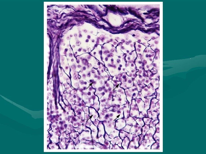
④ Plasma cell --- structure: LM: n n n round or ovoid Round eccentrically-located nucleus with more spotliked heterochromatin Basophilic cytoplasm
EM: rich in parallelly arranged RER, free ribosome and Golgi complex ---function: synthesize and secrete immunoglobulin, Ig-antibody
Arrows: plasma cells
⑤Fat Cell (adipocyte ) ---structure: n n large, round or polygonal flattened ovoid nucleus located on one side of cell thin layer of cytoplasm a large lipid droplet ---function: synthesize and store fat
⑥Undifferentiated Mesenchymal Cell ---structure: similar to fibrocyte ---function: multidifferentiating potential
⑦Leukocytes Granulocyte: neutrophil, eosinophil and basophil Agranulocyte: lymphocyte (B, T) and monocyte
Fibers v Collagen fiber Collagen (19 members) v Reticular fiber v Elastic fiber Elastin
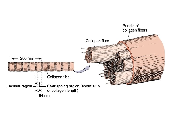
①Collagen fibers (white fibers) Collagen fibers made of collagen type I are the most numerous fibers in CT LM: • 1 -20 um in diameter • Belt-liked wave and branch to form a network • Eosinophilic
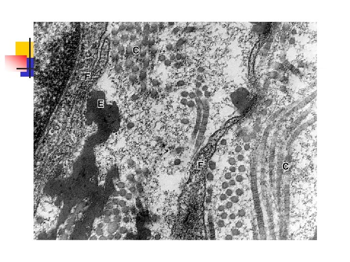
EM: parallel-arranged fibrils Fibril: • 20 -200 nm in diameter • Have periodic cross striation at 64 nm interval
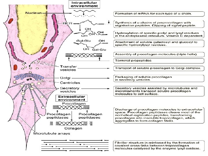
Synthesis of collagen fiber in three steps: a. synthesis of procollagen (RER) → process (Golgi) → out of cell b. procollagen→ tropocollagen → fibril c. fibril → collagenous fiber
Formation: Collagen fibril Collagen molecule (tropocollagen) Collagen (type I, III) triple helix α chain (procollagen)
Electron micrograph of human collagen fibrils
② Reticular fiber LM: • thin, with a diameter between 0. 5 and 2. 0 um • Branch to form network • Argyrophilic fiber (silver impregnation method) EM: • type III collagen • 64 nm cross striation
③ Elastic fiber (yellow fiber) LM: l thinner and less, 0. 1 -1. 0 um l Slight red (HE), purple (aldehyde fuchsin) or brown (orcein) l Branch and form a network EM: l core: elastin-low electron density l Peripheral: microfibril 10 -12 nm, electron dense ↑ fibrillin
Collagen fiber and Elastic fiber
Skin dermis, the elastic fibers are responsible for the skin elasticity.
Elastin molecules are joined by covalent bonds to generate an extensive cross-linked network. Abundant in vertebral ligaments, larynx, and elastic arteries.
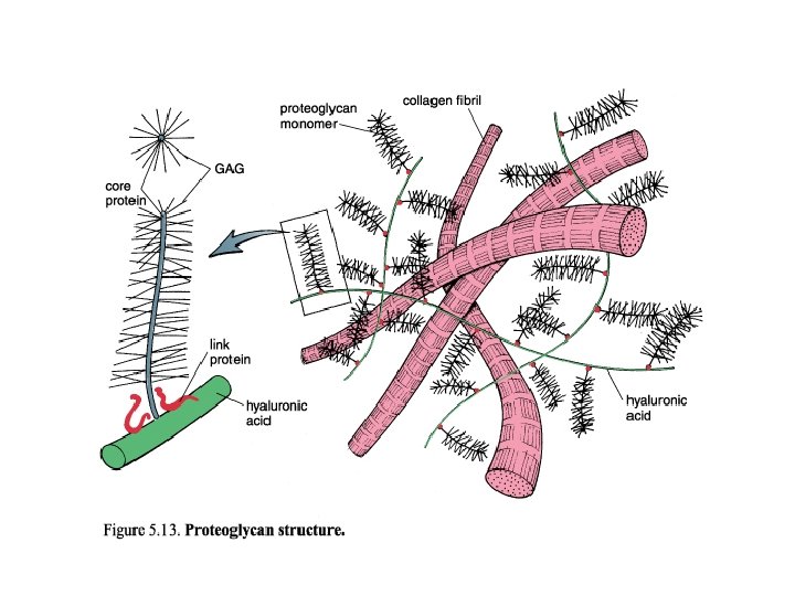
2. Ground substance ☻a highly hydrated colorless and transparent complex mixture of macromolecules occupies the space between the cells and fibers. ☻formed mainly of glycosaminoglycans, proteoglycans, and glycoproteins. ☻lubricant, permits O 2 and nutrients exchange
①proteoglycan large molecular complex ---glycosaminoglycans: n chondroitin sulfate n keratin sulfate n dermatan sulfate n heparin sulfate n hyaluronic acid --protein core
Tissue fluid
Dense connective tissue ---Abundant fibers and few cells ---Connection and supporting Dense regular CT Dense irregular CT
(1) irregular DCT: • Fiber arranged in bundles, running in different directions • Fibroblast • less ground substance Distribution: dermis, sclera and capsule of some organs
Dense irregular connective tissue from human dermis
(2) Regular DCT: • parallel-arranged collagen fibers • tendon cells /special fibroblast Distribution: tendons, ligament and cornea
Longitudinal section of tendon Cross section of tendon
Reticular tissue reticular cells reticular fibers ground substance
Distribution: Hemopoietic tissue and lymphatic tissue Reticular tissue provides the architectural framework that creates a special microenvironment for hematopoietic organs and lymphoid organs.
Section of an adrenal cortex, silver stained to show reticular fibers. This is a thick section made to emphasize the networks formed by these fibers. Nuclei are black, and cytoplasm is unstained.
Adipose tissue ---LCT+fat cells (1)Unilocular (common or yellow) adipose tissue n n single droplet in fat cell Distribution: in subcutaneous tissue throughout the body, mesenterium
Function: • Storage and mobilization of lipids The lipids stored in adipose cells are chiefly triglycerides
(2). Multilocular adipose tissue (brown fat) ♣ fat cells contain many small lipid droplets, ♣ centrally-located nucleus ♣ rich in mitochondria ♣ rich in capillaries. Distribution: in certain areas of neonate, greatly reduced in adulthood. Function: to produce heat
Multilocular fat cell Unilocular adipose tissue Multilocular adipose tissue Sympathetic nerve ending
Distribution of multilocular adipose tissue █: mixture of multilocular and unilocular adipose tissue
Summary • • • • A. Classification of CT B. Loose connective tissue C. 7 types of cell in CT 1. fibroblast, collagenous fiber synthesis 2. macrophage 3. plasma 4. mast cell 5. fat cell 6. undifferentiated mesenchymal cells 7. leukocyte D. 3 fibers E. Ground substance F. Tissue fluid
Questions • 1. Describe the characteristic , classification and function of connective tissue? • 2. What kind of cells present in connective tissue? And what are the functions of those cells? • 3. What is (are) the structure cell(s) of connective tissue?