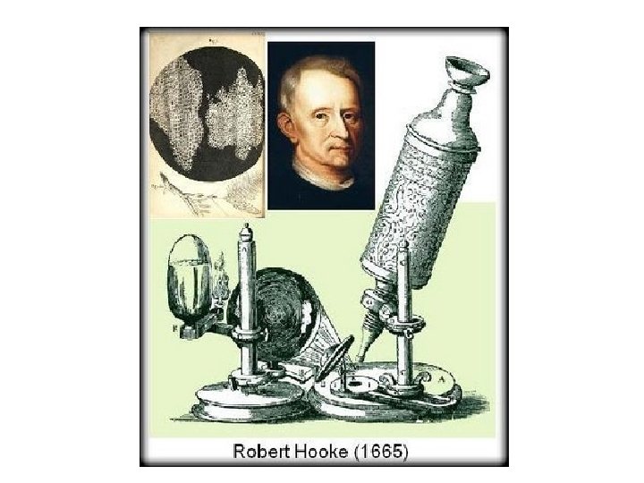
Chapter 3 Cell Structure Microscopes Light Microscope Electron


 Structural formula Choline Phosphate Glycerol Fatty")

 Glycoprotein Carbohydrate Glycolipid EXTRACELLULAR SIDE OF")


 Transport Receptor Signal transduction (b) Enzymatic")


- Slides: 73
Chapter 3 Cell Structure
Microscopes • Light Microscope • Electron Microscope
Microscopes • Light Microscope - light passes through one or more lenses to produce an image of a specimen. • Electron Microscope
Microscopes • Light Microscope - light passes through one or more lenses to produce an image of a specimen. • Electron Microscope – forms an image of a specimen using a beam of electrons.
Microscopes • Light Microscope - light passes through one or more lenses to produce an image of a specimen. • Electron Microscope – forms an image of a specimen using a beam of electrons. – Transmission Electron Microscope – Scanning Electron Microscope
Light Pros and Cons Pros Cons • Comparatively Inexpensive • Can observe living specimens • Can view biological processes happening in real time. • Slide prep is quick and easy • Magnification, depth of field, and resolution are not as great as in an Electron Microscope.
Electron Pros and Cons Pros • Higher resolution/magnificatio n. • Greater depth of field • 3 D images Cons • Expensive • Difficult and time consuming slide prep. • Specimens need to be dyed, which kills them, so no living organisms can be observed.
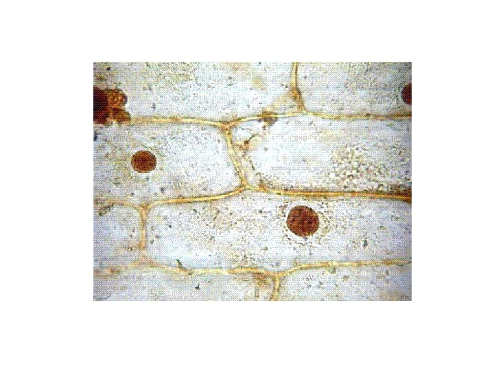
Compound Light Microscope
Onion Skin Cells
Single Celled Organisms Amoeba Paramecium Euglena
SECTION 2 Cell Features
THE CELL THEORY
The Cell Theory: 1. All organisms are made up of one or more cells.
The Cell Theory: 1. All organisms are made up of one or more cells. 2. The cell is the basic unit of all living things.
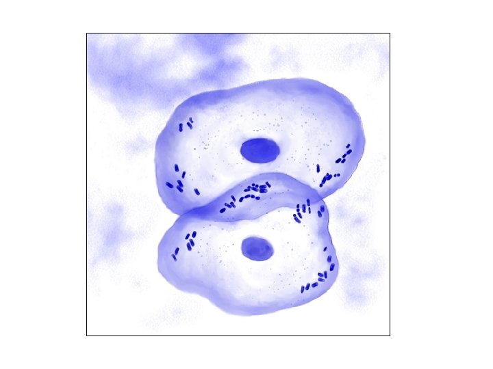
The Cell Theory: 1. All organisms are made up of one or more cells. 2. The cell is the basic unit of all living things. 3. All cells come from existing cells.
Cell Size Why are cells so small? Total surface area Total volume Surface area: volume ratio
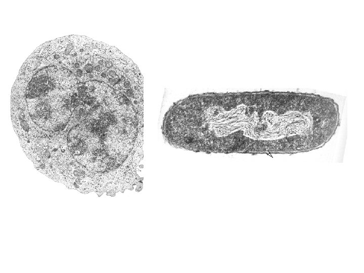
TWO KINDS OF CELLS 1. PROKARYOTES 2. EUKARYOTES
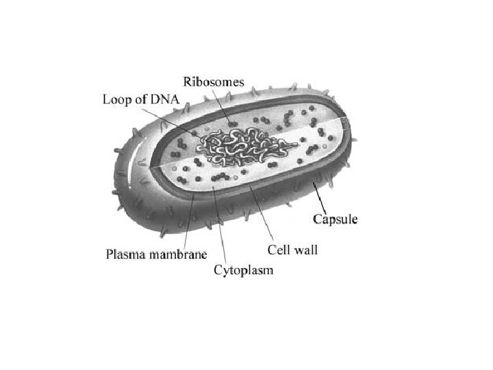
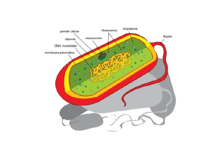
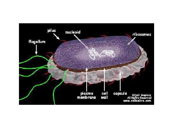
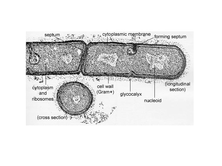
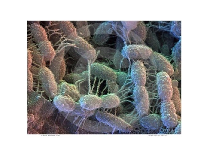
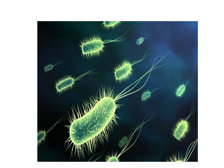
PROKARYOTES 1. 2. 3. 4. 5. 6. All single celled organisms Do not have a nucleus Can exist in a broad range of environmental conditions All have a cell wall made of strands of polysaccharides Sometimes have a capsule DNA is in a single circular ring
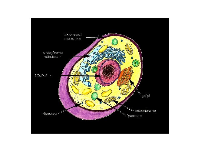
EUKARYOTES 1. Can be single or multicellular 2. Has a membrane bound nucleus which houses the cells DNA 3. Contains internal membrane bound compartments called organelles 4. DNA is in multiple chromosomes
Common Features of ALL cells: • Cell Membrane • Cytoplasm • DNA • Ribosomes
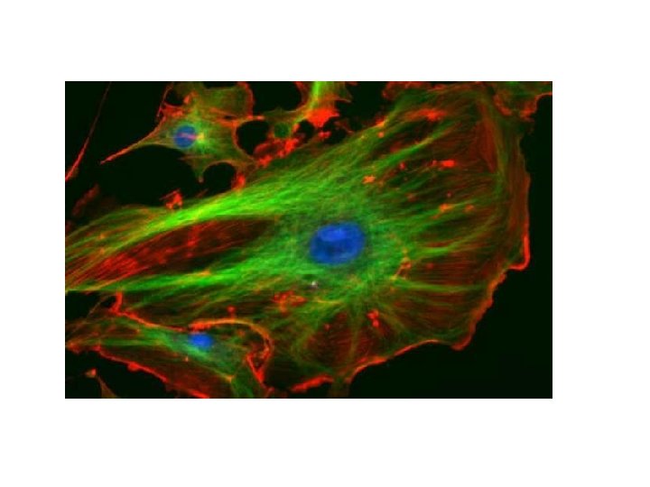
CYTOSKELETON • An intricate network of protein fibers anchored to the inside of the plasma membrane. • The network provides an internal framework for the cell which supports the cell shape and provides places for organelles to anchor.
CYTOSKELETON three different types of fibers: • Actin Fibers: Anchor to membrane proteins forming a network just beneath the cell surface.
CYTOSKELETON three different types of fibers: • Actin Fibers: Anchor to membrane proteins forming a network just beneath the cell surface. • Microtubules: act as a highway system for transportation of information from the nucleus throughout the cell.
CYTOSKELETON three different types of fibers: • Actin Fibers: Anchor to membrane proteins forming a network just beneath the cell surface. • Microtubules: act as a highway system for transportation of information from the nucleus throughout the cell. • Intermediate Fibers: help confine ribosomes and enzymes to particular regions of the cell.
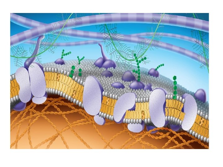
The Cell Membrane a phospholipid bilayer
A Selectively Permeable Barrier
Hydrophobic tails Hydrophilic head Fig. 5 -13 (a) Structural formula Choline Phosphate Glycerol Fatty acids Hydrophilic head Hydrophobic tails (b) Space-filling model (c) Phospholipid symbol
Fig. 7 -2 Hydrophilic head WATER Hydrophobic tail WATER
Fig. 7 -7 Fibers of extracellular matrix (ECM) Glycoprotein Carbohydrate Glycolipid EXTRACELLULAR SIDE OF MEMBRANE Cholesterol Microfilaments of cytoskeleton Peripheral proteins Integral protein CYTOPLASMIC SIDE OF MEMBRANE
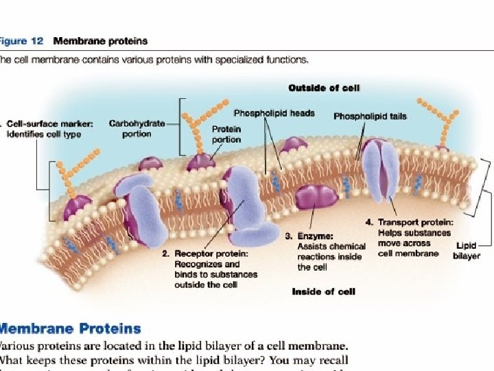
Cell Surface Proteins • Sit on the outer surface of the cell. • Attached to a carbohydrate which allows the cell to advertize what type it is. • Used in cell-cell recognition.
Receptor Proteins • Bind specific substances , such as single molecules, outside the cell.
Transport Proteins • Help substances move across the cell membrane.
Enzymes • Assists chemical reactions inside the cell.
Fig. 7 -9 Signaling molecule Enzymes ATP (a) Transport Receptor Signal transduction (b) Enzymatic activity (c) Signal transduction (e) Intercellular joining (f) Attachment to the cytoskeleton and extracellular matrix (ECM) Glycoprotein (d) Cell-cell recognition
Section 3 Cell Organelles are structures within a cell that preform a specific function.
NUCLEUS • Houses the cells DNA and controls many functions within the cell. • Surrounded by a double membrane called the nuclear envelope, which contains many small channels called nuclear pores.
RIBOSOMES • An organelle composed of RNA and protein that is the site of protein synthesis. • May be suspended in the cytoplasm, and make substances that will remain in the cell – “free”- ribosome. Or they may be attached to the ER, and make substances that will be exported from the cell.
ENDOPLASMIC RETICULUM A system of membranes that is found in the cells cytoplasm and that assists in the production, processing, and transport of proteins, and the production of lipids. ROUGH ER – attached ribosomes SMOOTH ER – no ribosomes
Vessicle • A small membrane-bound sac that transports substances in cells.
Golgi Apparatus • A set of flattened, membrane bound sacs that serve as the packaging and distribution center of the cell.
Lysosomes • Small spherical organelles that contain the cells digestive enzymes
Mitochondria • An organelle that harvests energy from organic compounds to make ATP (adenosine triphospahte- a cells energy source) • Has an inner and an outer membrane, and also contains DNA and ribosomes.
PLANTS • Plants have certain organelles unique to only them.
CELL WALL • Surround the cell membrane • Composed of proteins and carbohydrates, including the polysaccharide cellulose. • Helps support and maintain the cell shape, protects it from damage and connects it with neighboring cells.
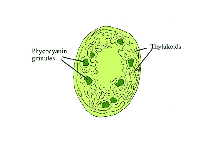
CHLOROPLAST The organelle in plants and algae where photosynthesis takes place. They also have two membranes and their own DNA.
VACUOLE Organelle that stores water and other materials.
Prokaryotic Cell
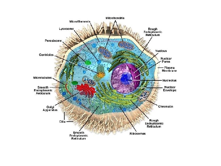
Animal Cell
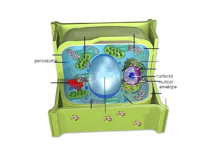
Plant Cell
Protein Packaging & Distribution • 1. Ribosomes make proteins on rough ER. The proteins are packaged into vesicles.
Protein Packaging & Distribution • 2. The vesicles transport newly made proteins from the ER to the Golgi Apparatus
Protein Packaging & Distribution • 3. In the golgi apparatus proteins are processed and packaged into new vesicles.
Protein Packaging & Distribution • 4. Many of these vesicles move to the cell membrane and release their content outside of the cell.
Protein Packaging & Distribution • 5. Other vesicles, including lysosomes, remain within the cytoplasm.