Chapter 3 Cell Structure and Function Processes of


 Glycocalyx (slime layer)")


- Slides: 50
Chapter 3 Cell Structure and Function
Processes of Life • • Growth Reproduction Responsiveness Metabolism © 2012 Pearson Education Inc.
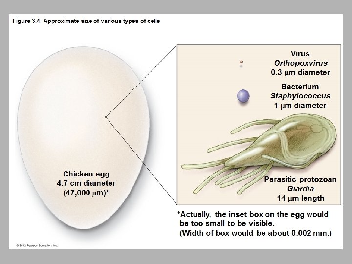
Prokaryotic and Eukaryotic Cells: An Overview • Prokaryotes – Lack nucleus – Lack various internal structures bound with phospholipid membranes – Are small (~1. 0 µm in diameter) – Have a simple structure – Include bacteria and archaea © 2012 Pearson Education Inc.
Prokaryotic and Eukaryotic Cells: An Overview • Eukaryotes – Have nucleus – Have internal membrane-bound organelles – Are larger (10– 100 µm in diameter) – Have more complex structure – Include algae, protozoa, fungi, animals, and plants © 2012 Pearson Education Inc.
External Structures of Bacterial Cells • Glycocalyces – Glycocalyx - gelatinous, sticky substance surrounding the outside of the cell – Composed of polysaccharides, polypeptides, or both • Two Types of Glycocalyces – Capsule – Composed of organized repeating units of organic chemicals – Firmly attached to cell surface – May prevent bacteria from being recognized by host – Slime layer – Loosely attached to cell surface – Water soluble – Sticky layer allows prokaryotes to attach to surfaces – Can help to form biofilm © 2012 Pearson Education Inc.
Figure 3. 5 Glycocalyces-overview Glycocalyx (capsule) Glycocalyx (slime layer)
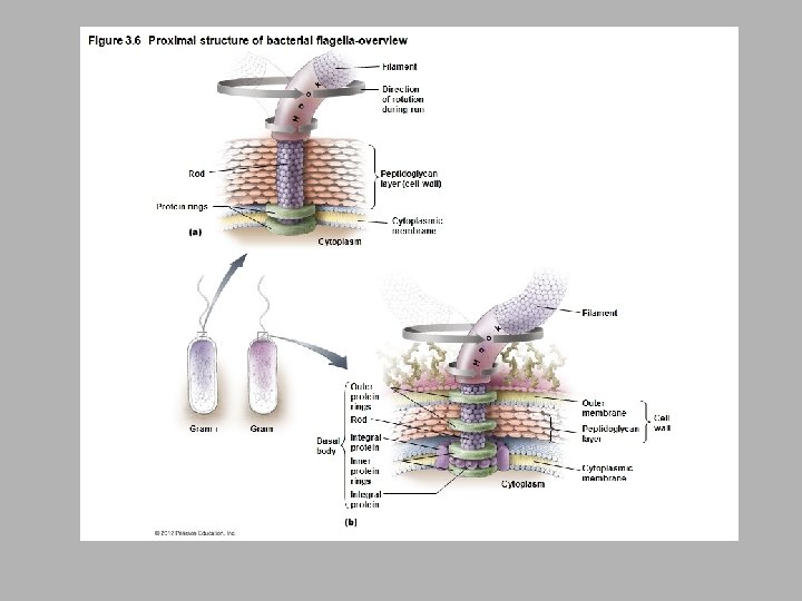
External Structures of Bacterial Cells • Flagella – Are responsible for movement – Have long structures that extend beyond cell surface – Are not present on all bacteria – Structure – Composed of filament, hook, and basal body – Basal body anchors filament and hook to cell wall by a rod and a series of either two or four rings of integral proteins © 2012 Pearson Education Inc.
External Structures of Bacterial Cells • Flagella – Function – Rotation propels bacterium through environment – Rotation reversible; can be counterclockwise or clockwise – Bacteria move in response to stimuli (taxis) – Runs – Tumbles © 2012 Pearson Education Inc.
External Structures of Bacterial Cells • Fimbriae – Sticky, bristlelike projections – Used by bacteria to adhere to one another, to hosts, and to substances in environment – Shorter than flagella – Serve an important function in biofilms © 2012 Pearson Education Inc.
External Structures of Bacterial Cells • Pili – Special type of fimbria – Also known as conjugation pili – Longer than other fimbriae but shorter than flagella – Bacteria typically have only one or two per cell – Mediate the transfer of DNA from one cell to another (conjugation) © 2012 Pearson Education Inc.
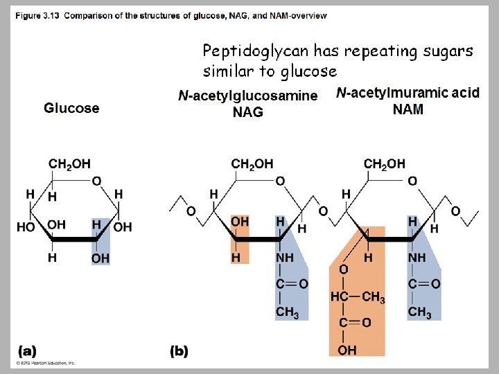
• Bacterial Cell Walls – Provide structure and shape and protect cell from osmotic forces – Assist some cells in attaching to other cells or in resisting antimicrobial drugs – Can target cell wall of bacteria with antibiotics – Give bacterial cells characteristic shapes – Composed of peptidoglycan – Scientists describe two basic types of bacterial cell walls, Gram-positive and Gram-negative © 2012 Pearson Education Inc.
Figure 3. 12 Bacterial shapes and arrangements-overview
Bacterial Cell Walls • Gram-Positive Bacterial Cell Walls – Relatively thick layer of peptidoglycan – Contain unique polyalcohols called teichoic acids – Appear purple following Gram staining procedure – Up to 60% mycolic acid in acid-fast bacteria helps cells survive desiccation © 2012 Pearson Education Inc.
Bacterial Cell Walls • Gram-Negative Bacterial Cell Walls – Have only a thin layer of peptidoglycan – Bilayer membrane outside the peptidoglycan contains phospholipids, proteins, and lipopolysaccharide (LPS) – May be impediment to the treatment of disease – Contains Lipid A – Appear pink following Gram staining procedure © 2012 Pearson Education Inc.
Bacterial Cytoplasmic Membranes • Structure – Referred to as phospholipid bilayer – Composed of lipids and associated proteins – Fluid mosaic model describes current understanding of membrane structure © 2012 Pearson Education Inc.
Bacterial Cytoplasmic Membranes • Function – Energy – Functions like the mitochondrial membrane in eukaryotes – Harvest light energy in photosynthetic bacteria – – – Functions like the chloroplast membrane in eukrayotes Selectively permeable Naturally impermeable to most substances Proteins allow substances to cross membrane Maintain concentration and electrical gradient © 2012 Pearson Education Inc.
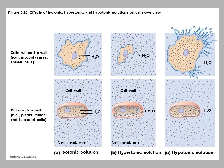
Bacterial Cytoplasmic Membranes • Function – Passive processes – Diffusion – Facilitated diffusion – Osmosis – Active processes – Active transport – Group translocation – Substance chemically modified during transport – © 2012 Pearson Education Inc.
Diffusion, Facilitated Diffusion, Osmosis
Active Transport Extracellular fluid Uniport Cytoplasmic membrane Symport Cytoplasm Uniport Antiport Coupled transport: uniport and symport
Figure 3. 22 Group translocation – modification for transport once modified it is trapped in the cell Glucose Extracellular fluid Cytoplasm Glucose 6 -PO 4
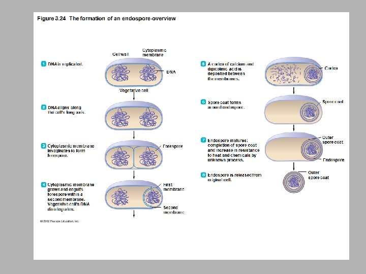
Cytoplasm of Bacteria • Cytosol – Liquid portion of cytoplasm • Inclusions – May include reserve deposits of chemicals – Stored for when nutrients are low – Specific inclusion bodies is diagnostic for some pathogenic bacteria • Endospores – Unique structures produced by some bacteria that are a defensive strategy against unfavorable conditions © 2012 Pearson Education Inc.
Cytoplasm of Bacteria • Nonmembranous Organelles – Ribosomes – Sites of protein synthesis – Cytoskeleton – Plays a role in forming the cell’s basic shape © 2012 Pearson Education Inc.
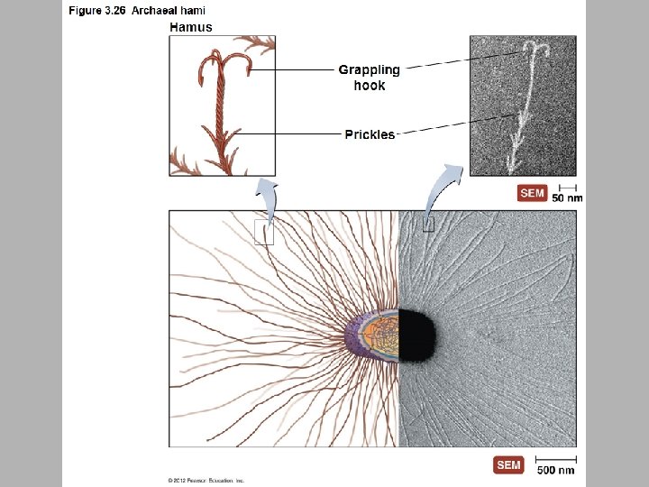
External Structures of Archaea • Glycocalyces – Function in the formation of biofilms – Adhere cells to one another and inanimate objects • Flagella – Consist of basal body, hook, and filament – Numerous differences with bacterial flagella • Fimbriae and Hami – Many archaea have fimbriae – Some make fimbriae-like structures called hami – Function to attach archaea to surfaces – Helical with tiny prickles sticking out – looks like barbed wire © 2012 Pearson Education Inc.
Archaeal Cell Walls and Cytoplasmic Membrane – Most archaea have cell walls – Do not have peptidoglycan – Contain variety of specialized polysaccharides and proteins – All archaea have cytoplasmic membranes – Maintain electrical and chemical gradients – Control import and export of substances from the cell © 2012 Pearson Education Inc.
Cytoplasm of Archaea – Archaeal cytoplasm similar to bacterial cytoplasm – Have 70 S ribosomes – Fibrous cytoskeleton – Circular DNA – Archaeal cytoplasm also differs from bacterial cytoplasm – Different ribosomal proteins – more similar to eukaryotes – Different metabolic enzymes to make RNA – Genetic code more similar to eukaryotes © 2012 Pearson Education Inc.
EUKARYOTES
External Structure of Eukaryotic Cells • Glycocalyces – – – Never as organized as prokaryotic capsules Help anchor animal cells to each other Strengthen cell surface Provide protection against dehydration Function in cell-to-cell recognition and communication © 2012 Pearson Education Inc.
Eukaryotic Cell Walls – Fungi, algae, plants, and some protozoa have cell walls – Composed of various polysaccharides – Plant cell walls composed of cellulose – Fungal cell walls composed of cellulose, chitin, and/or glucomannan – Algal cell walls composed of a variety of polysaccharides © 2012 Pearson Education Inc.
Eukaryotic Cytoplasmic Membranes – – All eukaryotic cells have cytoplasmic membrane Are a fluid mosaic of phospholipids and proteins Contain steroid lipids to help maintain fluidity Contain regions of lipids and proteins called membrane rafts – Control movement into and out of cell
Figure 3. 30 Endocytosis-overview Pseudopodium
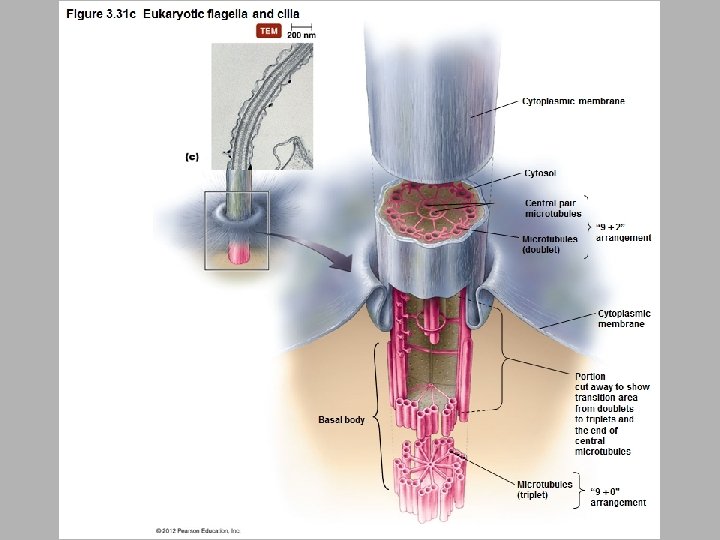
• Eukaryotic Flagella – Structure and arrangement – Differ structurally and functionally from prokaryotic flagella – Within the cytoplasmic membrane – Shaft composed of tubulin arranged to form microtubules – Filaments anchored to cell by basal body – May be single or multiple – Function – Do not rotate but undulate rhythmically © 2012 Pearson Education Inc.
• Eukaryotic Cilia – Shorter and more numerous than flagella – Coordinated beating propels cells through their environment – Also used to move substances past the surface of the cell © 2012 Pearson Education Inc.
Cytoplasm of Eukaryotes • Other Nonmembranous Organelles – Ribosomes – Larger than prokaryotic ribosomes (80 S versus 70 S) – Composed of 60 S and 40 S subunits – Cytoskeleton – – Extensive network of fibers and tubules Anchors organelles Produces basic shape of the cell Made up of tubulin microtubules, actin microfilaments, and intermediate filaments © 2012 Pearson Education Inc.
Cytoplasm of Eukaryotes • Other Nonmembranous Organelles – Centrioles and centrosome – Centrioles play a role in mitosis, cytokinesis, and formation of flagella and cilia – Centrosome is region of cytoplasm where centrioles are found © 2012 Pearson Education Inc.
Cytoplasm of Eukaryotes • Membranous Organelles – Nucleus – Often largest organelle in cell – Contains most of the cell’s DNA – Nucleoplasm – Contains chromatin – Nucleoli present in nucleoplasm; RNA synthesized in nucleoli – Surrounded by nuclear envelope © 2012 Pearson Education Inc.
Cytoplasm of Eukaryotes • Membranous Organelles – Endoplasmic reticulum – Netlike arrangement of flattened, hollow tubules continuous with nuclear envelope – Functions as transport system – Two forms – Smooth endoplasmic reticulum (SER) – Rough endoplasmic reticulum (RER) © 2012 Pearson Education Inc.
Cytoplasm of Eukaryotes • Membranous Organelles – Golgi body – Flattened hollow sacs surrounded by phospholipid bilayer – Receives, processes, and packages large molecules for export from cell – Packages molecules in secretory vesicles – Not in all eukaryotic cells © 2012 Pearson Education Inc.
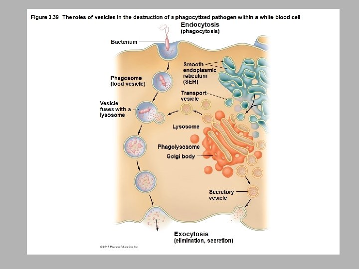
Cytoplasm of Eukaryotes • Membranous Organelles – Lysosomes, peroxisomes, vacuoles, and vesicles – – Store and transfer chemicals within cells May store nutrients in cell Lysosomes contain catabolic enzymes Peroxisomes contain enzymes that degrade poisonous wastes © 2012 Pearson Education Inc.
Cytoplasm of Eukaryotes • Membranous Organelles – Mitochondria – Have two membranes composed of phospholipid bilayer – Produce most of cell’s ATP – Interior matrix contains 70 S ribosomes and molecule of DNA – Just like you find in bacteria © 2012 Pearson Education Inc.
Cytoplasm of Eukaryotes • Membranous Organelles – Chloroplasts – Light-harvesting structures found in photosynthetic eukaryotes – Have two phospholipid bilayer membranes and DNA and 70 S ribosomes – Just like you find in bacteria © 2012 Pearson Education Inc.
• Endosymbiotic Theory – Eukaryotes formed from union of small aerobic prokaryotes with larger anaerobic prokaryotes – Smaller prokaryotes became internal parasites – Parasites lost ability to exist independently – Larger cell became dependent on parasites for aerobic ATP production – Aerobic prokaryotes evolved into mitochondria – Similar scenario for origin of chloroplasts – Does not explain everything though, there are some missing pieces to this story © 2012 Pearson Education Inc.