Chapter 3 Antigens and Antibodies and T cell

Chapter 3 Antigens and Antibodies and T cell receptors Dr. Capers IMMUNOLOGY

Hallmark molecules of adaptive immunity Antibody and T-cell receptor Antibody is part of the B cell receptor Innate immunity recognizes patterns, whereas antibodies and T cell receptors have high degree of specificity
 and/or cell-mediated (T cell) immune")
Immunogenicity Ability to induce humoral (B cell antibody) and/or cell-mediated (T cell) immune response Immunogen is substance that induces response Antigenicity Ability to combine specifically with Abs or T- cell receptor/MHC Not all antigens are immunogenic Haptens – will talk about later in powerpoint

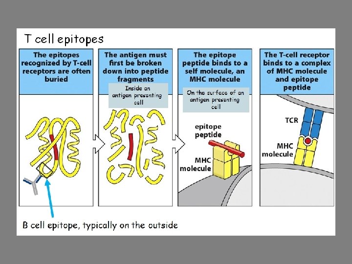
Antibodies and T cell receptors ○ Recognize epitopes Immunologically active regions of immunogen that bind to antigen-specific antibodies or T-cell receptors

Epitopes Antigenic determinants recognized by B cells and T cells B cell epitopes tend to be on the outside of the antigen For example, the hydrophilic amino acids on a protein’s surface T cell epitopes from proteins derived from enzymatic digestion of peptide and then association with MHC

Properties of Immunogen contribute to Immunogenicity 4 Properties ○ Foreignness ○ Molecular size ○ Chemical composition and complexity ○ Ability to be processed and presented on MHC

Foreignness Lymphocytes that do not bind to self antigens are allowed to further develop Therefore they will later only recognized nonself antigens For example: ○ Bovine serum albumin (BSA) is not immunogenic when injected into cow but is when injected into chicken ○ Some macromolecules are highly conserved throughout evolution and display little immunogenicity - Cytochrome c, collagen
 immunogens - > 100, 000 Daltons ○ Poor")
Molecular Size ○ Active (good) immunogens - > 100, 000 Daltons ○ Poor immunogens - < 5, 000 -10, 000 Daltons

Chemical Composition Polymers composed of multiple copies of same amino acid or sugar tend to be poor immunogens Lipids are haptens and need to be congugated with carrier to produce antibodies Important for assays for detection of some steroids, vitamins

Susceptibility to antigen processing Large, insoluble macromolecules are more likely to be phagocytized for processing

The biological system contributes to immunogenicity Host Genetic make-up Manner in which material is presented Use of agents (adjuvants) to enhance immunogenicity

Genotype of recipient animal Genes of MHC Genes in coding for specific antibodies

Material presentation – immunogen dosage and route of administration ○ Too low or high of dosage can induce tolerance ○ Single dose is often not enough – booster is needed ○ Route Intravenous (iv) Intradermal (id) Subcutaneous (sc) Intramuscular (im) Intraperitoneal (ip) - Antigen administered iv would travel to spleen; administered sc would travel to lymph nodes

Adjuvants Enhance immunogenicity Not exactly sure how they work but are recognized by Toll-like receptors Water-in-oil adjuvants Freund’s incomplete adjuvant – antigen in aqueous solution, mineral oil, and emulsifying agent - Antigen is then released very slowly from injection site - Based on Freund’s complete adjuvant - also contained heat –killed Mycobacteria

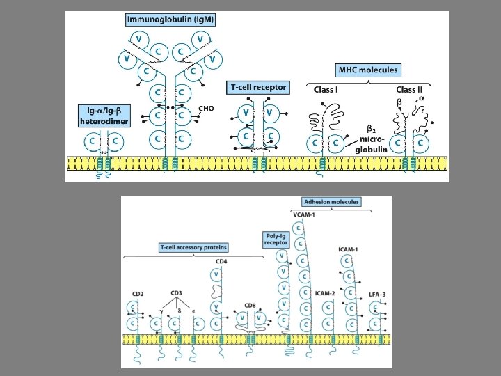
Immunoglobulin Superfamily All have similar structures Examples: ○ Antibodies ○ T-cell receptors ○ Class I and II MHC molecules ○ Part of B cell receptor Most members of immunoglobulin superfamily cannot bind antigen
 Epitope binding proteins ○ Membrane bound on B cells OR ○ Secreted")
Antibodies (Abs) Epitope binding proteins ○ Membrane bound on B cells OR ○ Secreted in blood - Humoral immunity Share structural features, bind to antigen, and participate in number of effector functions Known collectively as Immunoglobulins (Igs) Abs don’t kill anything, their job is to plant the “kiss of death” on an invader

Basic Structure of Antibodies Known since late 19 th century that antibodies are in serum ○ Serum is fluid phase that remains after plasma is allowed to clot ○ Antibodies are also found in other secretions

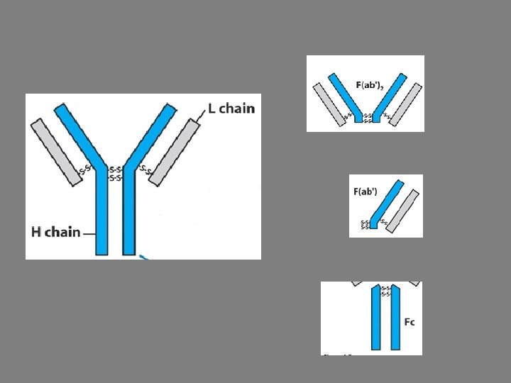
Antibodies are heterodimers 2 light chains ○ ~ 22, 000 daltons each 2 heavy chains ○ ~ 55, 000 daltons each First 110 aa of amino- terminal end of heavy and light chain vary depending on antibody specificity
2 still shows antigen binding capability")
Different digestion procedures reveal different fragments F(ab’)2 still shows antigen binding capability

Light Chains When aa sequences of light chains from several individuals were sequenced, pattern emerged: Amino-terminal end (110 aa) varied Other part remained constant Were found to be either kappa (κ) OR Lambda (λ) - In mice and humans, different lambda subtypes have been found
")
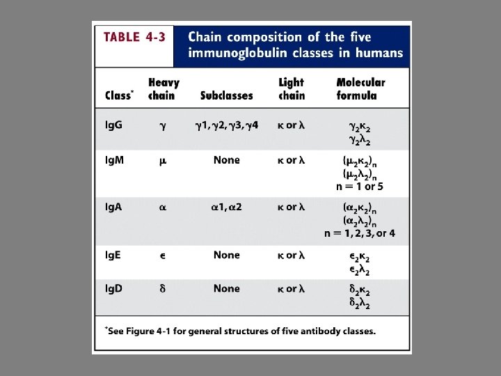
Heavy Chains Amino-terminal end also shows variability 5 different heavy chain constant regions (isotypes) ○ Ig. M – μ ○ Ig. G – γ ○ Ig. A – α ○ Ig. D – δ ○ Ig. E – ε Some subisotypes have been discovered in some species Each antibody has 2 identical heavy chains, 2 identical light chains

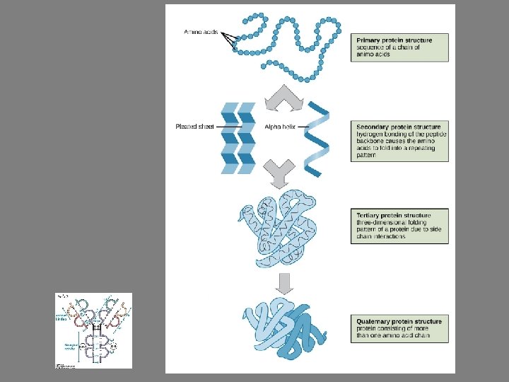
Overall structure of immunoglobulin Primary – sequence of amino acids Secondary – folding into series of β pleated sheets Tertiary – compact globular domains Quarternary – adjacent light and heavy chains interact
 ○ Complimentary to epitopes that they will")
Hypervariable regions = complimentarity-determining regions (CDRs) ○ Complimentary to epitopes that they will bind
, δ (delta), and α (alpha) heavy chains have extended")
Hinge Region γ (gamma), δ (delta), and α (alpha) heavy chains have extended peptide sequence ○ Rich in proline and cysteine ○ Gives flexibility Immunoglobulins can be secreted or membranebound ○ Membrane-bound differ in the carboxyl-terminal end: - Extracellular “spacer” of 26 aa - Hydrophobic transmembrane sequence - Cytoplasmic tail
 Membrane bound antibody + accessory membrane bound molecules = BCR")
B Cell Receptor (BCR) Membrane bound antibody + accessory membrane bound molecules = BCR Heavy chain portion of membrane-bound antibody does not extend far enough through the cell membrane for signaling ○ Membrane bound antibody is accompanied by Igα and Igβ

Antibody-mediated Effector Functions Remember, they plant “kiss of death” on an invader In addition to binding antigen, Abs can: ○ Promote phagocytosis (opsonization) ○ Activate complement ○ Antibody dependent cell mediated cytotoxicity (ADCC) Natural killer cells have receptor for Fc portion of antibody ○ Some can cross epithelial layers to be excreted through mucous or across placenta

Changing the constant region of an antibody is class switching ○ Makes an Ig. G different from an Ig. M ○ Watch this video: https: //www. youtube. com/watch? v=Iqw. RGyk. Q_ OE
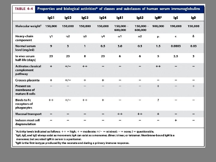
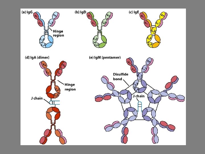

Monomeric Ig. M expressed on B cells Secreted is pentameric 1 st class produced in primary response Activates complement Very good at agglutination

Membrane bound on B cells

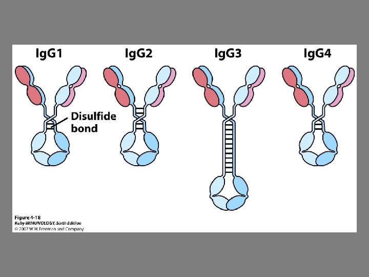
Most abundant 4 human subclasses Crosses placenta Involved in complement

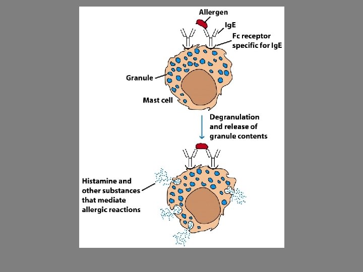
Involved in allergic reactions Involvement in parasitic infections

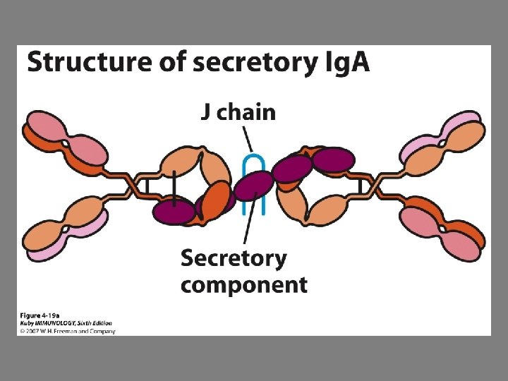
Predominant class in secretions J chain and secretory component helps with transport across intestinal wall J chain makes Ig. A more resistant to acids and enzymes found in digestive tract Ig. A and macrophages restrict commensal bacteria that occasionally enter the tissues from the intestines ○ Better for Ig. A to interact than Ig. G – this is because the Fc portion of Ig. G has high affinity for receptors of immune cells and would constantly trigger inflammatory responses

Multiple noncovalent bonds between antibody and antigen ○ Hydrogen bonds ○ Ionic bonds ○ Van der Waals ○ Hydrophobic interactions

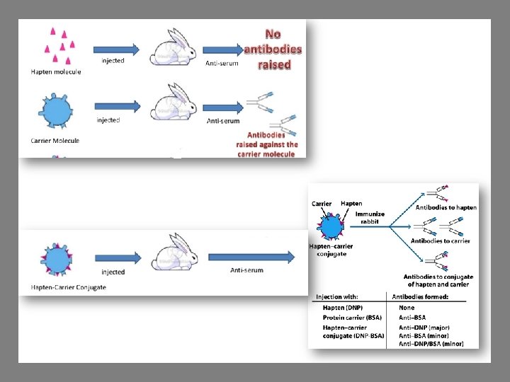
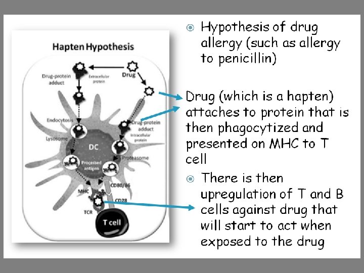
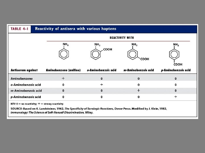
Haptens Hapten – too small, lack immunogenicity ○ If hapten is coupled to carrier protein, immune response can be induced ○ Hapten-carrier conjugate Produces 3 types of antigenic determinants - Antibodies to hapten - Antibodies to carrier - Antibodies to hapten-carrier conjugate

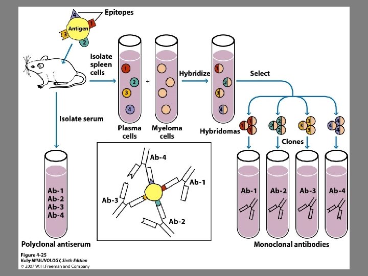
Monoclonal Antibodies Most antigens offer multiple epitopes However, a single B cell will only produce antibody specific to single epitope Antibodies found in serum are from many different B cells ○ Polyclonal antibodies However, for diagnostic uses, monoclonal antibodies are needed We’ll discuss this more later in the semester
 T Cell Receptor ○ Expressed on surface of T cells")
T cell Receptor (TCR) T Cell Receptor ○ Expressed on surface of T cells ○ Recognize processed antigen complexed with MHC molecules

T cell receptor vs B cell receptor T cell receptor is only membrane bound ○ Doesn’t appear in soluble form like antibodies so more difficult to assess it’s structure Antigen binding of T cell receptor is weaker than that of antibodies Antigen recognized by T cells is not antigen alone but antigen associated with MHC molecules
 T cell receptor (TCR) is specific for peptide A (b) Right MHC")
(a) T cell receptor (TCR) is specific for peptide A (b) Right MHC haplotype but wrong antigen (peptide B) (c) Right antigen (peptide A) but wrong haplotype
 ○ TCR heterodimers are similar to immunoglobulins Therefore they")
T cell receptor (TCRs) ○ TCR heterodimers are similar to immunoglobulins Therefore they are classified in immunoglobulin superfamily Resembles Fab fragment

TCR-CD 3 Complex Accessory molecules help in signal transduction after interaction of T cell with antigen

T cell accessory molecules T cells can be divided into 2 populations: ○ CD 4+ Recognize antigen associated with Class II MHC OR ○ CD 8+ Recognize antigen associated with Class I MHC ○ CD 4 and CD 8 function as coreceptors and assist with signal transduction

Affinity of TCR for peptide-MHC complexes is enhanced by coreceptors

Allogenic – genetically different individuals of same species Alloreactivity of T cells is puzzling: ○ Evidence supports that T cells can only respond to antigen+MHC ○ However, T cells can recognize a foreign MHC molecule alone - As with transplants
- Slides: 58