
Chapter 28 Protists Key components Introduction Most eukaryotes

- Slides: 54
Chapter 28 Protists Key components: Introduction Most eukaryotes are single-celled organisms Page 581, Trypansoma sp. Page 583, Plasmodium sp. Brown algae Oomycetes Dinoflagellates (red tide) Red & Green algae Slime molds Protists play key roles in ecological relationships
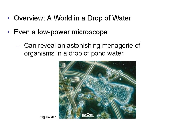
• Overview: A World in a Drop of Water • Even a low-power microscope – Can reveal an astonishing menagerie of organisms in a drop of pond water Figure 28. 1 50 m
• These amazing organisms – Belong to the diverse kingdoms of mostly single-celled eukaryotes informally known as protists • Advances in eukaryotic systematics – Have caused the classification of protists to change significantly
• Concept 28. 1: Protists are an extremely diverse assortment of eukaryotes • Protists are more diverse than all other eukaryotes – And are no longer classified in a single kingdom • Most protists are unicellular – And some are colonial or multicellular
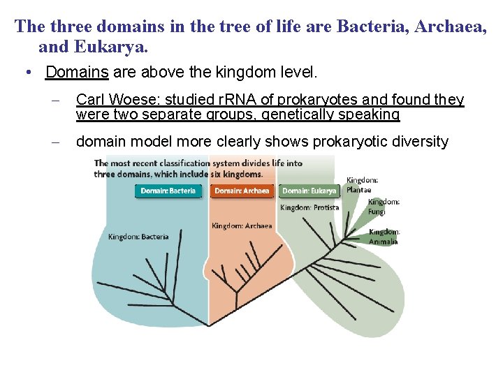
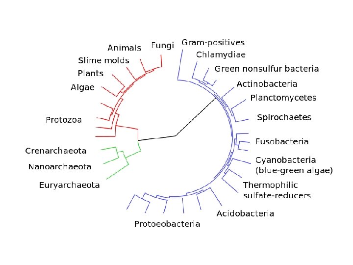
The three domains in the tree of life are Bacteria, Archaea, and Eukarya. • Domains are above the kingdom level. – Carl Woese: studied r. RNA of prokaryotes and found they were two separate groups, genetically speaking – domain model more clearly shows prokaryotic diversity
http: //tolweb. org/tree/ http: //eol. org/
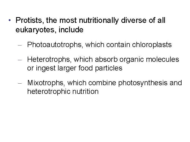
• Protists, the most nutritionally diverse of all eukaryotes, include – Photoautotrophs, which contain chloroplasts – Heterotrophs, which absorb organic molecules or ingest larger food particles – Mixotrophs, which combine photosynthesis and heterotrophic nutrition
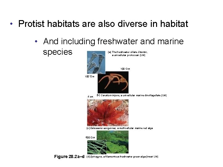
• Protist habitats are also diverse in habitat • And including freshwater and marine species (a) The freshwater ciliate Stentor, a unicellular protozoan (LM) 100 m 4 cm (b) Ceratium tripos, a unicellular marine dinoflagellate (LM) (c) Delesseria sanguinea, a multicellular marine red alga 500 m Figure 28. 2 a–d (d) Spirogyra, a filamentous freshwater green alga (inset LM)
• Reproduction and life cycles – Are also highly varied among protists, with both sexual and asexual species
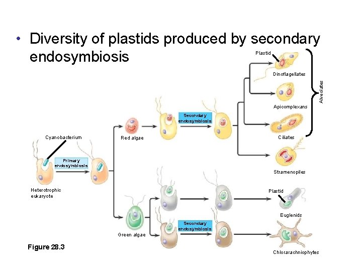
Endosymbiosis in Eukaryotic Evolution • There is now considerable evidence – That much of protist diversity has its origins in endosymbiosis
• The plastid-bearing lineage of protists – Evolved into red algae and green algae • On several occasions during eukaryotic evolution – Red algae and green algae underwent secondary endosymbiosis, in which they themselves were ingested
• Diversity of plastids produced by secondary endosymbiosis Plastid Alveolates Dinoflagellates Apicomplexans Secondary endosymbiosis Cyanobacterium Ciliates Red algae Primary endosymbiosis Stramenopiles Heterotrophic eukaryote Plastid Euglenids Secondary endosymbiosis Green algae Figure 28. 3 Chlorarachniophytes
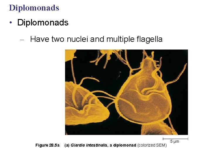
Diplomonads • Diplomonads – Have two nuclei and multiple flagella Figure 28. 5 a (a) Giardia intestinalis, a diplomonad (colorized SEM) 5 µm
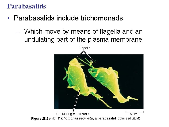
Parabasalids • Parabasalids include trichomonads – Which move by means of flagella and an undulating part of the plasma membrane Flagella Undulating membrane 5 µm Figure 28. 5 b (b) Trichomonas vaginalis, a parabasalid (colorized SEM)
• Concept 28. 3: Euglenozoans have flagella with a unique internal structure • Euglenozoa is a diverse clade that includes – Predatory heterotrophs, photosynthetic autotrophs, and pathogenic parasites
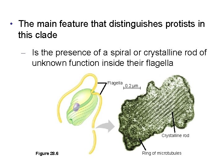
• The main feature that distinguishes protists in this clade – Is the presence of a spiral or crystalline rod of unknown function inside their flagella Flagella 0. 2 µm Crystalline rod Figure 28. 6 Ring of microtubules
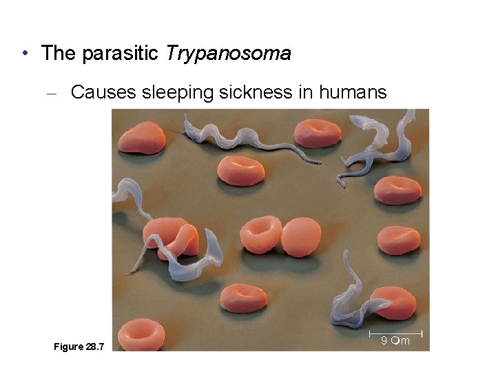
• The parasitic Trypanosoma – Causes sleeping sickness in humans Figure 28. 7 9 m
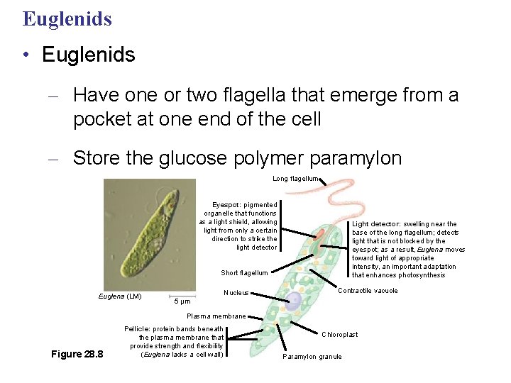
Euglenids • Euglenids – Have one or two flagella that emerge from a pocket at one end of the cell – Store the glucose polymer paramylon Long flagellum Eyespot: pigmented organelle that functions as a light shield, allowing light from only a certain direction to strike the light detector Light detector: swelling near the base of the long flagellum; detects light that is not blocked by the eyespot; as a result, Euglena moves toward light of appropriate intensity, an important adaptation that enhances photosynthesis Short flagellum Euglena (LM) Nucleus Contractile vacuole 5 µm Plasma membrane Figure 28. 8 Pellicle: protein bands beneath the plasma membrane that provide strength and flexibility (Euglena lacks a cell wall) Chloroplast Paramylon granule
Dinoflagellates • Dinoflagellates – Are a diverse group of aquatic photoautotrophs and heterotrophs – Are abundant components of both marine and freshwater phytoplankton
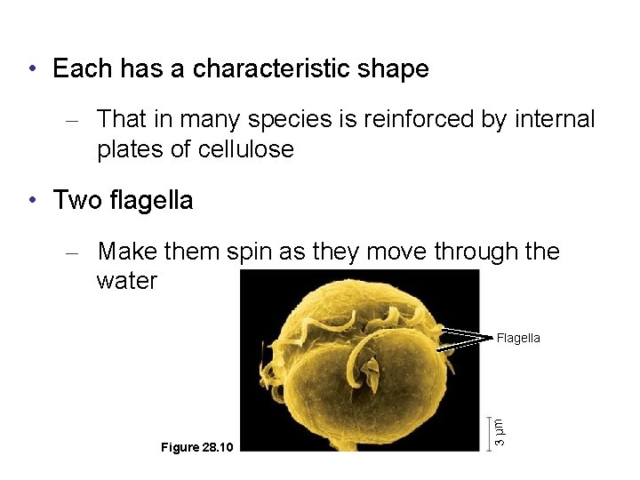
• Each has a characteristic shape – That in many species is reinforced by internal plates of cellulose • Two flagella – Make them spin as they move through the water Figure 28. 10 3 µm Flagella
• Rapid growth of some dinoflagellates – Is responsible for causing “red tides, ” which can be toxic to humans
• Most apicomplexans have intricate life cycles – With both sexual and asexual stages that often require two or more different host species for completion
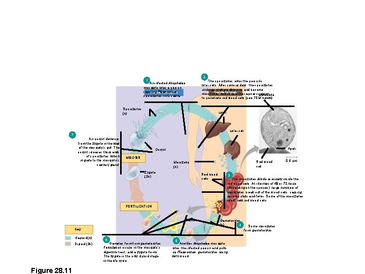
2 1 The sporozoites enter the person’s liver cells. After several days, the sporozoites undergo multiple divisions and become Inside human merozoites, which use their apical complex Merozoite to penetrate red blood cells (see TEM below). An infected Anopheles mosquito bites a person, injecting Plasmodium Inside mosquito sporozoites in its saliva. Sporozoites (n) Liver cell 7 An oocyst develops from the zygote in the wall of the mosquito’s gut. The oocyst releases thousands of sporozoites, which migrate to the mosquito’s salivary gland. Apex Oocyst MEIOSIS Red blood cell Merozoite (n) Zygote (2 n) Red blood cells 3 The merozoites divide asexually inside the red blood cells. At intervals of 48 or 72 hours (depending on the species), large numbers of merozoites break out of the blood cells, causing periodic chills and fever. Some of the merozoites infect new red blood cells. FERTILIZATION Gametes Gametocytes (n) 4 Key Haploid (n) Diploid (2 n) Figure 28. 11 6 Gametes form from gametocytes. Fertilization occurs in the mosquito’s digestive tract, and a zygote forms. The zygote is the only diploid stage in the life cycle. 0. 5 µm 5 Another Anopheles mosquito bites the infected person and picks up Plasmodium gametocytes along with blood. Some merozoites form gametocytes.
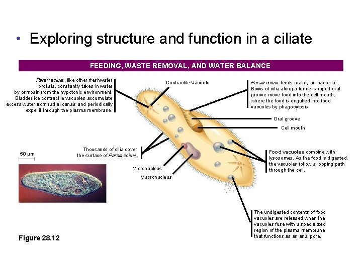
• Exploring structure and function in a ciliate FEEDING, WASTE REMOVAL, AND WATER BALANCE Paramecium, like other freshwater protists, constantly takes in water by osmosis from the hypotonic environment. Bladderlike contractile vacuoles accumulate excess water from radial canals and periodically expel it through the plasma membrane. Contractile Vacuole Paramecium feeds mainly on bacteria. Rows of cilia along a funnel-shaped oral groove move food into the cell mouth, where the food is engulfed into food vacuoles by phagocytosis. Oral groove Cell mouth 50 µm Thousands of cilia cover the surface of Paramecium. Micronucleus Food vacuoles combine with lysosomes. As the food is digested, the vacuoles follow a looping path through the cell. Macronucleus Figure 28. 12 The undigested contents of food vacuoles are released when the vacuoles fuse with a specialized region of the plasma membrane that functions as an anal pore.
Oomycetes (Water Molds and Their Relatives) • Oomycetes – Include water molds, white rusts, and downy mildews – Were once considered fungi based on morphological studies
• Most oomycetes – Are decomposers or parasites – Have filaments (hyphae) that facilitate nutrient uptake
• The ecological impact of oomycetes can be significant – Phytophthora infestans causes late blight of potatoes
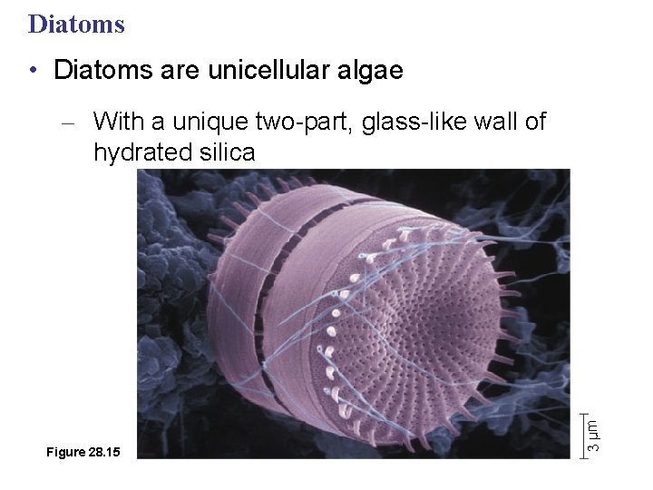
Diatoms • Diatoms are unicellular algae Figure 28. 15 3 µm – With a unique two-part, glass-like wall of hydrated silica
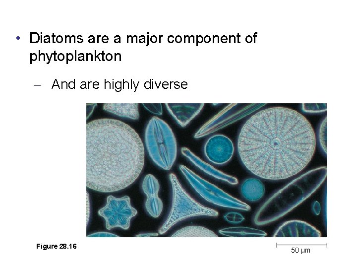
• Diatoms are a major component of phytoplankton – And are highly diverse Figure 28. 16 50 µm
• Accumulations of fossilized diatom walls – Compose much of the sediments known as diatomaceous earth
Golden Algae • Golden algae, or chrysophytes – Are named for their color, which results from their yellow and brown carotenoids • The cells of golden algae – Are typically biflagellated, with both flagella attached near one end of the cell
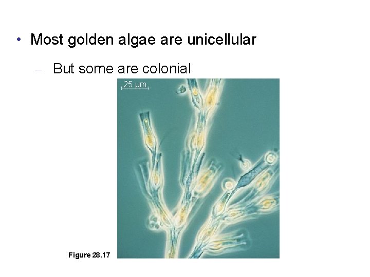
• Most golden algae are unicellular – But some are colonial 25 µm Figure 28. 17
Brown Algae • Brown algae, or phaeophytes – Are the largest and most complex algae – Are all multicellular, and most are marine
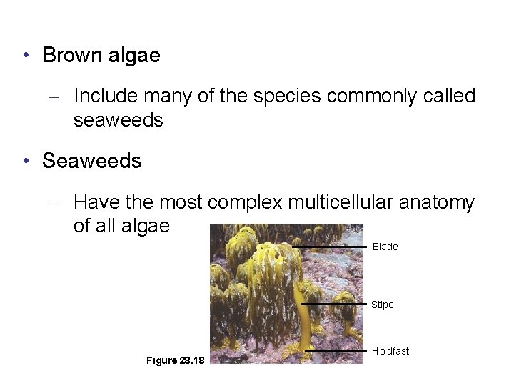
• Brown algae – Include many of the species commonly called seaweeds • Seaweeds – Have the most complex multicellular anatomy of all algae Blade Stipe Figure 28. 18 Holdfast
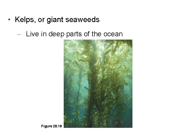
• Kelps, or giant seaweeds – Live in deep parts of the ocean Figure 28. 19
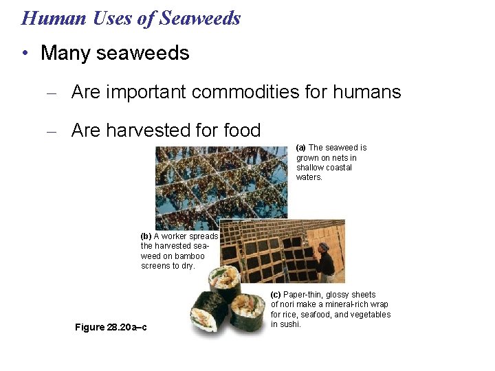
Human Uses of Seaweeds • Many seaweeds – Are important commodities for humans – Are harvested for food (a) The seaweed is grown on nets in shallow coastal waters. (b) A worker spreads the harvested seaweed on bamboo screens to dry. Figure 28. 20 a–c (c) Paper-thin, glossy sheets of nori make a mineral-rich wrap for rice, seafood, and vegetables in sushi.
Alternation of Generations • A variety of life cycles – Have evolved among the multicellular algae • The most complex life cycles include an alternation of generations – The alternation of multicellular haploid and diploid forms
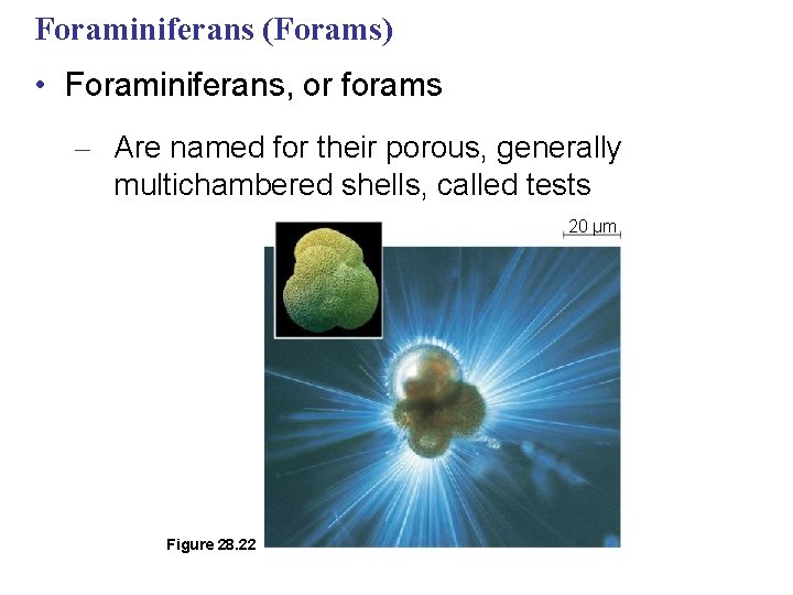
Foraminiferans (Forams) • Foraminiferans, or forams – Are named for their porous, generally multichambered shells, called tests 20 µm Figure 28. 22
Radiolarians • Radiolarians are marine protists – Whose tests are fused into one delicate piece, which is generally made of silica – That phagocytose microorganisms with their pseudopodia
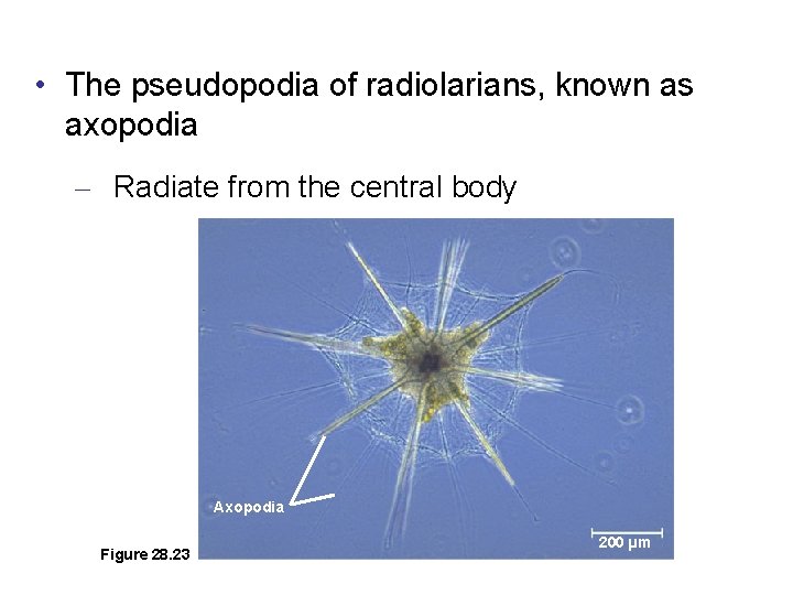
• The pseudopodia of radiolarians, known as axopodia – Radiate from the central body Axopodia Figure 28. 23 200 µm
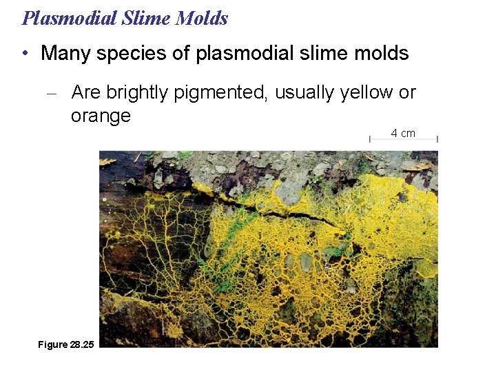
Plasmodial Slime Molds • Many species of plasmodial slime molds – Are brightly pigmented, usually yellow or orange 4 cm Figure 28. 25
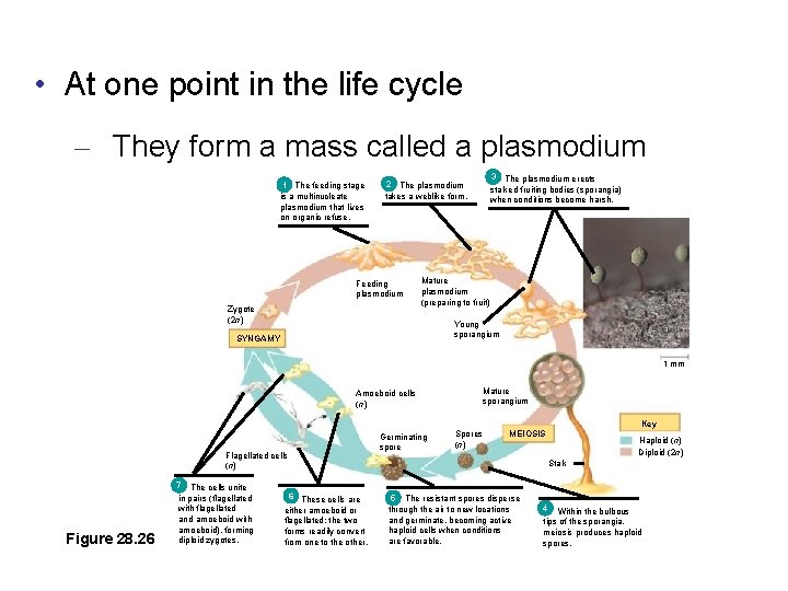
• At one point in the life cycle – They form a mass called a plasmodium 1 The feeding stage is a multinucleate plasmodium that lives on organic refuse. 2 The plasmodium takes a weblike form. Feeding plasmodium Zygote (2 n) 3 The plasmodium erects stalked fruiting bodies (sporangia) when conditions become harsh. Mature plasmodium (preparing to fruit) Young sporangium SYNGAMY 1 mm Mature sporangium Amoeboid cells (n) Flagellated cells (n) Figure 28. 26 7 The cells unite in pairs (flagellated with flagellated and amoeboid with amoeboid), forming diploid zygotes. 6 These cells are either amoeboid or flagellated; the two forms readily convert from one to the other. Germinating spore Spores (n) Key MEIOSIS Haploid (n) Diploid (2 n) Stalk 5 The resistant spores disperse through the air to new locations and germinate, becoming active haploid cells when conditions are favorable. 4 Within the bulbous tips of the sporangia, meiosis produces haploid spores.
• The plasmodium – Is undivided by membranes and contains many diploid nuclei – Extends pseudopodia through decomposing material, engulfing food by phagocytosis
Cellular Slime Molds • Cellular slime molds form multicellular aggregates – In which the cells remain separated by their membranes
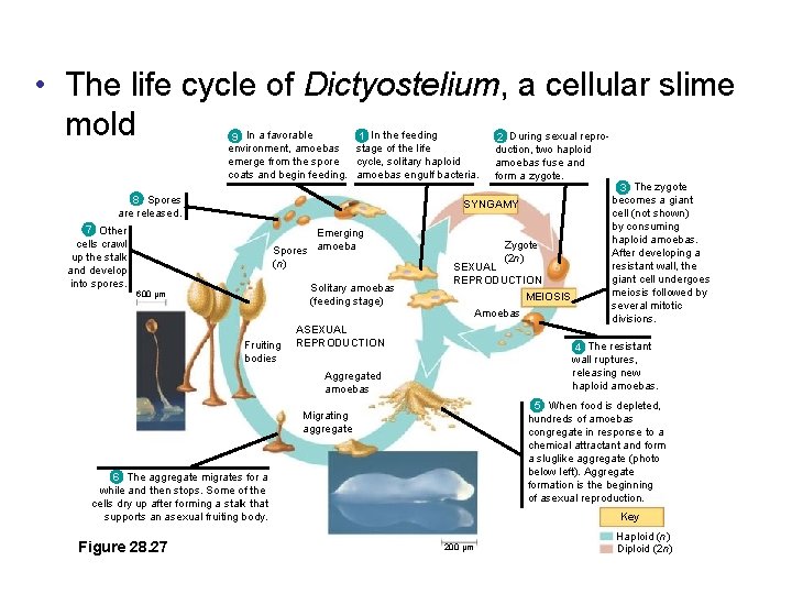
• The life cycle of Dictyostelium, a cellular slime mold 1 In the feeding 9 In a favorable environment, amoebas stage of the life cycle, solitary haploid emerge from the spore coats and begin feeding. amoebas engulf bacteria. 8 Spores are released. 2 During sexual reproduction, two haploid amoebas fuse and form a zygote. SYNGAMY 7 Other cells crawl up the stalk and develop into spores. Emerging Spores amoeba (n) Solitary amoebas (feeding stage) 600 µm Zygote (2 n) SEXUAL REPRODUCTION MEIOSIS Amoebas Fruiting bodies ASEXUAL REPRODUCTION 4 The resistant wall ruptures, releasing new haploid amoebas. Aggregated amoebas 5 When food is depleted, hundreds of amoebas congregate in response to a chemical attractant and form a sluglike aggregate (photo below left). Aggregate formation is the beginning of asexual reproduction. Migrating aggregate 6 The aggregate migrates for a while and then stops. Some of the cells dry up after forming a stalk that supports an asexual fruiting body. Figure 28. 27 3 The zygote becomes a giant cell (not shown) by consuming haploid amoebas. After developing a resistant wall, the giant cell undergoes meiosis followed by several mitotic divisions. Key 200 µm Haploid (n) Diploid (2 n)
• Dictyostelium discoideum – Has become an experimental model for studying the evolution of multicellularity
• Concept 28. 8: Red algae and green algae are the closest relatives of land plants • Over a billion years ago, a heterotrophic protist acquired a cyanobacterial endosymbiont – And the photosynthetic descendants of this ancient protist evolved into red algae and green algae
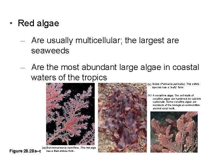
Red Algae • Red algae are reddish in color – Due to an accessory pigment call phycoerythrin, which masks the green of chlorophyll
• Red algae – Are usually multicellular; the largest are seaweeds – Are the most abundant large algae in coastal waters of the tropics (b) Dulse (Palmaria palmata). This edible species has a “leafy” form. (c) A coralline alga. The cell walls of coralline algae are hardened by calcium carbonate. Some coralline algae are members of the biological communities around coral reefs. Figure 28. 28 a–c (a) Bonnemaisonia hamifera. This red alga has a filamentous form.
Green Algae • Green algae – Are named for their grass-green chloroplasts – Are divided into two main groups: chlorophytes and charophyceans – Are closely related to land plants
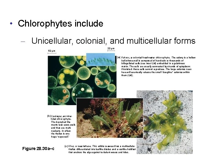
• Chlorophytes include – Unicellular, colonial, and multicellular forms 20 µm 50 µm (a) Volvox, a colonial freshwater chlorophyte. The colony is a hollow ball whose wall is composed of hundreds or thousands of biflagellated cells (see inset LM) embedded in a gelatinous matrix. The cells are usually connected by strands of cytoplasm; if isolated, these cells cannot reproduce. The large colonies seen here will eventually release the small “daughter” colonies within them (LM). (b) Caulerpa, an intertidal chlorophyte. The branched filaments lack cross-walls and thus are multinucleate. In effect, the thallus is one huge “supercell. ” Figure 28. 30 a–c (c) Ulva, or sea lettuce. This edible seaweed has a multicellular thallus differentiated into leaflike blades and a rootlike holdfast that anchors the alga against turbulent waves and tides.
Protists Eukaryotes Most unicellular, some colonial and multicellular (algae, kelp) Reproduction – Asexual – Sexual – meiosis or syngamy Nutrition – Photoautotrophs - chloroplasts – Heterotrophs - absorb organic molecules or ingest larger food particles – Mixotrophs – both
Slime molds are protists that use spores to reproduce. During part of their life cycle, they look like gelatinous "slime". Come in many colors, more than 900 species. Feed on microorganisms that live in dead plant material. Found in soil, lawns, forest floors, old logs, tropical areas, mulch, gutters, air conditioners When food is abundant, a slime mold is a single-celled. When food is in short supply, slime molds congregate and start moving as a single body. In this state they are sensitive to airborne chemicals and can detect food sources. They can readily change the shape and function of parts and may form stalks that produce fruiting bodies, releasing countless spores, light enough to be carried on the wind or hitching a ride on passing animals. Video