Chapter 26 Development of cardiovascular system Establishment of

Chapter 26 Development of cardiovascular system

Ⅰ. Establishment of the primitive cardiovascular system Blood island Primitive blood vessels Hemopoietic stem cells Extraembryonic capillary networks Intraembryonic capillary networks

capillary networks fuse or disappear to form primitive cardiovascular system

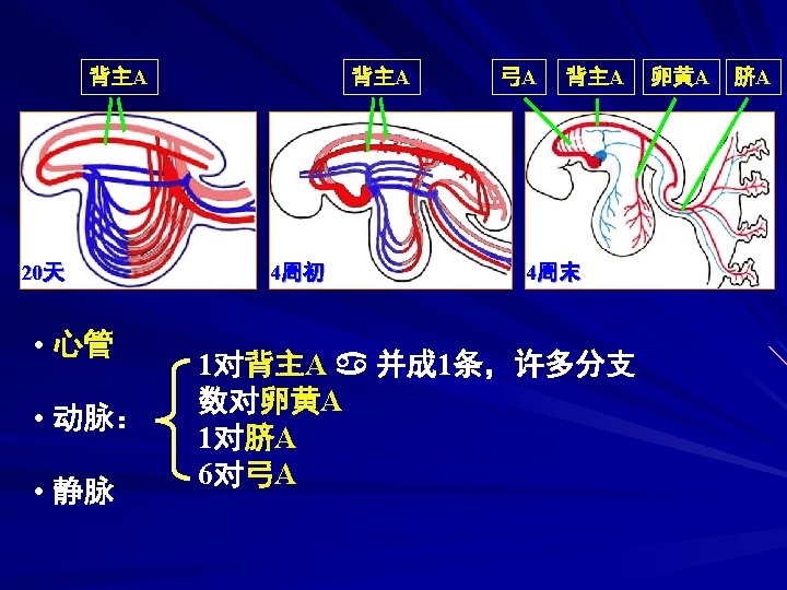
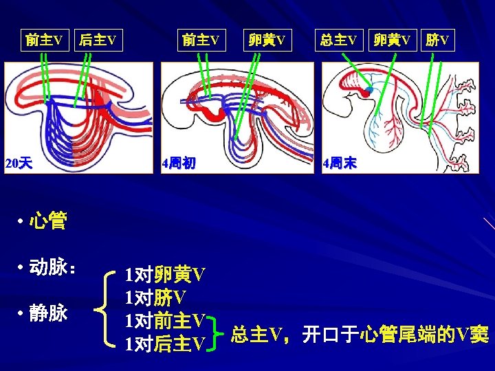
Primitive cardiovascular system a pair of heart tubes a pair of ventral aortae a pair of dorsal aortae the first pair of aortic arches Vitelline arteries Vitelline veins allantoic( umbilical) A Sinus venosus allantoic ( umbilical) V

Aortic sac intersegmental artery anterior cardinal veins posterior cardinal veins common cardinal veins 2 nd~6 th pairs of aortic arches
 circulation embryonic circulation")
vitelline circulation allantoic(umbilical ) circulation embryonic circulation


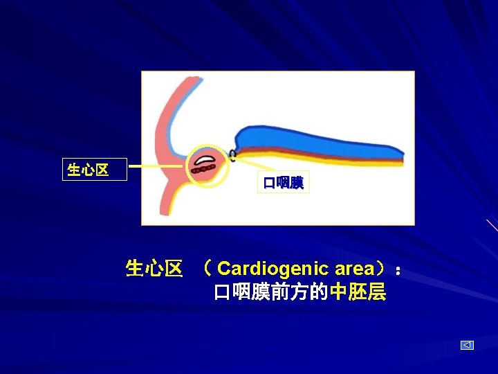
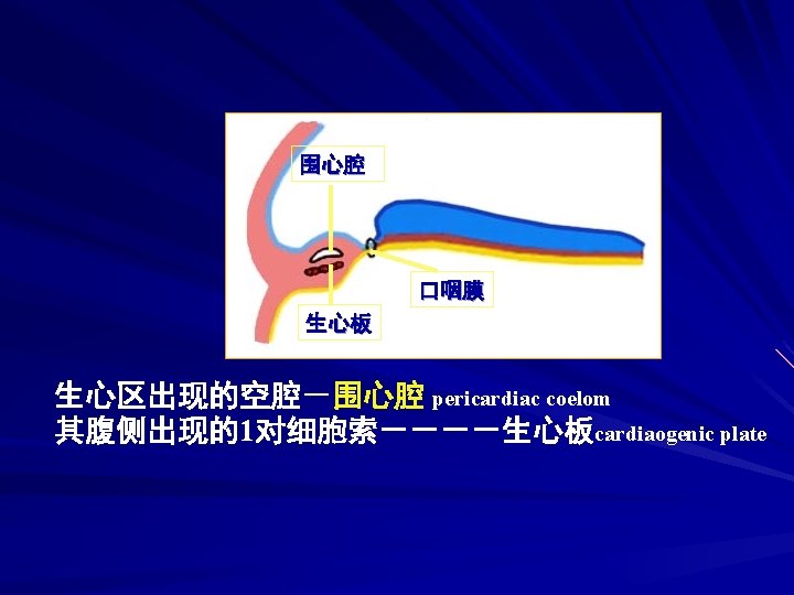
Ⅱ. Development of the heart 1. Development of the Cardiac tube Cardiogenic area Cardiogenic plate (cord) Pericardiac coelom Cardiac tube

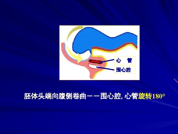
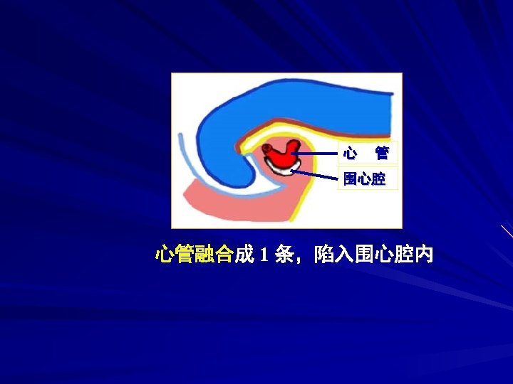

---lateral folds of the embryo make the two cardiac tube approach and fuse with each other

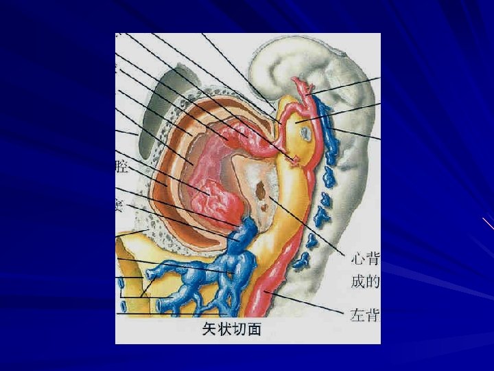
心背系膜 dorsal mesocardium: cardiac tube invaginate into pericardiac coelom and connect to it by dorsal mesocardium

心包横窦 Pericardial sinus Myoepicardial mantle myocardium epicardium cardiac jelly Subendothelium subendocardium 心包横窦 心肌膜 心胶貭 heart wall: endocardium ,myocardium and epicardium
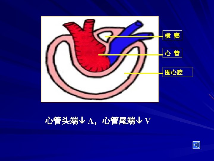

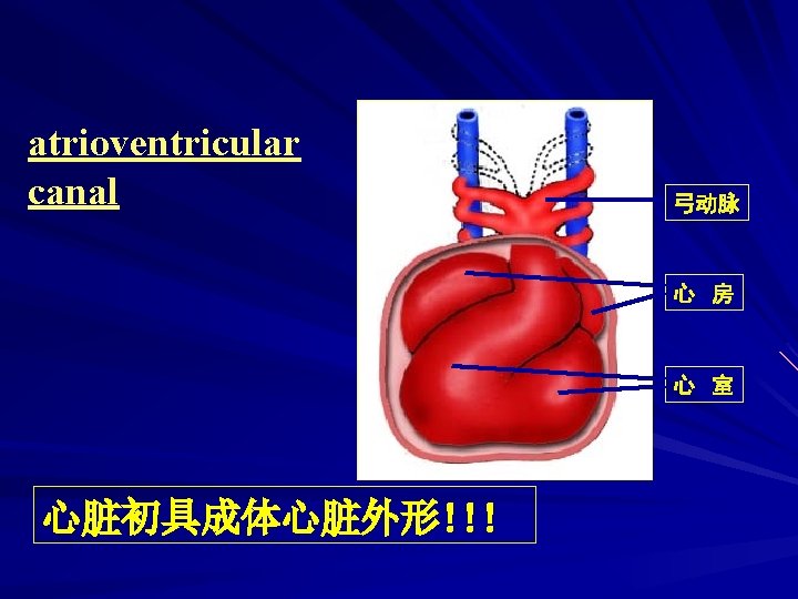
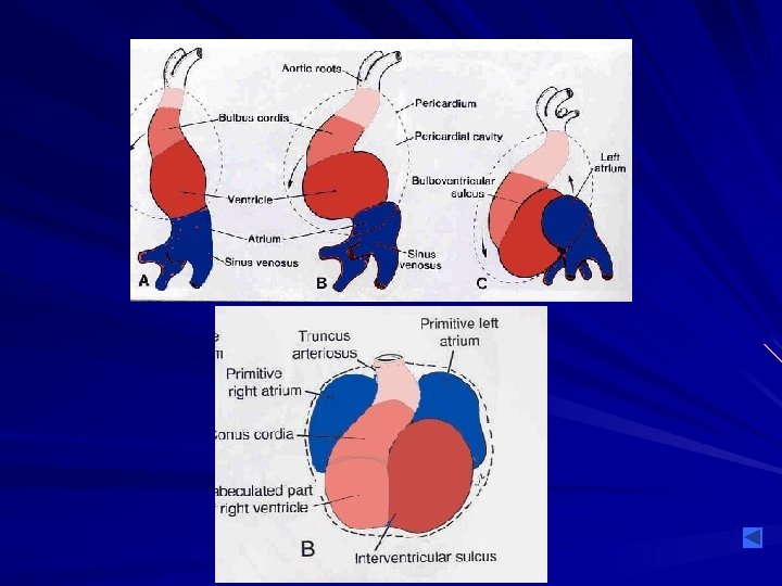
2. Establishment of the external appearance of the heart 心脏外形的演变 动脉端 心 管 静脉端

出现三个膨大 three dilatations 心球 bulbus cordis 心室 Primitive ventircle 心房 Primitive atrium 心 球 心 室 心 房

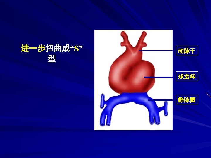
静脉窦 sinus venosus 动脉干 truncus arteriosus 心管扭曲成“U”型 球室袢 bulboventricular loop 动脉干 心 球 心 室 心 房 静脉窦
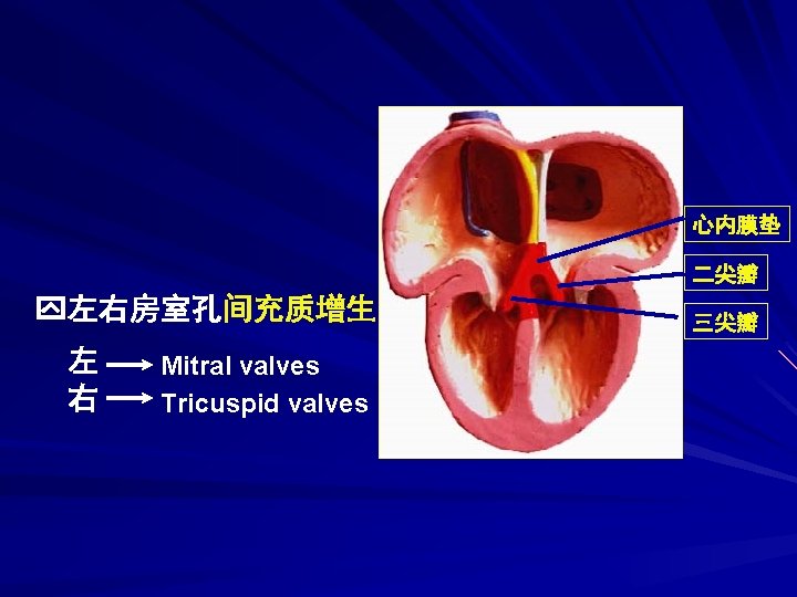
房室管的分隔 Separation of the atrioventricular canal 心内膜垫 endocardiac")
3. Formation of the cardiac septa (1)房室管的分隔 Separation of the atrioventricular canal 心内膜垫 endocardiac cushion Left atrioventricular canal Right atrioventricular canal

Separation of the atrioventricular canal 心内膜垫 腹、背心内膜垫融合 左房室孔 右房室孔
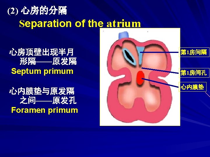

Separation of the atrium 小穿孔 第 1房间孔 心内膜垫

Separation of the atrium 小穿孔融合成大孔—— 继发孔 Foramen secundum 原发孔闭合 心房顶壁又出现新月形 隔---继发隔 Septum secundum appears in the right of the septum primum 第 2房间隔 第 2房间孔 第 1房间隔 心内膜垫
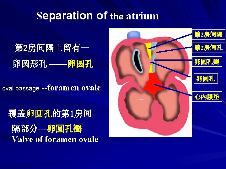

Separation of the atrium blood can flow from right atrium toward the left atrium before 血液经右房 birth 左房 after birth 左房 右房 卵圆孔关闭 左、右心房完全分隔 two septums fuse to separate atrium completely
 Separation of the ventricles 心室底壁出现肌性嵴 -------室间隔肌部 Muscular part of interventricular septum 心内膜垫 左心室")
(3) Separation of the ventricles 心室底壁出现肌性嵴 -------室间隔肌部 Muscular part of interventricular septum 心内膜垫 左心室 右心室 室间隔肌部
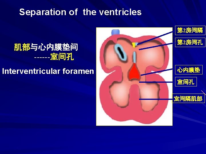

Separation of the ventricles 肌部与心内膜垫间形 成隔膜---室间隔膜部 Membranous part of interventricular septum —— made up of bulbar ridge , endocardial cushion and muscular part of interventricular septum 心内膜垫 室间隔膜部 室间隔肌部

Separation of the ventricles
静脉窦的演变和永久性左、右心房的形成 Evolution of the sinus venosus and formation of the permanent atrium right sinus")
(4)静脉窦的演变和永久性左、右心房的形成 Evolution of the sinus venosus and formation of the permanent atrium right sinus horns left sinus horns vitelline vein umbilical vein common cardinal vein

distal part: oblique vein left sinus horn proximal portion: coronary sinus right sinus horn: right atrium right auricle

left auricle
 动脉干与心球的分隔 Separation of the truncus arteriosus & bulbus cordis 第 5周 动脉干嵴 动脉干")
(5) 动脉干与心球的分隔 Separation of the truncus arteriosus & bulbus cordis 第 5周 动脉干嵴 动脉干 心球嵴 心球 动脉干嵴 truncal ridge 心球嵴 bulbar ridge

two spiral ridges grow , twist around each other and fuse to form a spiral aortico-pulmonary septum 主动脉 肺动脉隔 嵴融合------主动脉肺动脉隔 aortico-pulmonary septum

Pulmonary trunk Ascending aorta

半月瓣 隆起 半月瓣 aortic valves pulmonary valves

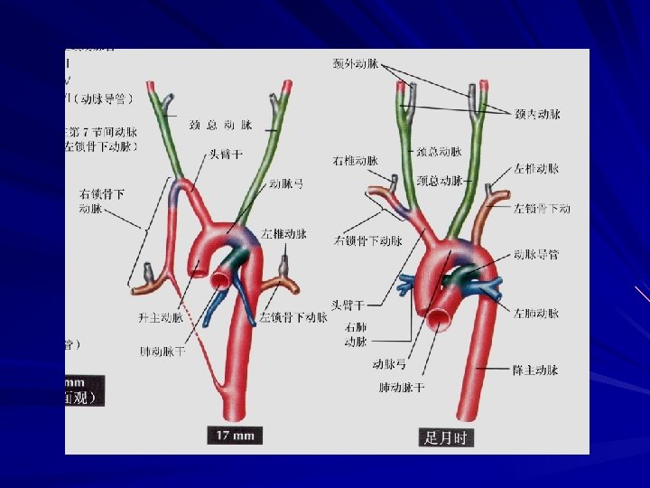
Ⅳ. Development and derivative of the aortic arches

3 rd common carotid artery internal carotid artery 4 th Left: arch of the aorta (7 th intersegmental A ---left subclavian A) Right: brachiocephalic trunk (with 7 th intersegmental A) subclavian artery 6 th Proximal part: pulmonary artery Distal part: Right: disappear Left: ductus arteriosus

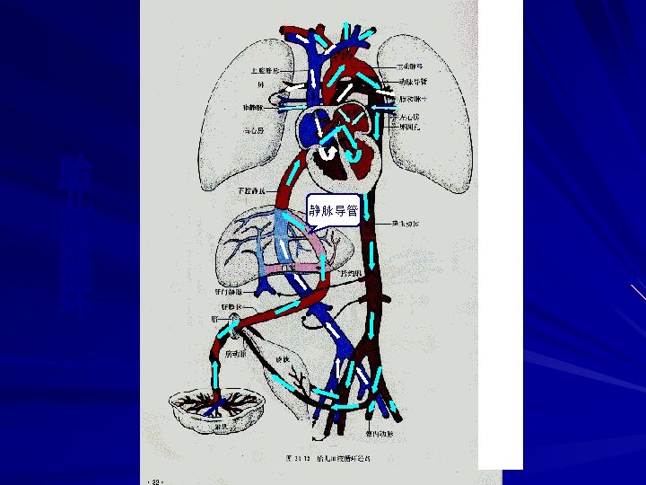
Ⅴ. Blood circulation of fetus and its changes after birth 1. Fetal circulation umbilical V→ducts venosus →inferior vena cava → right atrium →foramen ovale →left atrium →left ventricle → Ascending aorta → head and neck region →superior vena cava →right atrium →right ventrical → ductus arteriosus → descending aorta gut and lower region of the body placenta umbilical A

head veins from neck upper limb superior vena cava right atrium L subclavian A A L common carotid A innominate A oval foramen aorta left atrium left ventricle right ventricle pulmonary A pulmonary V ductus arteriosus inferior vena cava descending aorta trunk V from lower limb A of abdomen, pelvis, lower limb hepatic V ductus venosus portal V V from digestive tract liver umbilical vein placenta umbilical A
Umbilical A: – lateral umbilical ligaments –Umbilical V: –")
2. Circulatory changes after birth: (1)Umbilical A: – lateral umbilical ligaments –Umbilical V: – ligamentum teres hepatis (2)Ductus venosus: – ligamentus venosum (3) Ductus arteriosus: – ligamentum arteriosum (4) Oval foramen: –oval pit

Ⅴ. Congenital malformations of the cardiovascular system 1. Atrial septal defect patency of the foramen ovale

2. Ventricular septal defect
Transposition of aortic &")
3. Abnormal division of the truncus arteriosus and bulbus cordis (1)Transposition of aortic & pulmonary arteries 主 A 肺 A 右心室 左心室
 Stenosis of Aorta or Pulmonary Atery 主A狭窄 肺A狭窄 unequal division of aortico-pulmonary septum")
(2) Stenosis of Aorta or Pulmonary Atery 主A狭窄 肺A狭窄 unequal division of aortico-pulmonary septum
 Persistent truncus arteriosus")
(3) Persistent truncus arteriosus
Tetralogy of Fallot pulmonary stenosis Overriding aorta ventricular septal defect hypertrophy of the right")
(4)Tetralogy of Fallot pulmonary stenosis Overriding aorta ventricular septal defect hypertrophy of the right ventricle 肺A狭窄 主A骑跨 室间隔缺损 右心室肥大

4. Patent ductus arteriosus 正常 异常
- Slides: 60