
CHAPTER 24 Diagnostic Microbiology and Immunology GrowthDependent Diagnostic
CHAPTER 24 Diagnostic Microbiology and Immunology
Growth-Dependent Diagnostic Methods Isolation of Pathogens from Clinical Specimens

• Proper sampling and culture of a suspected pathogen is the most reliable way to identify an organism that causes a disease (Figure 24. 1).
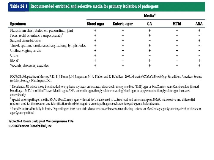
• Most clinical samples are first grown on generalpurpose media, media such as blood agar that support the growth of most aerobic and facultatively anaerobic organisms. • Enrichment culture, the use of selected culture media and incubation conditions to isolate microorganisms from samples, is an important part of clinical microbiology. • Table 24. 1 shows recommended enriched media and selective media for primary isolation of pathogens.

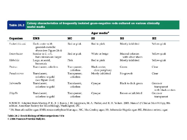
• Differential media are specialized media that allow identification of organisms based on their growth and appearance on the media. • Experienced clinical microbiologists may make a tentative identification of an isolate by observing the color and morphology of colonies of the suspected pathogen growth on various media, as described in Table 24. 2.
• Bacteremia is the presence of bacteria in the blood. • Septicemia is a blood infection resulting from the growth of a virulent organism entering the blood from a focus of infection, multiplying, and traveling to various body tissues to initiate new infections. • The selection of appropriate sampling and culture conditions requires knowledge of bacterial ecology, physiology, and nutrition.

Growth-Dependent Identification Methods • Traditional methods for identifying pathogens depend on observing metabolic changes induced as a result of growth. These growth-dependent methods provide rapid and accurate pathogen identification.
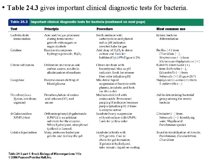
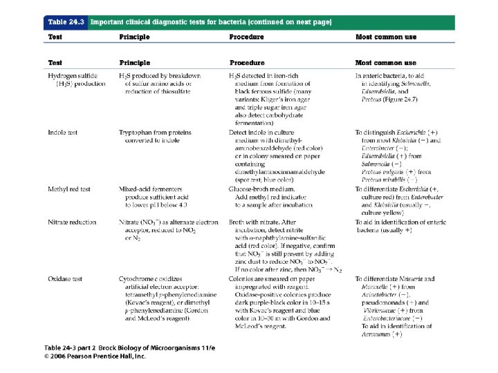
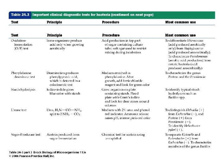
• Table 24. 3 gives important clinical diagnostic tests for bacteria.
Antimicrobial Drug Susceptibility Testing • Antimicrobial drugs are widely used for the treatment of infectious diseases. • Pathogens should be tested for susceptibility to individual antibiotics to ensure appropriate chemotherapy. This rigorous approach to antimicrobial drug treatment is usually applied only in health care settings.
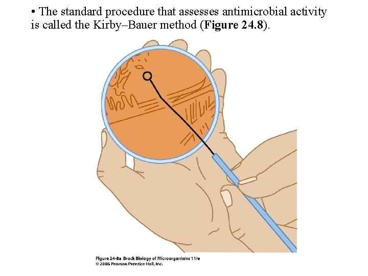
• The standard procedure that assesses antimicrobial activity is called the Kirby–Bauer method (Figure 24. 8).
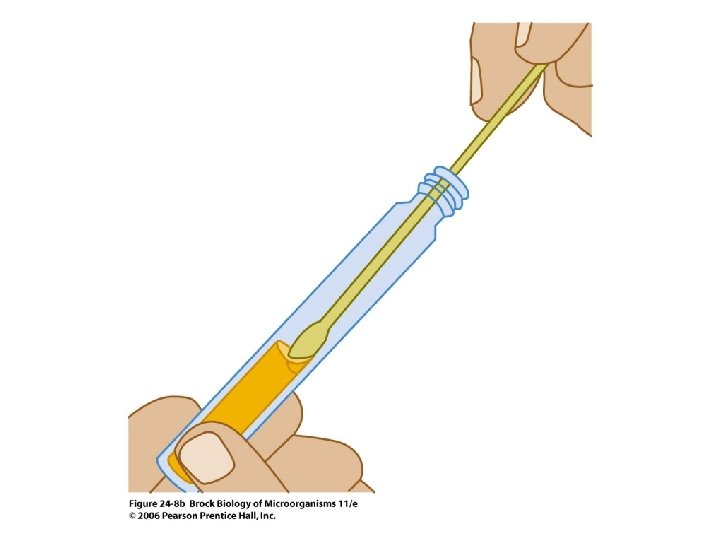
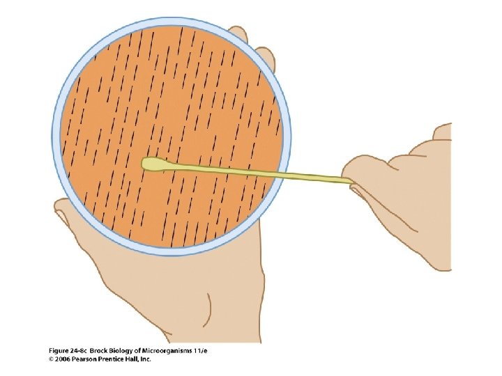
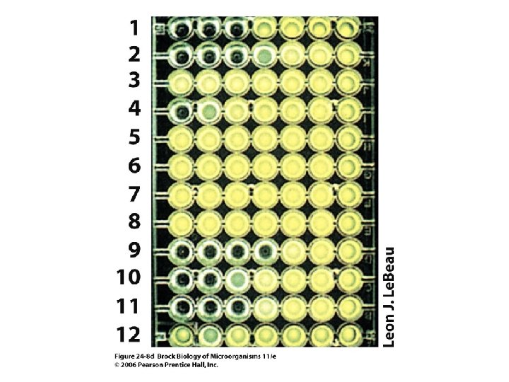
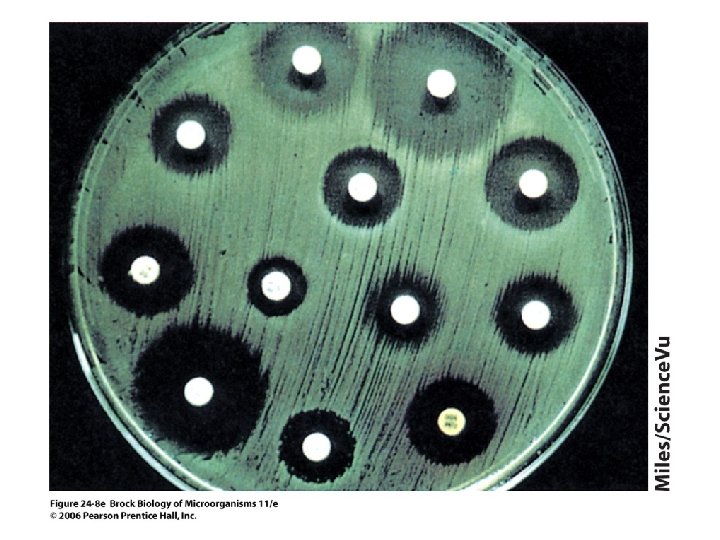
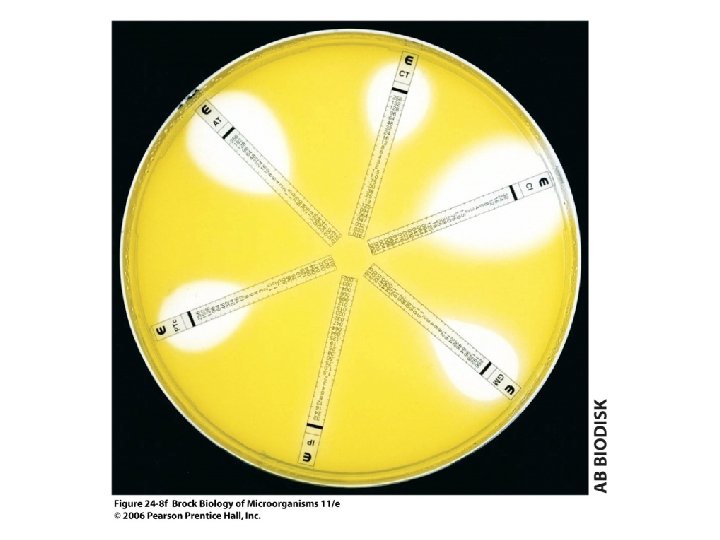
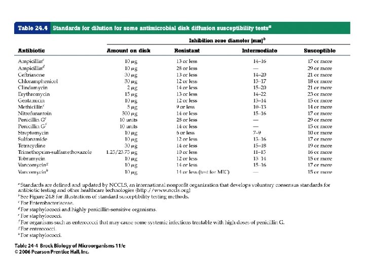
• Agar media are inoculated by evenly spreading a defined density of a suspension of the pure culture on the agar surface. Filter paper disks containing a defined quantity of the antimicrobial agents are then placed on the inoculated agar. • After a specified period of incubation, the diameter of the inhibition zone around each disk is measured. Table 24. 4 presents zone sizes for several antibiotics.
• Antibiograms are periodic reports that indicate the susceptibility of clinically isolated organisms to the antibiotics in current local use.
Safety in the Microbiology Laboratory • Safety in the clinical laboratory requires effective training, planning, and care to prevent the infection of laboratory workers with pathogens. • Materials such as live cultures, inoculated culture media, used hypodermic needles, and patient specimens require specific precautions for safe handling.
Immunology and Clinical Diagnostic Methods Immunoassays for Infectious Disease

• An immune response is a natural outcome of infection. The major aspects of immunity are summarized in Figure 24. 10.
• Specific immune responses, particularly antibody titers and skin tests, can be monitored to provide information about past infections, current infections, and convalescence (Figure 24. 11).

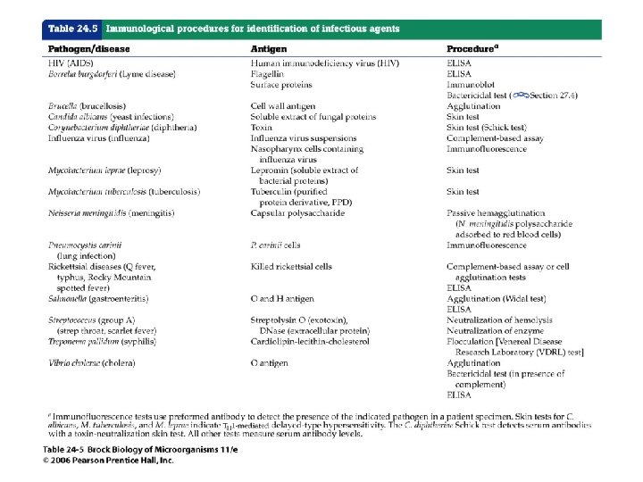
• Common immunodiagnostic tests for pathogens are shown in Table 24. 5.
Polyclonal and Monoclonal Antibodies • Polyclonal and monoclonal antibodies are used for research and clinical applications.
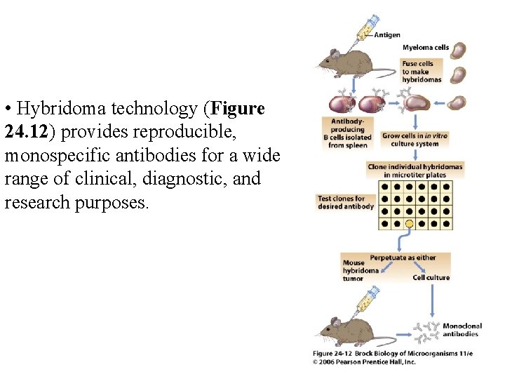
• Hybridoma technology (Figure 24. 12) provides reproducible, monospecific antibodies for a wide range of clinical, diagnostic, and research purposes.
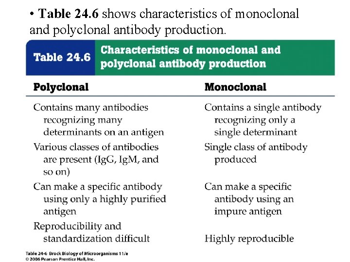
• Table 24. 6 shows characteristics of monoclonal and polyclonal antibody production.
In Vitro Antigen-Antibody Reactions: Serology • The study of antigen-antibody reactions in vitro is called serology.
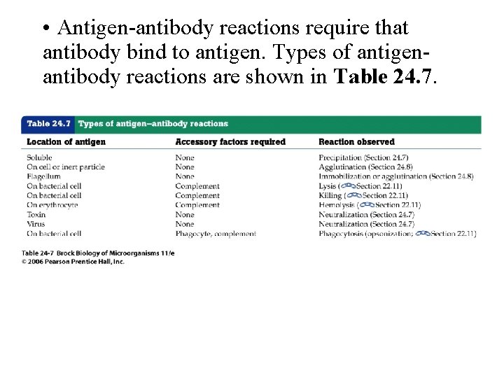
• Antigen-antibody reactions require that antibody bind to antigen. Types of antigenantibody reactions are shown in Table 24. 7.
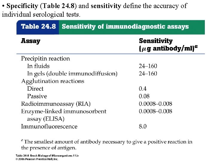
• Specificity (Table 24. 8) and sensitivity define the accuracy of individual serological tests.
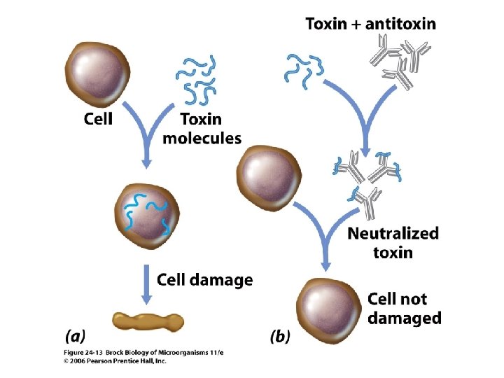
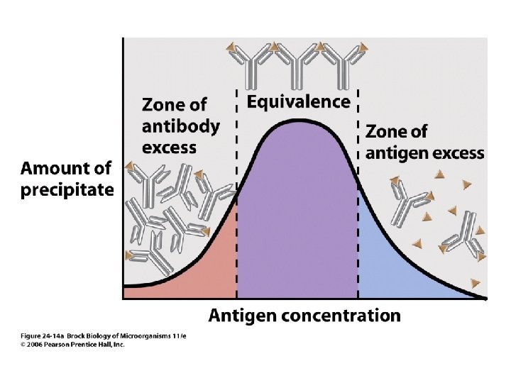
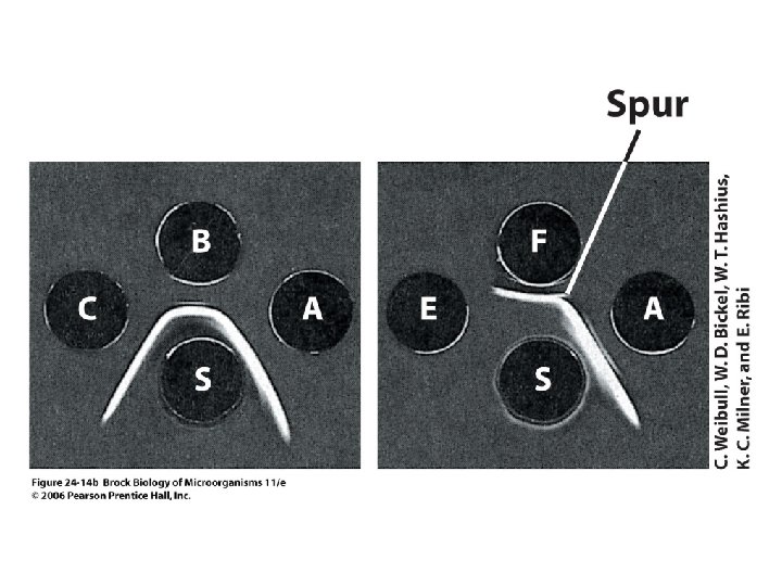
• Neutralization (Figure 24. 13) and precipitation (Figure 24. 14) reactions are examples of antigen-binding tests that produce visible results involving antigen-antibody interactions.
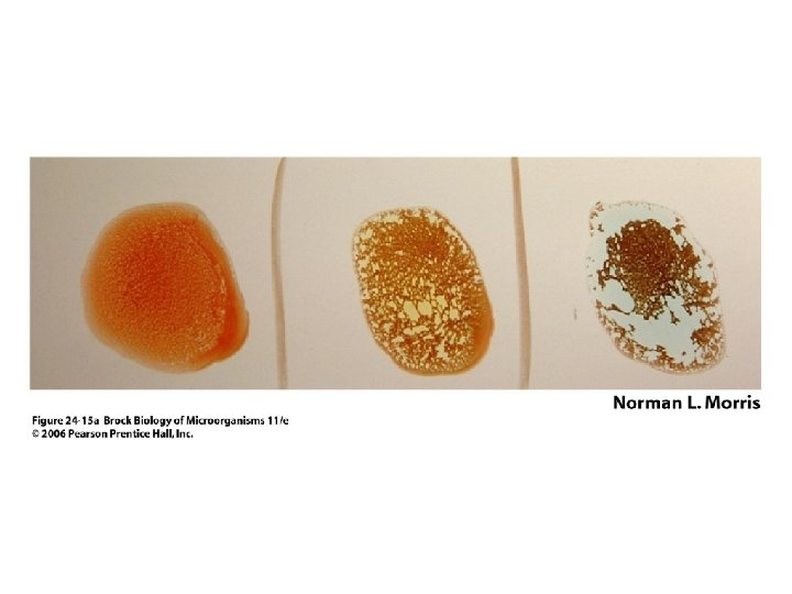
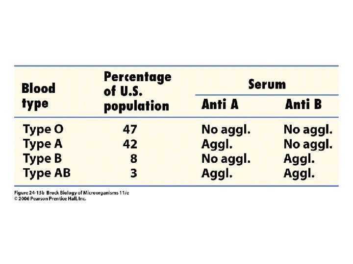
Agglutination • Direct agglutination tests are widely used for determination of blood types (Figure 24. 15).
• A number of passive agglutination tests are available for identification of a variety of pathogens and pathogen-related products. Agglutination tests are rapid, relatively sensitive, highly specific, simple to perform, and inexpensive.
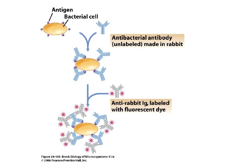
Fluorescent Antibodies • Fluorescent antibodies are used for quick, accurate identification of pathogens and other antigenic substances in tissue samples and other complex environments.
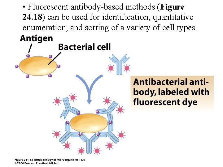
• Fluorescent antibody-based methods (Figure 24. 18) can be used for identification, quantitative enumeration, and sorting of a variety of cell types.
Enzyme-Linked Immunosorbent Assay and Radioimmunoassay
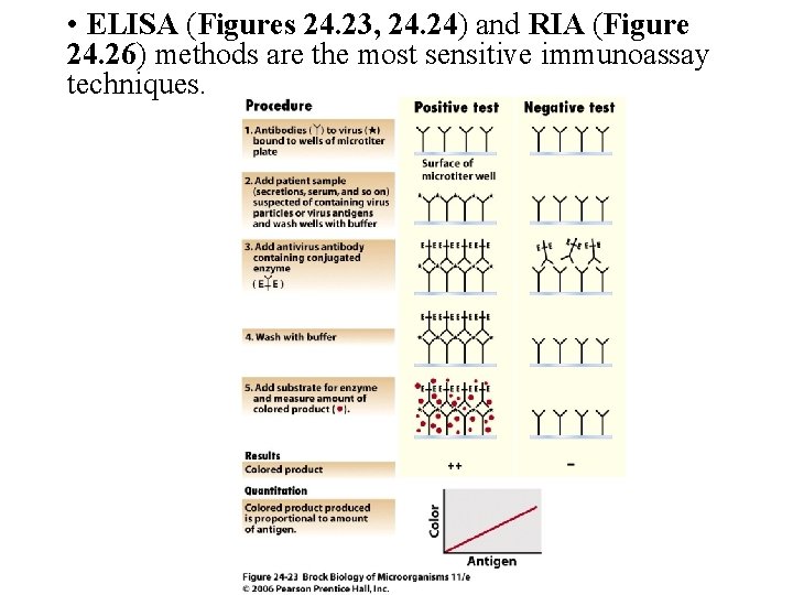
• ELISA (Figures 24. 23, 24. 24) and RIA (Figure 24. 26) methods are the most sensitive immunoassay techniques.
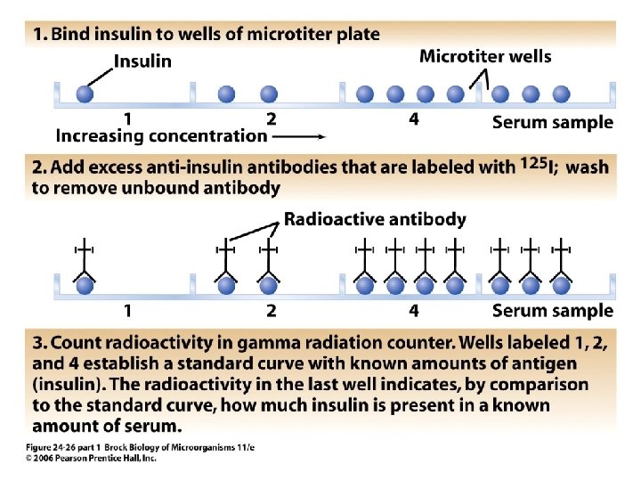
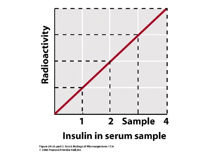
• Both involve linking a detection system, either an enzyme or a radioactive molecule, to an antibody or antigen, significantly enhancing sensitivity. • ELISA and RIA are used for clinical and research work; tests have been designed to detect either antibody or antigen in many applications.
Immunoblot Procedures • Immunoblot (Western blot) procedures (Figure 24. 27) are used to detect antibodies to specific antigens or to detect the presence of the antigens themselves.

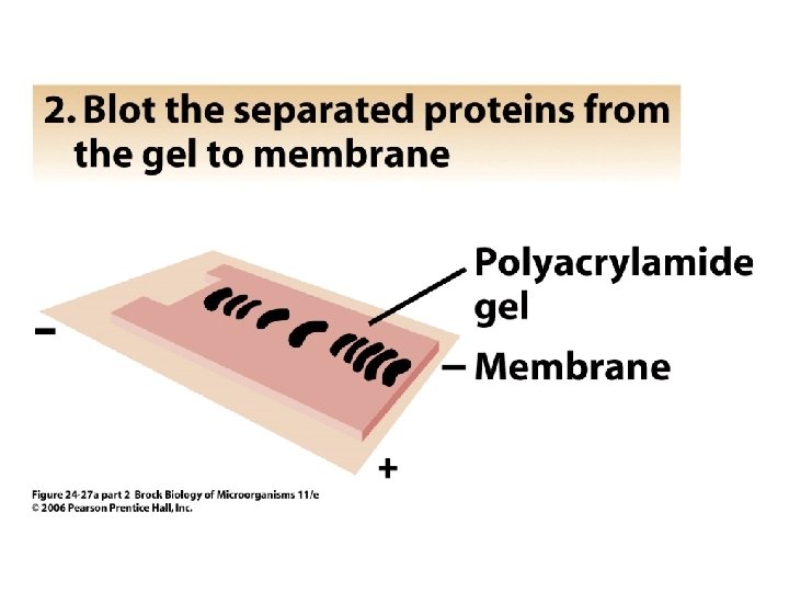
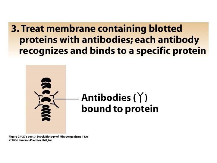
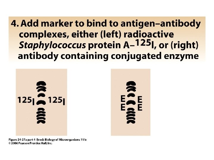
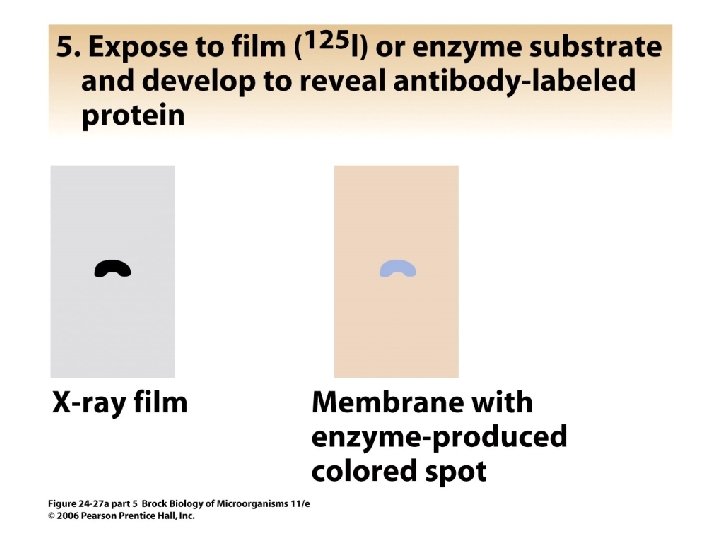
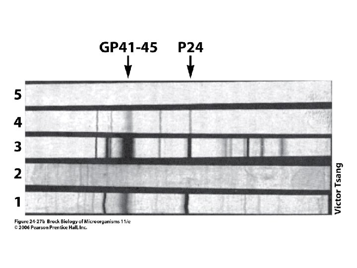
• The antigens are separated by electrophoresis, transferred (blotted) to a membrane, and exposed to antibody. • Immune complexes are visualized with enzyme-labeled or radioactive secondary antibodies. Immunoblots are extremely specific, but procedures are complex and timeconsuming.
Molecular and Visual Diagnostic Methods Nucleic Acid Methods
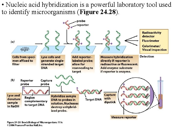
• Nucleic acid hybridization is a powerful laboratory tool used to identify microorganisms (Figure 24. 28).
• To design a nucleic acid probe, a nucleic acid sequence specific for the microorganism of interest must be available. • Perhaps the most widespread use of probebased technology is in the application of gene amplification (PCR) methods. Various DNAbased methodologies are currently used in clinical, food, and research laboratories.
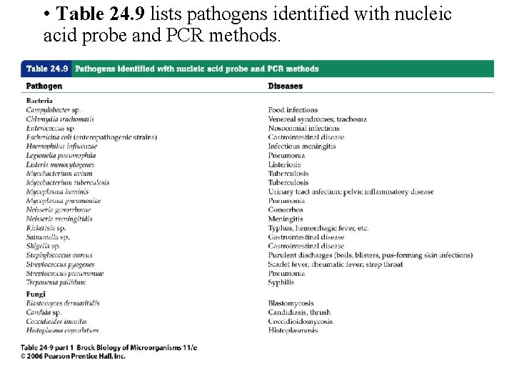
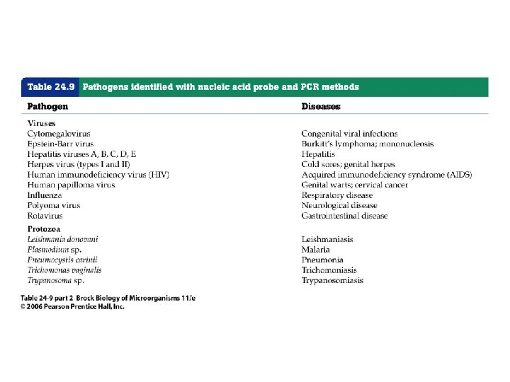
• Table 24. 9 lists pathogens identified with nucleic acid probe and PCR methods.
Diagnostic Virology • Virus propagation in vitro can be accomplished only in tissue culture. Therefore, most diagnostic techniques for viral identification are not growth-dependent but routinely rely on immunoassays and nucleic acid–based techniques. • Electron microscopy techniques are useful for direct observation of viruses in host samples.
- Slides: 65