CHAPTER 20 THE KNEE ANATOMY OF THE KNEE


, gastrocnius, gracilis, sartorius, plantaris and")


- Slides: 63
CHAPTER 20 THE KNEE
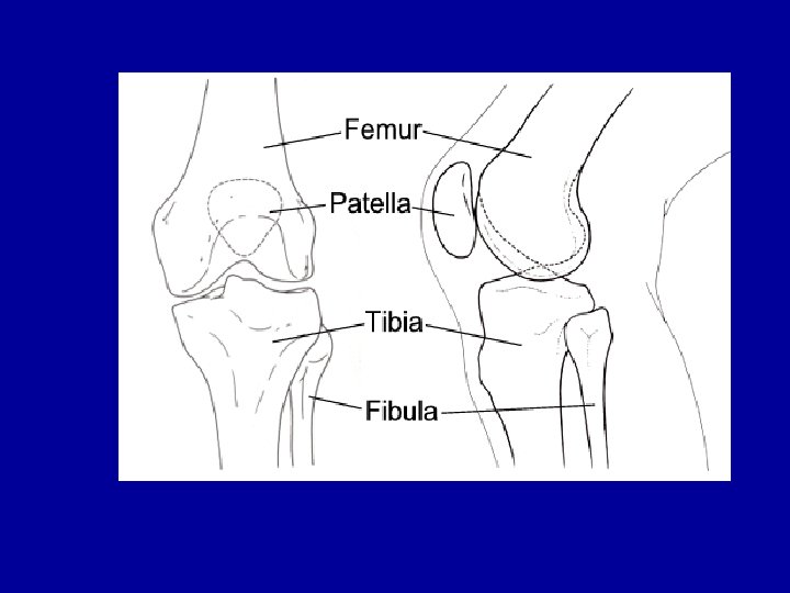
ANATOMY OF THE KNEE • BONE: - Femur: superior, largest bone in the body - Patella: sesamoid bone - Tibia: inferior and medial to the knee - Fibula: inferior and lateral to the knee
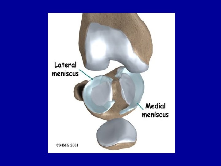
MENISCI • The menisci are two oval fibrocartilages • Cushion the head of the tibia and the head of the fibula • Medial meniscus: C-shaped • Lateral meniscus: O-shaped
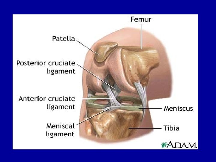
STABILIZING LIGAMENTS • Include cruciate and collateral ligaments • Cruciate: -ACL: Anterior Cruciate ligament * attaches below and in front of the tibia; then passes backward, attachin to lateral condyle. *Commonly injured in females *Surgically repaired
PCL • PCL: Posterior Cruciate Ligament *Stronger of the 2, crosses from the back of tibia in the upward, forward and medial direction and attaches to the medial condyle of the femur. * Is not usually surgically repaired - lack of blood supply makes repairing difficult
THE COLLATERAL LIGAMENTS • MCL: Medial Collateral Ligament -Attaches on medial side of femur to medial head of tibia. - Heals on its own, not usually surgically repaired • LCL: Lateral Collateral Ligament -Attaches to the lateral condyle and lateral head of fibula
JOINT CAPSULE • Includes, bones, ligaments, meniscus, muscles, tendons, bursa, fat pads and synovial fluid • Largest joint capsule in the body
KNEE MUSCULATURE • FLEXION: Hamstrings (biceps femoris, semitendonosus, semimembranosus), gastrocnius, gracilis, sartorius, plantaris and politeus. - all muscles and posterior side of body * EXTENSION: Quadriceps (rectus femoris, vastus medialis, vastus intermedius, and vastus lateralis). All on anterior side
Anterior muscles extension
Posterior muscles. . flexion
NERVE AND BLOOD SUPPLY • Nerves: Tibial nerve, Femoral nerve and the peroneal nerve • Blood supply: Popliteal artery and femoral artery.
ACTIONS OF THE KNEE • • • FLEXION EXTENSION ROTATION GLIDING ROLLING
HOPS • HISTORY: What, where and when • OBSERVATION: What do you see? - Walking - Leg alignment - Patellar orientation: * Genu Valgum: knocked knee * Genu Varum: bowlegged * Genu recurvation: hyperextended knees
Genu valgum Knocked knee Genu varum bowlegs Genu recurvatum hyperextended knees
HOPS • PALPATION: BONEY PALPATION - BONES: patella tibial plateau head of the fibula condyls of femur - SOFT TISSUE: Quads, Hamstring, Gastroc Ligaments (ACL, PCL, MCL, LCL) Meniscus
HOPS • SPECIAL TESTS: ROM - Anterior/posterior Draw test: cruciate ligaments - Lachman’s Test: Cruciate ligaments - Pivot shift test: ACL injury - Valgus/varus stress test: Collateral ligaments - Meniscal test: Mc. Murrays test, Apley’s compression and distraction tests
Anterior draw test Posterior draw test
Lachman’s Test
Lachman’s Test
Mc. Murray’s Meniscus test
Apley’s Compression and Distraction test
FUNCTIONAL EXAMINATION • Patellar Exam: -Q angle: angle between the hips and the patella. Normal is 10 degrees for males 15 degrees for females - If angle is in excess of 20 degrees it is considered to be abnormal - A angle: Measures the patellar orientation of the tibial tubercle. 35 degrees or greater causes injury.
Q ANGLE A ANGLE
FUNCTIONAL EXAM • Palpation of patella:
FUNCTIONAL EXAM • Patellar compression, Patellar grinding and Apprehension Tests Patellar compression Patellar grinding
Apprehension Test
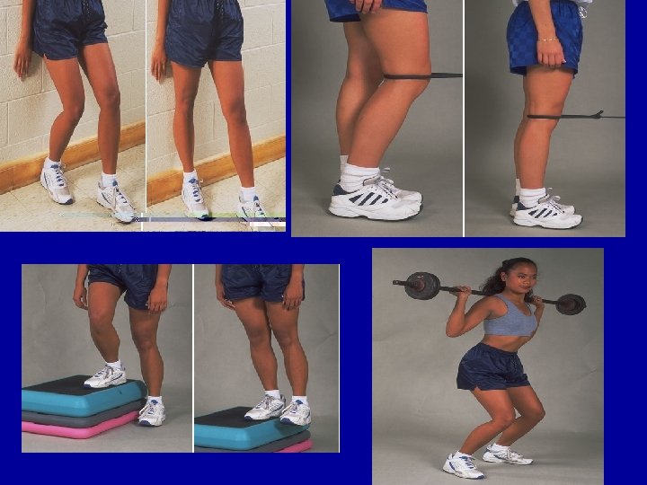
PREVENTION OF KNEE INJURIES • Physical conditioning and rehabilitation • ACL injury prevention program - strengthening of Quad, Ham and Gastroc - Proprioceptive balance board training - Neuromuscular training: wt lifting, landing cues, stretching and plyometric training - Intervention programs: strength, balance, and technique training. lilly
KNEE INJURIES • LIGAMENT INJURIES: -ACL, PCL, MCL AND LCL SPRAINS -Special tests: Draw tests - Lachman’s Test - Valgus/varus stress tests - ROM
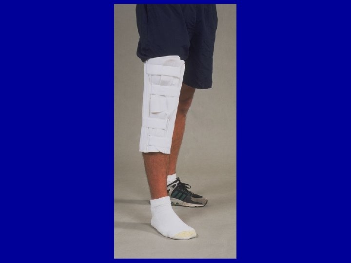
INJURY PREVENTION • Shoe type • Functional and prophylactic knee braces
GRADES • GRADE 1 SPRAIN: - a few ligamentous fibers are torn and stretched - the joint is stable during valgus stress test - there is little or no joint effusion(swelling) - There may be some joint stiffness and point tendernesslilly
Grade 1 sprain of MCL
GRADE 2 SPRAIN • Complete tear of the deep capsular ligament and a partial tear of the superficial layer of the MCL or ligament • No gross instability, min. laxity during full extension • Moderate swelling and severe joint tightness • Definite loss of ROMlilly
GRADE 3 • Complete loss of stability • Min. to moderate swelling • Immediate severe pain followed by a dull ache • Loss of motion
Grade 3 rupture of the ACL
INJURIES TO THE KNEE • Meniscal Lesions: tears to cartilage • Knee plica: fetus has 3 synovial cavities in the knee. . Adults have 1. When the body fails to absorb the cavities leftover septa (dividers) form synovial folds (plica) • Osteochondral knee fractures: ligament tears it shears off part of the chondyl of the femur
MENISCAL TEAR
KNEE PLICA
Osteochondral knee fractures
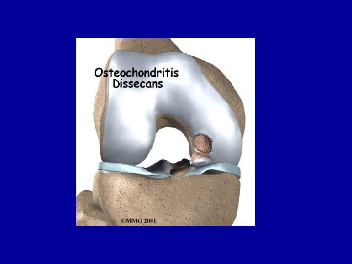
JOINT INJURIES • Osteochondritis Dissecans: partial or complete separation of a piece of articular cartilage and subchondral bone. • Loose bodies within the knee • Joint contusions • Bursitis
Loose bodies within the knee
Patella bursitis
PATELLAR CONDITIONS • Patellar fracture • Acute patellar subluxation or dislocation: -sublux: patella goes in and out on its own -dislocation: patella goes out and stays out *Chondromalacia: softening of articular cartilage. *Patellaofemoral stress syndrome: inflam of the tendon above and below the patella
PATELLAR FRACTURE
PATELLA SUBLUXATION/DISLOCATION
CHRONDOMALACIA OF PATELLA
PATELLOFEMORAL STRESS SYNDROME
OTHER INJURIES • Osgood-Schlatter disease: occurs in teens between 12 -15 years of age • Larsen-Johansson Disease • Patellar tendonitis: jumper’s or kicker’s knee • Patellar tendon rupture • Runner’s knee: cyclist’s kneelilly
OSGOOD-SCHLATTER’S DISEASElilly
LARSEN-JOHANSSON DISEASElilly
JUMPER’S OR KICKER’S KNEElilly
PATELLAR TENDON RUPTURElilly
ITB FRICTION SYNDROME OR RUNNER’S KNEE
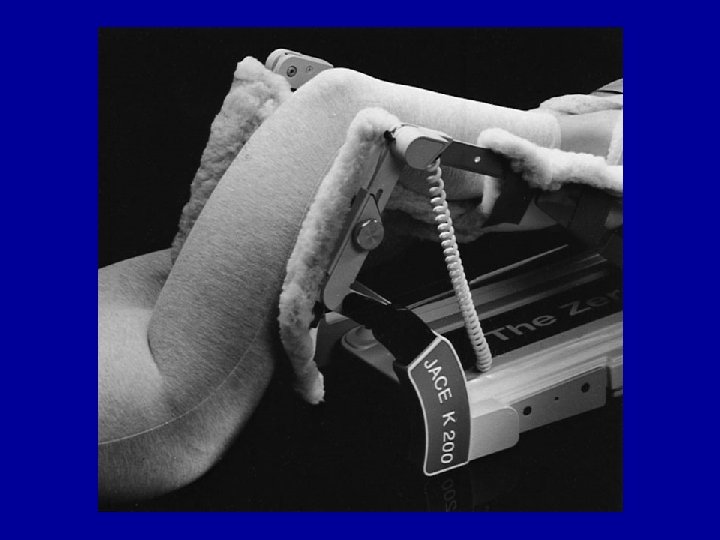
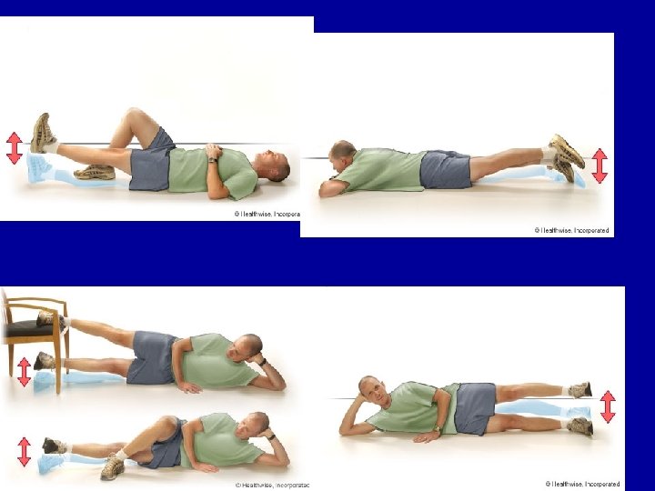
KNEE JOINT REHAB • • • General body conditioning Weight bearing Knee Joint Mobilization Flexibility: ROM Muscular strength: flexion, extension, abduction, adduction… • Neuromuscular control
FUNCTIONAL TESTING • Walking: forward, backward, straight line, curve. • Progress to jogging: forward, backward, uphill, downhill, and curves • Running • Sprinting • Figure 8’s • RETURN TO ACTIVITYlilly