
Chapter 2 pgs 27 35 Chapter 7 Chromosomes
- Slides: 44
Chapter 2 pgs. 27 -35 & Chapter 7 Chromosomes and Congenital and Hereditary Diseases
Chromosomes • Present in the nucleus and control cell activities • Exist in pairs, one derived from the male parent and one from the female parent – With the exception of sex cells, both members of the pair are similar in size, shape, and appearance and are called: homologous chromosomes • Humans normally have 23 pairs of chromosomes (46 total) – 22 pairs of autosomes & one pair of sex chromosomes • Composed of double coils of DNA combined with protein
Anatomy and Physiology • Sex chromosomes: determine genetic sex – Normal male: XY chromosomes • Large X chromosome contains genes that: direct cell activities • Small Y chromosome mainly contains genes that direct: male sexual differentiation – Normal female: XX chromosomes • X-inactivation or lyonization: only one of the two X chromosomes in females is genetically active – occurs so female doesn’t have twice as many X chromosome gene products as the male – Which x is inactivated is random – Inactive X chromosome called: Barr Body
Cell Division • Two types of cell division – Mitosis – Meiosis
Mitosis • Cell division of somatic cells – Not all mature cells are able to divide • Cardiac, skeletal muscle, & nerve cells cant divide – Connective tissue and liver cells divide to replace lost or damaged cells – Cells lining testicular tubules that produce sperm cells divide continually – Blood-forming cells in bone marrow divide continually to replace circulating cells in bloodstream
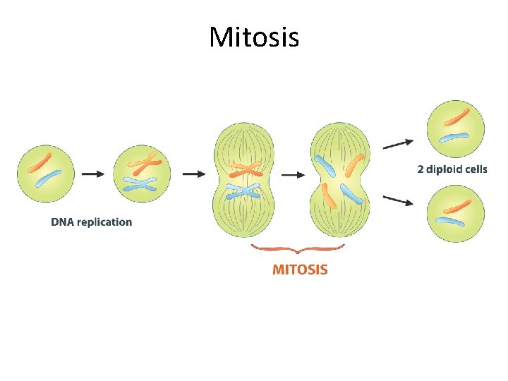
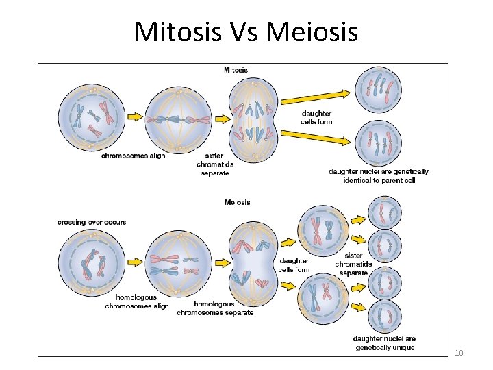
Mitosis • A single cell will divide into two identical daughter cells • There will be no reduction in the number of chromosomes – Every cell created has 23 pairs of chromosomes (46 total) – Each of two new daughter cells receives same number of chromosomes as in the parent cell – Each chromosome duplicates itself before beginning cell division
Mitosis
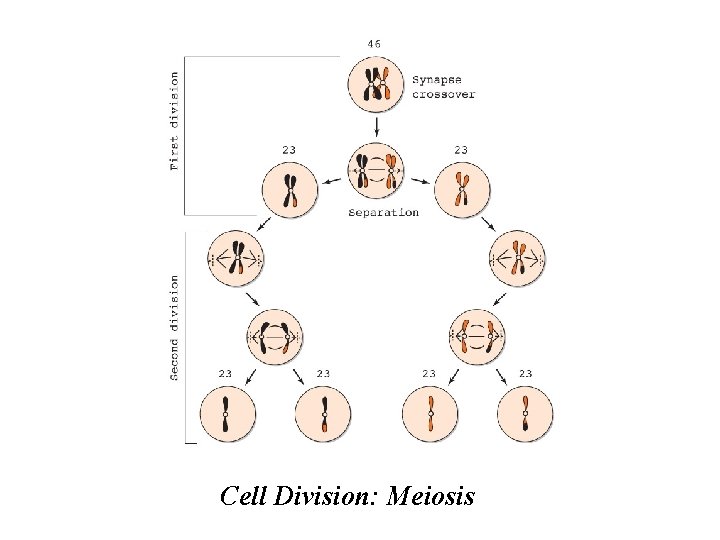
Meiosis • Characteristic of germ cells (sperm & egg) • Intermixing of genetic material between homologous chromosomes • The number of chromosomes is reduced by half • Entails two separate divisions – First meiotic division: reduces number of chromosomes by half • Daughter cells receive only half of number of chromosomes by the parent cell • Chromosomes are not exact duplicates of those in parent cell – Second meiotic division: similar to mitosis, but each cell contains only 23 chromosomes
Cell Division: Meiosis
Mitosis Vs Meiosis 10
Gametogenesis • Process of forming gametes (mature germ cells) – Gonads (testes and ovaries): contain precursor cells called germ cells capable of developing into mature sperm or ova • Spermatogenesis: development of sperm – Spermatogonia: precursor cells in the testicular tubes • Oogenesis: development of ova – Oogonia: precursor cells • Both processes have similarities and differences
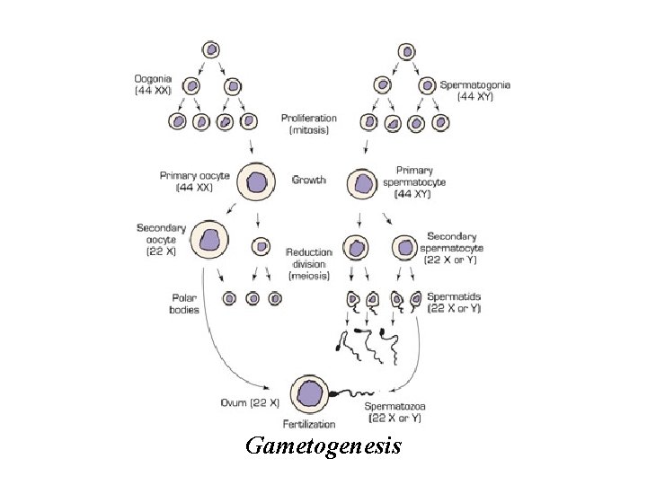
Gametogenesis
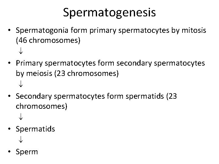
Spermatogenesis • Spermatogonia form primary spermatocytes by mitosis (46 chromosomes) • Primary spermatocytes form secondary spermatocytes by meiosis (23 chromosomes) • Secondary spermatocytes form spermatids (23 chromosomes) • Spermatids • Sperm
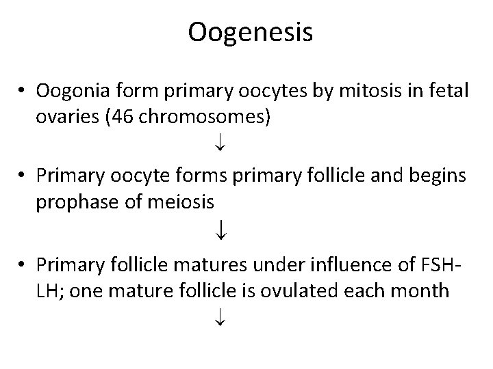
Oogenesis • Oogonia form primary oocytes by mitosis in fetal ovaries (46 chromosomes) • Primary oocyte forms primary follicle and begins prophase of meiosis • Primary follicle matures under influence of FSHLH; one mature follicle is ovulated each month
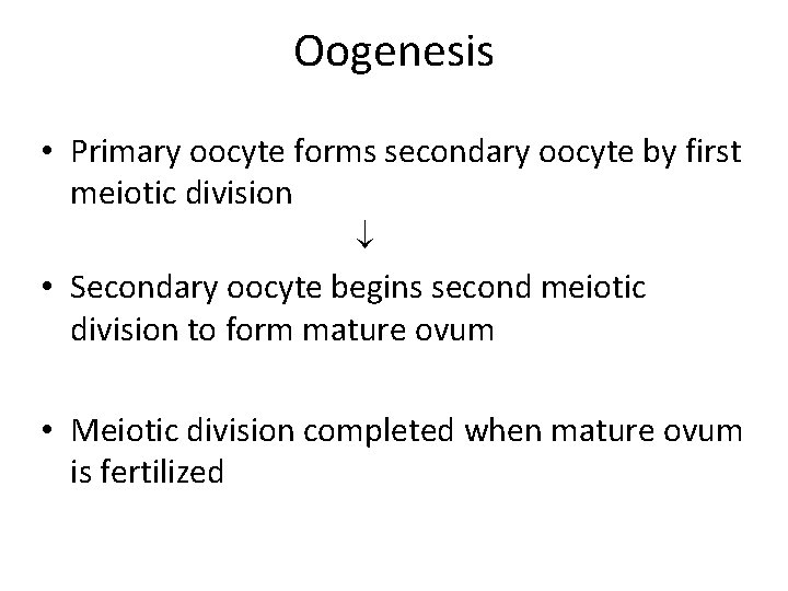
Oogenesis • Primary oocyte forms secondary oocyte by first meiotic division • Secondary oocyte begins second meiotic division to form mature ovum • Meiotic division completed when mature ovum is fertilized
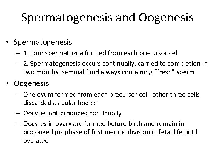
Spermatogenesis and Oogenesis • Spermatogenesis – 1. Four spermatozoa formed from each precursor cell – 2. Spermatogenesis occurs continually, carried to completion in two months, seminal fluid always containing “fresh” sperm • Oogenesis – One ovum formed from each precursor cell, other three cells discarded as polar bodies – Oocytes not produced continually – Oocytes in ovary are formed before birth and remain in prolonged prophase of first meiotic division in fetal life until ovulated
Spermatogenesis and Oogenesis • Congenital abnormalities from abnormal separation of chromosomes more frequent in older women • Ova released late in woman’s reproductive life have been held in prophase for a long time before assuming meiosis at time of ovulation (about 45 years) • Ova have been exposed for years to potentially harmful radiation, chemicals, and injurious agents • Predisposes to abnormal chromosome separation when cell division resumes at ovulation = excess or deficient number of chromosomes
Causes of Congenital Abnormalities • Approximately 2 percent of newborns born with congenital anomalies – Clinical manifestations can range from minor to incompatible with life • Additional 2 -3% defects are NOT recognized at birth – Developmental defects demonstrated later as infants grow older • 25% to 50% spontaneously aborted embryos, fetuses, and stillborn infants have major malformations • 4 major causes: – – 1. Chromosomal abnormalities 2. Abnormalities of individual genes (genetic) 3. Intrauterine injury to embryo or fetus 4. Environmental factors
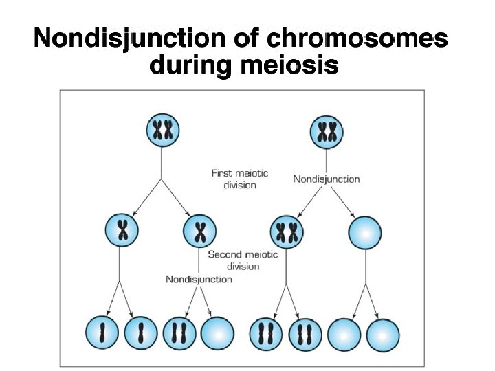
1. Chromosomal Abnormalities • Nondisjunction: failure of homologous chromosomes in germ cells to separate in first or second meiotic division – May involve either sex chromosomes or autosomes – Causes abnormalities in distribution of chromosomes between germ cells – One of two germ cells has an extra chromosome while the other lacks a chromosome
1. Chromosomal Abnormalities • Monosomy: absence of a chromosome in a cell • Trisomy: presence of an extra chromosome in a cell • Deletions: chromosome breaks during meiosis and broken piece is lost • Translocations: misplaced chromosome or part of it attaches to another chromosome
1. Chromosome Abnormalities (sex chromosomes) • Variations in normal number of sex chromosomes are often associated with: some reduction of intelligence – Y chromosome: • directs masculine sexual differentiation, associated with male body configuration regardless of number of X chromosomes present – Extra Y: • no significant effect as it mainly carries genes concerned with male sexual differentiation – Absent Y: • body configuration is female – Extra X in female: • has little effect (one X chromosome is inactivated) – Extra X in male: • has adverse effects on male development
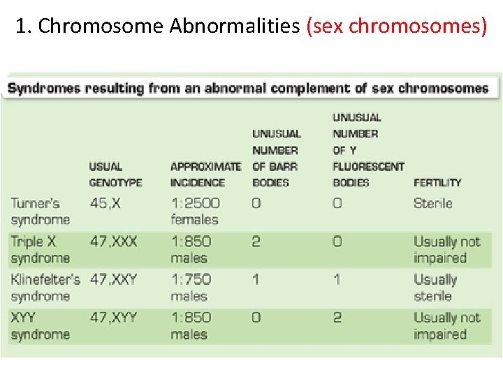
1. Chromosome Abnormalities (sex chromosomes) • Turner syndrome – Occurs in about 1 in 2500 female births – Cause: • Nondisjunction resulting in monosomy • Lack an X chromosome: designated as 45, X • Cells will lack a Barr body
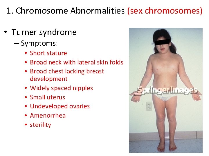
1. Chromosome Abnormalities (sex chromosomes) • Turner syndrome – Symptoms: • Short stature • Broad neck with lateral skin folds • Broad chest lacking breast development • Widely spaced nipples • Small uterus • Undeveloped ovaries • Amenorrhea • sterility
1. Chromosome Abnormalities (sex chromosomes) • Klinefelter syndrome – Occurs in about 1 in 750 male births – Cause: • Nondisjunction resulting in trisomy • Extra X chromosome: designated as 47, XXY
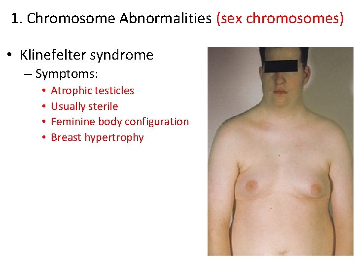
1. Chromosome Abnormalities (sex chromosomes) • Klinefelter syndrome – Symptoms: • • Atrophic testicles Usually sterile Feminine body configuration Breast hypertrophy
1. Chromosome Abnormalities (sex chromosomes)
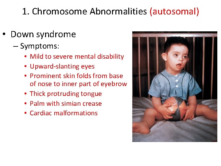
1. Chromosome Abnormalities (autosomal) • Down syndrome – Most common chromosomal abnormality – Occurs in about 1 in 600 births – Cause: • Most common cause is nondisjunction resulting in trisomy of chromosome 21 • Increased frequency with advancing maternal age – 1 in 50 if mother is > 40 years old
1. Chromosome Abnormalities (autosomal) • Down syndrome – Symptoms: • Mild to severe mental disability • Upward-slanting eyes • Prominent skin folds from base of nose to inner part of eyebrow • Thick protruding tongue • Palm with simian crease • Cardiac malformations
2. Genetically Transmitted Diseases • Autosomal dominant inheritance – Achondroplasia – Osteogenesis Imperfecta
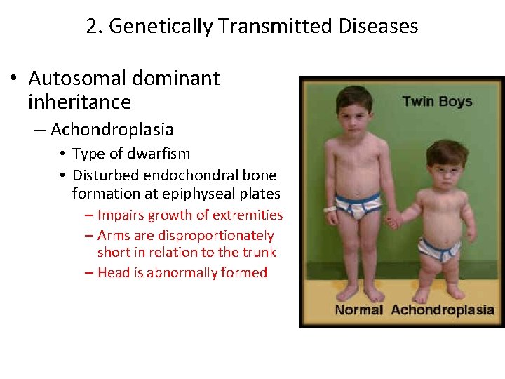
2. Genetically Transmitted Diseases • Autosomal dominant inheritance – Achondroplasia • Type of dwarfism • Disturbed endochondral bone formation at epiphyseal plates – Impairs growth of extremities – Arms are disproportionately short in relation to the trunk – Head is abnormally formed
2. Genetically Transmitted Diseases • Autosomal dominant inheritance – Osteogenesis Imperfecta • Most often caused by genetic transmission but also by gene mutation • Means imperfect bone formation • Don’t form collagen correctly in bone – Form thin delicate bones – Bones are easily broken – In severe cases infants will be born with fractures – Intrauterine fractures can heal in poor alignment causing bones to look bent
2. Genetically Transmitted Diseases • Autosomal recessive inheritance – Expressed only in a homozygous individual – Both parents must carry the abnormal gene • Phenylketonuria (PKU) • Tay-Sachs Disease • Cystic Fibrosis
2. Genetically Transmitted Diseases • Autosomal recessive inheritance – Phenylketonuria (PKU) • Caused by a deficiency of the enzyme phenylalanine hydroxylase – Required to break down the amino acid phenylalanine – Causes an issue after birth when infant drinks milk – Phenylalanine build up in blood & is found in urine – Elevated blood levels is toxic to brain cells & causes mental deficiency – Can be prevented if detected early (restricted diet) – Mandatory testing occurs within 72 hours of birth in the U. S.
2. Genetically Transmitted Diseases • Autosomal recessive inheritance – Tay-Sachs disease • Primarily affects families of Eastern Jewish origin • Caused by the absence of a lysosomal enzyme called hexosaminidase A – Causes a specific lipid to accumulate in lysosomes of nerve cells in brain, spinal cord, ANS, & retina – Lipid is toxic to cells causing cell dysfunction & eventually degeneration – Will lead to mental deterioration, neurologic dysfunction, & blindness – Symptoms start at about 6 months and children usually die by 3 -4 years old
2. Genetically Transmitted Diseases • Autosomal recessive inheritance – Cystic fibrosis • Occurs in 1 in 3000 white people • Caused by the mutation of gene which regulates the movement of salt & water in & out of epithelial cells – Water secretion is deficient in mucus production causing abnormally thick mucus – Obstructs pancreatic ducts, bronchioles, & bile ducts – Poor digestion, weight loss, & increased risk of respiratory infection occur – Sweat contains large amounts of sodium – Respiratory treatments, antibiotics, postural drainage, chestclapping, and meds are used to treat – Average life expectancy is about 35 years old
2. Genetically Transmitted Diseases • Codominant inheritance – Sickle cell trait • Heterozygous individuals have a gene for both hemoglobin S (sickle) & hemoglobin A (normal) – RBC’s will have equal amounts of normal & sickled hemoglobin • 8% of African American population – Asymptomatic unless engage in vigorous activities & high altitudes – Sickle cell anemia • Homozygous individuals have 2 hemoglobin S genes – – – RBC’s become sickled within capillaries when oxygen is low Sickled cells obstruct blood flow Heart, kidneys, spleen, & other organs become damaged Pain will occur May live to be 40 -50 years old
2. Genetically Transmitted Diseases • X-linked recessive inheritance – Females are carriers – Males will have disorder: X-linked gene functions like a dominant gene when paired with the Y chromosome • Hemophelia – Deficiency of protein required for normal blood coagulation • Duchenne’s muscular dystrophy – Degenerative muscles will be seen – Muscle tissue will look normal but are really filled with fat – Onset between 2 -5 years of age – Pelvic & leg muscles usually affected first – Life expectancy is about 20 years old
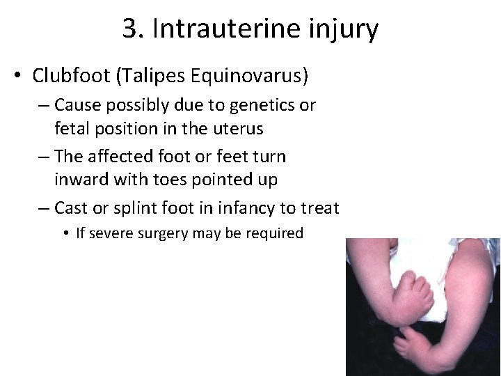
3. Intrauterine injury • Clubfoot (Talipes Equinovarus) – Cause possibly due to genetics or fetal position in the uterus – The affected foot or feet turn inward with toes pointed up – Cast or splint foot in infancy to treat • If severe surgery may be required
3. Intrauterine injury • Cerebral Palsy – Bilateral paralysis – Results from inadequate blood or oxygen to brain during development or delivery – Cause: • Maternal rubella, toxemia, prolonged labor, asphyxia from umbilical cord around neck, head trauma, meningitis – Symptoms: • Affects motor or muscle performance – Noticed if baby has trouble sucking or swallowing • Mental challenges may also be present
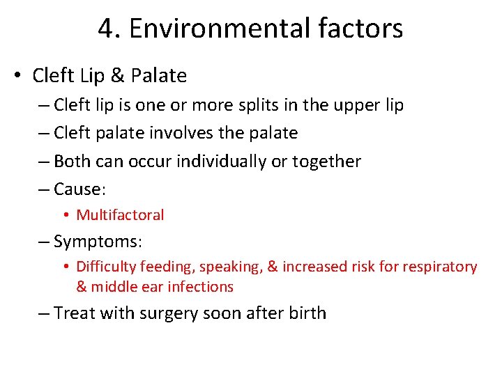
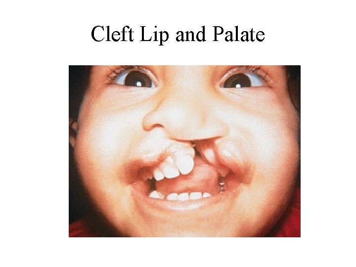
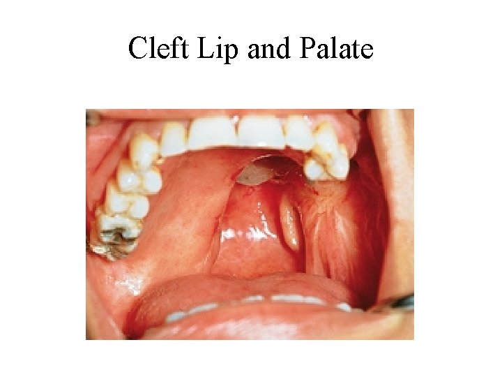
4. Environmental factors • Cleft Lip & Palate – Cleft lip is one or more splits in the upper lip – Cleft palate involves the palate – Both can occur individually or together – Cause: • Multifactoral – Symptoms: • Difficulty feeding, speaking, & increased risk for respiratory & middle ear infections – Treat with surgery soon after birth
Cleft Lip and Palate
Cleft Lip and Palate
? ? Questions ? ?