Chapter 2 Anatomy of Neurons and the Nervous

 = Brain and Spinal Cord")


 v v Lipids – a")


 routing sensory info. 2. Substantia Nigra –")

 in Latin v v")


- Slides: 30
Chapter 2: Anatomy of Neurons and the Nervous System
General Organization of the Nervous System 1. Central (CNS) = Brain and Spinal Cord 2. Peripheral (PNS) has two branches a) b) Somatic NS – whole body movement (skeletal muscles) Autonomic NS – basic bodily functions i. ii. Sympathetic NS – activating Parasympathetic NS – calming
Generic Slide
Autonomic Nervous System 1. 2. à Sympathetic – activation of this system prepares the body for action Parasympathetic – has the opposite effect, returns the body to normal, baseline homeostatic function Anatomical differences between the two systems: à Parasym. includes cranial and sacral nerves, while the sympath. consists of thoracic and lumbar nerves
Protection for your CNS v Protective membranes: meninges v Protective ‘buoy’ system in the CNS, it is made up of 5 fluid-filled parts v v The ventricles & cerebrospinal fluid The Blood-Brain-Barrier (BBB) is a protective mechanism of tightly joined endothelial cells lining all vessels that serve CNS tissue. v This barrier shields the CNS from exposure to many chemicals, bacteria and viruses.
What is a neuron, exactly? v Neurons are cells that send and receive information (this action is different than other organ systems!) And, where are they located? v Your brain and spinal cord, which together make up your CENTRAL NERVOUS SYSTEM (CNS). v The end points (I’ll tell you what that’s called later) of some neurons are outside of the CNS
Important Structures of a Neural Cell
The Outside 1. Plasma Membrane (aka the phospholipid bilayer) v v Lipids – a nice way to say fat Protein channels – the revolving doors for ions
The Inside 1. 2. 3. 4. Nucleus – the prize possession Mitochondria- the cafeteria workers and janitors Ribosomes – the protein factories Endoplasmic Reticulum (ER) – the cellular superhighway
Three Types of Neurons 1. 2. 3. Motor Neurons: the focus is on the ‘output’ in this kind of cell – information from these cells is received by muscles and glands Sensory Neurons: the focus is on the ‘input’ here – these are the cells that allow us to experience stimuli like light, sound, touch, etc. Inter-neurons: these are ‘messenger’ boys and are contained entirely within the CNS. v v Local – short messages over short distances Relay - connect circuits of neurons with other circuits in different parts of the brain
Features of a Neural Cell v Neurons have all the structures on a normal cell, with a funny shape and a few extras: 1. Soma = the cell body- all cells have a ‘body’, but because of the unique shape of neural cells, their cell bodies get a special name. 2. 3. 4. 5. Dendrites = the ‘incoming/ receiving’ end of the cell Axons – the ‘outgoing’ end of the cell ( at least on motor neurons)– electrical conduction occurs along the axon. Presynaptic terminals – the end-point of the axon. Electrical stimulation here causes chemical release. Synapses = spaces between cells – chemical communication between cells occurs here.
Example of a Unipolar Motor Neuron
More about Axons 1. Most axons are covered in a myelin sheath v 2. Nodes of Ranvier (breaks/ spaces in the myelin) Information traveling down an Axon can be described two ways: a) as moving towards a structure (afferent) v b) All sensory neurons are afferent to the CNS (i. e. information flows into the CNS from the sensory organs) as moving away from a structure (efferent) v All motor neurons are efferent to the CNS (i. e. information only flows out from the CNS to the muscles)
Helper Cells in the CNS: GLIA 1. Astrocytes – star shaped cells that help with chemical ‘reuptake’ of several axon terminals v Radial Glia – embryonic astro’s that guide neuron growth 2. Microglia – little janitors, act like immune cells 3. Oligodenrocytes (in CNS) & Schwann cells (in the periphery) build myelin sheaths around neurons/nerves, respectively.
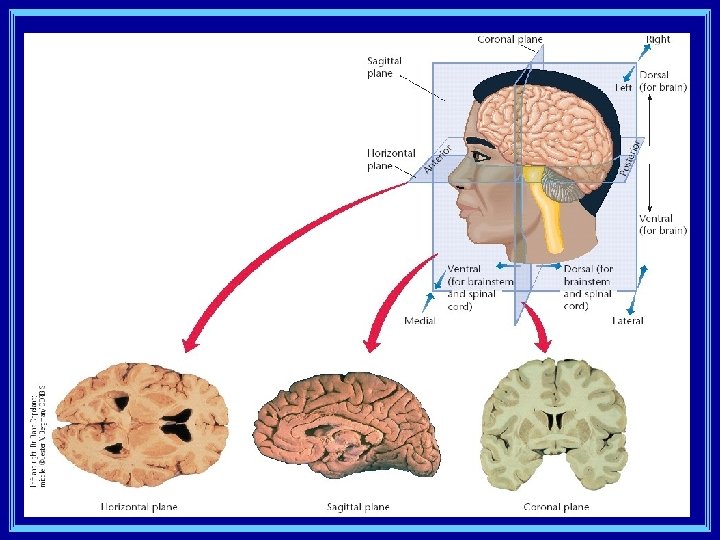
Directions in the CNS v There are 3 spatial planes the CNS is described on: a) Coronal plane = front to back v b) Sagital plane = side to side v c) Medial vs. Lateral Horizontal place = top to bottom v v Ventral vs. Dorsal Anterior vs. Posterior Location of structures are often described in relation to one another: anterior/posterior, superior/inferior, proximal/distal, ipsalateral/contralateral
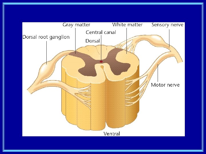
Spinal Cord Anatomy 1. Axons that enter the dorsal side of the spinal cord are on their way IN, carrying sensory information 2. Axon that leave the ventral side of the spinal cord are on their way OUT to give information from the brain to the muscles & glands.
More Fun Facts about the Cord 1. The center of your spinal cord contains the soma and dendrites belonging to ganglia and other neurons in the cord= gray matter 2. The white matter surrounding the gray matter is the myelinated axons of neurons sending their info up to the brain.
Moving on to Brain Anatomy v Before we start, I want you to know there are two ways we talk about brain anatomy: 1. We talk about structures – physical landmarks 2. And we also talk about systems – circuits of neurons that go a particular place or deliver a particular kind of information to many other places
The whole brain… v v v We spend a lot of time dividing up the different parts of the brain, but it is very important to remember that all parts work in concert, constantly exchanging information. The study of how different sensations lead to a unified perception of something is called the binding problem The idea that we use only 10% of our brain is absolutely ridiculous! Which 90% would you like to give up?
Structures in the Brain Stem 1. Medulla – considered the transition point from the cord to the brain, controls many autonomic, ‘vegetative’ functions 2. Pons – crossover point for contralateral control 3. Cerebellum – ‘little brain’, muscle coordination & perceptual tasks
More about the Stem à Two systems in the stem: à à à the reticular formation (arousal/ attention) raphe system (responsiveness to stimuli) The cranial nerves (except #1) all exit the brain from the stem
12 Cranial Nerves I. III. IV. V. VI. Olfactory = smell Optic = vision Oculomotor = pupil Trochlear = eye movement Trigeminal = eating/ feeling face Abducens = eye movement VII. Facial – taste front 2/3 of tongue VIII. Statoacoustic – hearing, balance IX. Glossopharyngeal – taste back of tongue, muscle control X. Vagus – sensation and motor control of digestive organs XI. Accessory – neck and shoulders XII. Hypoglossal - tongue
Midbrain 1. The colliculi (inferior and superior) routing sensory info. 2. Substantia Nigra – very important in voluntary movement v Cell death occurs here in Parkinson’s Disease
Forebrain 1. The limbic system resides in the forebrain: v v Olfactory bulb – smell information Hypothalamus – ‘drive’ control Amygdala – emotion & fear responding Hippocampus - memory 2. Thalamus – relay station for (almost) all sensory information before it heads to other parts of the brain 3. Pituitary gland
Finally: The Cortex 1. Cortex means ‘bark’ (as in tree) in Latin v v v 2. 3. 4. Appearance Outer layer The wrinkled, grey matter surface of the brain The cortex is made up of 6 layers of cells (laminae) Neurons are also arranged in columns that go through several laminae and contain cells of similar function. Two hemispheres (right and left) – connected by the corpus callosum
More Organization of the cortex v The cortex is divided into 4 lobes (by general function) v v Frontal lobe – prefrontal cortex (planning & judging), motor cortex, language production (Broca’s area) Parietal lobe – sensory cortex & language production Occipital lobe – visual center Temporal lobe – auditory center & language understanding (Wernicke’s area).
Examining Brain Function 1. 2. 3. 4. 5. 6. First efforts by Gall - Phrenology Computerized Imaging a) CT/CAT scan = X-ray b) PET scan = radiolabled glucose c) regional cerebral blood flow d) f. MRI Study the functional effects of brain injury Brain stimulation EEG – task related ERPs (very imprecise) Examining brains of extraordinary people (posthumously) and comparing to normals
Next Week: CHAPTER 3 Neural Communication