Chapter 18 The Skin Overview of Major Diseases


 •")
 • Vesicle—fluid-filled elevation of epidermis, smaller than 1 cm (e.")
 • Crust—skin defect covered with coagulated plasma (“scab”; e. g.")


—most common epithelial skin tumor; benign • Basal")


 • Lentigo maligna—“malignant freckle, ” flat macule, usually")


 • Alopecia – Alopecia areata – Diffuse")

- Slides: 41
Chapter 18 The Skin
Overview of Major Diseases • Traumatic lesions caused by mechanical, chemical, or thermal injuries • Infectious diseases • Immune diseases and diseases of presumptive immune etiology • Metabolic diseases and those secondary to diseases of internal organs • Tumors
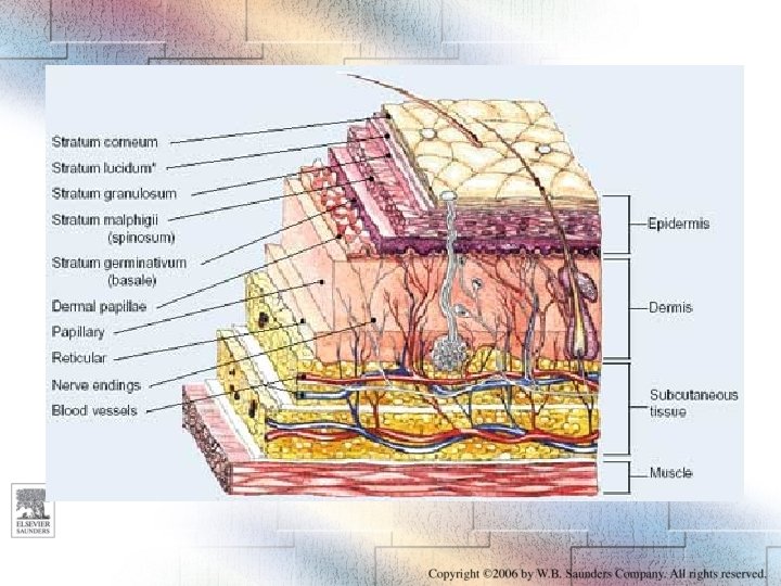
Basic Skin Lesions • Macule—flat, smaller than 2 cm (e. g. , freckle) • Patch—similar to macule but larger (e. g. , childhood rash caused by measles) • Papule—slightly elevated, smaller than 1 cm (e. g. , eczema caused by allergy) • Nodule—similar to papule but greater than 1 cm (e. g. , nevus [mole]) • Tumor—nodule greater than 5 cm (e. g. , melanoma)
Basic Skin Lesions (Cont’d) • Vesicle—fluid-filled elevation of epidermis, smaller than 1 cm (e. g. , herpesvirus lesion on the lip) • Bulla—vesicle that measures more than 1 cm (e. g. , burns) • Pustule—vesicle filled with pus (e. g. , impetigo [bacterial infection]) • Ulcer—defect of the epidermis (e. g. , syphilitic chancre)
Basic Skin Lesions (Cont’d) • Crust—skin defect covered with coagulated plasma (“scab”; e. g. , healing wound) • Scales—keratin layers covering the skin as flakes or sheets that can be scraped away (e. g. , seborrheic keratosis, psoriasis) • Squames—large scales (e. g. , as in ichthyosis) • Excoriation—superficial skin defect caused by scratching • Fissure—sharp-edged defect extending deeper into dermis (e. g. , athlete’s foot)
Skin Lesions Figure 18 -02
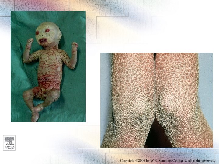
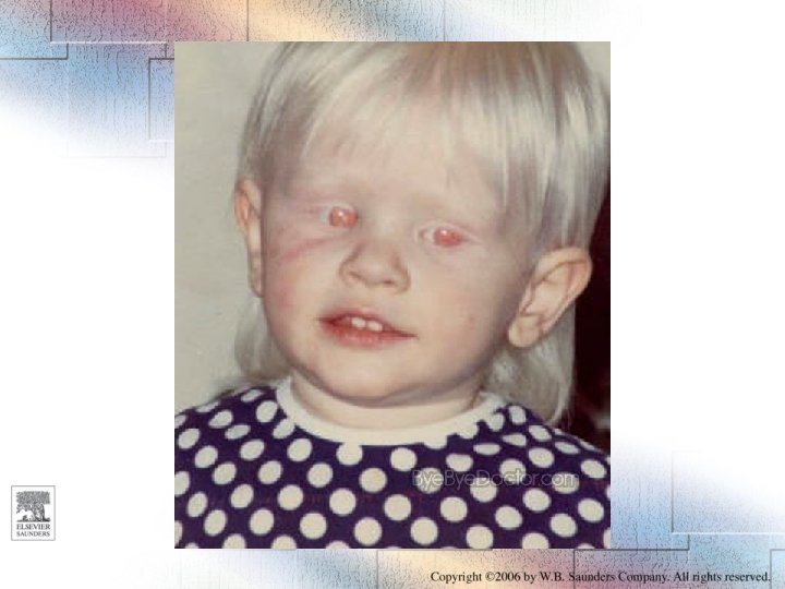
Congenital Disorders • • Nevus Ichthyosis congenita Albinism Epidermolysis bullosa
External Injury • Mechanical trauma • Thermal injury – Burns – Cold injury • Electrical injury • Radiation injury
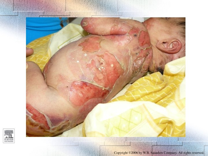
Burns—Grading • First-degree burns – Erythema and swelling, transitory and reversible • Second-degree burns – Blisters involving the epidermis; hair follicles and adnexa in the dermis spared • Third-degree burns – Full-thickness burns with massive necrosis of epidermis and parts of dermis and subcutis; cannot heal spontaneously
Burns—“Rule of Nines” From Applegate EJ: The Anatomy and Physiology Learning System: Textbook. Philadelphia, WB Saunders, 1995. Figure 18 -03
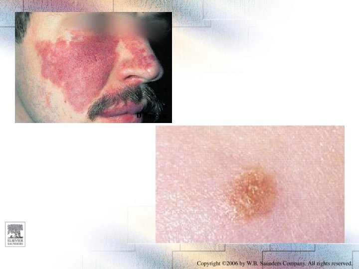
Sunlight Injury Figure 18 -04
Infections of the Skin • • Bacterial Fungal Viral Insect infestations and bites
Bacterial Infectious Diseases of the Skin • • • Impetigo Folliculitis Furuncle Carbuncle Secondary infection of other skin diseases or wounds • Septic lesions in bacteremia
Bacterial Skin Infections Figure 18 -05
Viral Skin Infections • • • Measles—maculopapular rash Chickenpox—vesicles Herpes labialis or genitalis—vesicles Herpes zoster—vesicles Human papillomavirus (HPV)—wart
Insect Infestations and Bites • Bites by blood-sucking insects – Flees – Mosquitoes – Bed bugs – Lice • Scabies—caused by Sarcoptes scabiei, which burrow into the epidermis
Acne Figure 18 -06
Eczema or Dermatitis • These terms are used to denote many forms of inflammatory skin diseases that have the following characteristics: – Present with superficial lesion such as localized edema, papules, and vesicles – Are uniformly accompanied by pruritus (itching) • They are divided into two groups: – Exogenous eczema—infection or irritants – Endogenous eczema—cause unknown
Seborrheic Dermatitis • Widespread multifactorial disease of unknown origin affecting 10% to 20% of the population • Presents with reddening, scaling, and itching and a lot of dandruff • Localized most often to nasolabial folds, eyebrows, the upper chest, and scalp • Treatment—corticosteroids, shampoos
Psoriasis • Common disease of unknown origin affecting 1% to 2% of the population • Familial incidence • Slightly elevated papules and patches with scaling, mostly on extensor surface of knees and elbows but also the face and scalp • Papules erupt in crops, which may be related to emotional stress or trauma
Neoplasms • • Tumors of epithelial cells Tumors of pigmentary cells Tumors of dermal connective tissue Tumors of blood-borne “immigrant” cells
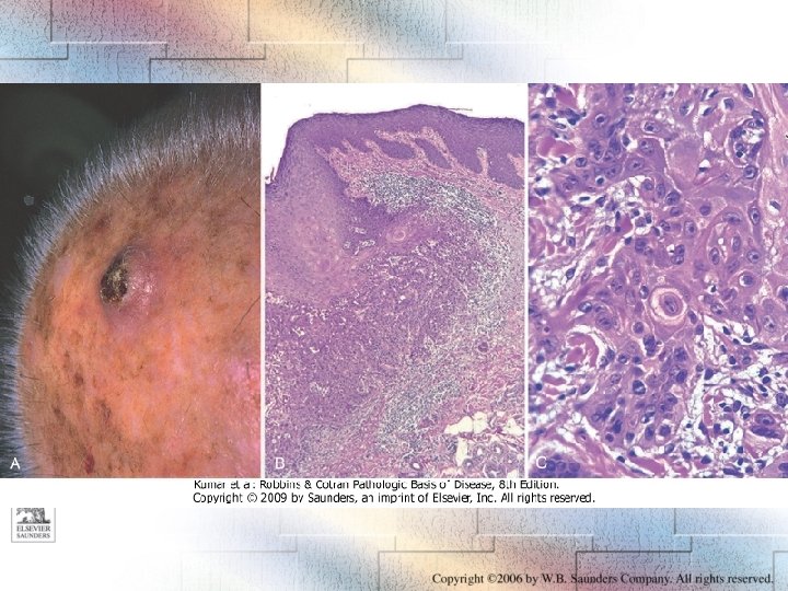
Epithelial Tumors • Seborrheic keratosis (“senile wart”)—most common epithelial skin tumor; benign • Basal cell carcinoma—most common malignant skin tumor; good prognosis • Squamous cell carcinoma—worst prognosis of tumors in this group; often preceded by actinic keratosis and carcinoma in situ _________________ Note: Basal cell and squamous carcinoma are related to sun exposure!
Basal Cell Carcinoma Figure 18 -07 A
Squamous Cell Carcinoma Figure 18 -08
Telltale Signs of Skin Cancer • Persistent, nonhealing ulcer • Ulcer or nodule of irregular shape or indistinct margins • Friable, multicolored, bleeding tissue in an ulcer • Indurated margins of an ulcer or the tissue around it • Indurated skin surrounded by atrophic and keratotic skin typical of sunlight injury
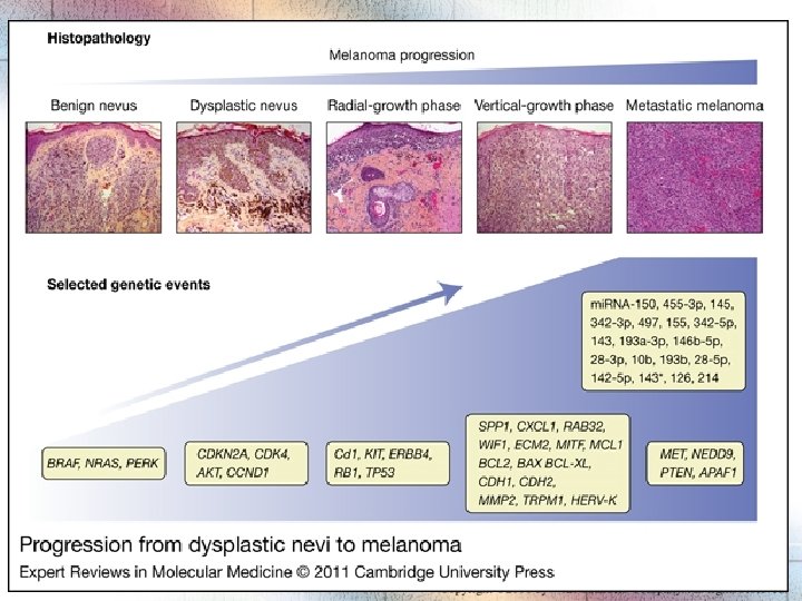
Pigmented Skin Lesions • Freckle—flat macule that responds to sunlight • Lentigo—macule or papule that is pigmented but does not respond to sun • Nevus (mole)—congenital or acquired pigmentation in form of a macule or papule or even a nodule • Malignant melanoma—malignant tumor of melanocytes
Pigmented Skin Lesions Figure 18 -09
Clinicopathologic Classification of Malignant Melanomas (MMs) • Lentigo maligna—“malignant freckle, ” flat macule, usually seen in elderly persons • Superficial spreading melanoma— 70% of MMs are diagnosed in this form; irregularly pigmented macules that enlarge • Nodular melanoma—nodule or tumor, rapidly growing, cells invading the dermis • Acral-lentiginous—on palms and plantae
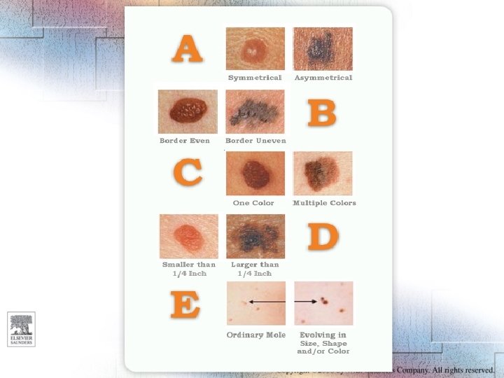
A-B-C-D of Diagnosis of Malignant Melanoma • A—asymmetry of the pigmented lesion • B—borders that are irregular • C—color; varies from dark black to dark brown to red • D—diameter of the lesion; one should worry if the lesion exceeds 6 mm in diameter
Kaposi’s Sarcoma Figure 18 -11
Diseases of the Nails • • Onychogryphosis Koilonychia Onychomycosis Paronychia
Hair Diseases • Hirsutism (hormonal or idiopathic) • Alopecia – Alopecia areata – Diffuse alopecia (males > females) • Tinea capitis (fungal infection!) • Trichotillomania (psychological urge to pull hair)
Thank you for the attention!