
Chapter 18 Gastrointestinal Alterations Copyright 2017 Elsevier Inc
- Slides: 84
Chapter 18 Gastrointestinal Alterations Copyright © 2017 Elsevier Inc. All rights reserved.
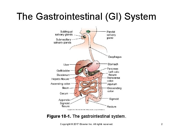
The Gastrointestinal (GI) System Figure 18 -1. The gastrointestinal system. Copyright © 2017 Elsevier Inc. All rights reserved. 2
Anatomy and Physiology Alimentary canal Accessory organs Gut wall Mucosa Ø Gastric mucosal barrier Ø Submucosa Ø Copyright © 2017 Elsevier Inc. All rights reserved. 3
Anatomy and Physiology (Cont. ) Oropharyngeal cavity Mouth Ø Salivary glands Ø Pharynx Ø Esophagus Copyright © 2017 Elsevier Inc. All rights reserved. 4
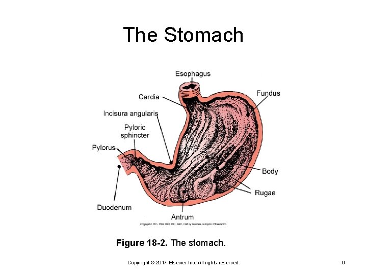
Anatomy and Physiology (Cont. ) Stomach Cardia, fundus, body, and antrum Ø Gastric glands and pyloric glands Ø Hydrochloric acid (p. H 0. 8) Ø Small intestine Duodenum, jejunum, and ileum Ø Digestion and absorption of foodstuff Ø Water, electrolyte, and vitamin absorption Ø Copyright © 2017 Elsevier Inc. All rights reserved. 5
The Stomach Figure 18 -2. The stomach. Copyright © 2017 Elsevier Inc. All rights reserved. 6
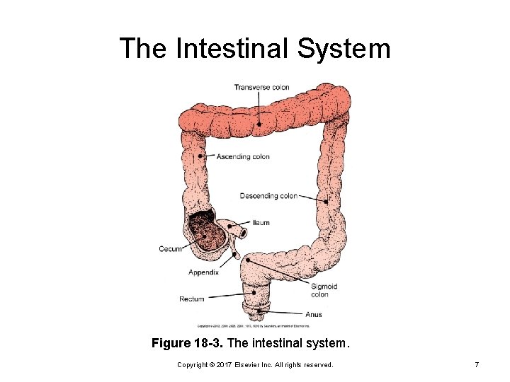
The Intestinal System Figure 18 -3. The intestinal system. Copyright © 2017 Elsevier Inc. All rights reserved. 7
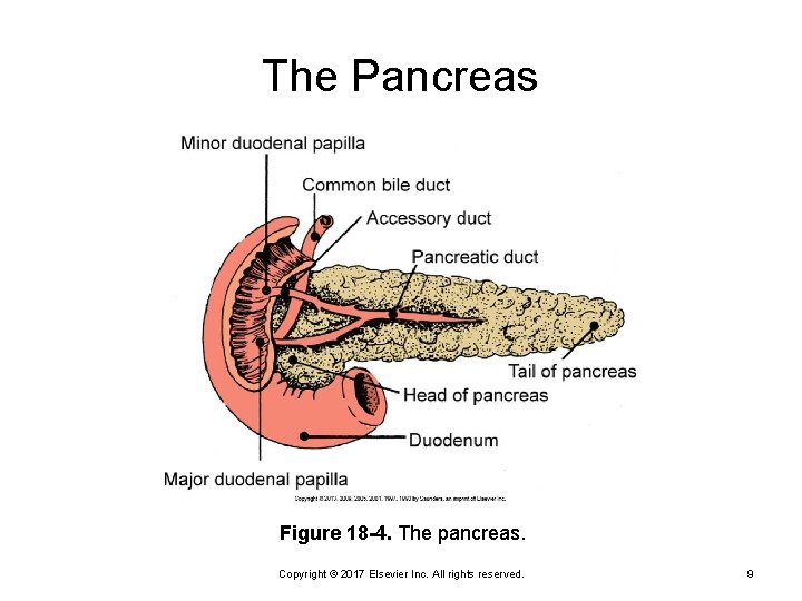
Anatomy and Physiology Large intestine Ascending, transverse, descending, and rectum Ø Absorption of water and electrolytes Ø Pancreas Exocrine = digestive enzymes Ø Endocrine = insulin and glucagon Ø Gallbladder Ø Bile Copyright © 2017 Elsevier Inc. All rights reserved. 8
The Pancreas Figure 18 -4. The pancreas. Copyright © 2017 Elsevier Inc. All rights reserved. 9
Anatomy and Physiology Liver: 400 different functions Receives 25% of cardiac output Ø Liver vascular functions • Blood storage • Filtration Ø Liver secretory function • Bile production • Bilirubin metabolism Ø Copyright © 2017 Elsevier Inc. All rights reserved. 10
Anatomy and Physiology (Cont. ) Liver: 400 different functions (Cont. ) Ø Liver metabolic functions • Carbohydrate, fat, and protein • Clotting factors • Detoxification • Vitamin and mineral storage Copyright © 2017 Elsevier Inc. All rights reserved. 11
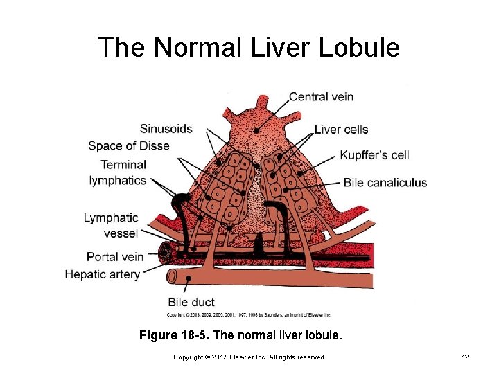
The Normal Liver Lobule Figure 18 -5. The normal liver lobule. Copyright © 2017 Elsevier Inc. All rights reserved. 12
Blood Supply of the Gastrointestinal System Splanchnic circulation Superior and inferior mesenteric and celiac arteries Copyright © 2017 Elsevier Inc. All rights reserved. 13
GI History History of swallowing, digestive, and elimination problems Nonspecific symptoms, such as anorexia and fatigue Weight changes and diet Pain assessment Surgical procedures Medications—prescription and over-the-counter Copyright © 2017 Elsevier Inc. All rights reserved. 14
GI Assessment Inspection Skin color and texture Ø Symmetry and contour of abdomen Ø Masses and pulsation Ø Peristalsis and movement Ø Copyright © 2017 Elsevier Inc. All rights reserved. 15
GI Assessment (Cont. ) Auscultate before percussion and palpation Ø Auscultate for 5 minutes before determining absence of bowel sounds Percussion Palpation Last Ø Assess rebound tenderness, rigidity, and guarding Ø Copyright © 2017 Elsevier Inc. All rights reserved. 16
Quick Quiz! Which assessment component is done last when assessing the abdomen? A. Auscultation B. Inspection C. Palpation D. Percussion Copyright © 2017 Elsevier Inc. All rights reserved. 17
Acute Upper GI Bleeding Etiology Peptic ulcer disease Ø Stress ulcer Ø Mallory-Weiss tear Ø Esophageal varices Ø Potential for hypovolemic shock Copyright © 2017 Elsevier Inc. All rights reserved. 18
Peptic Ulcer Disease Duodenal versus peptic Risk factors Smoking Ø Helicobacter pylori bacteria Ø Drugs: Nonsteroidal antiinflammatory drugs (NSAIDs), aminosalicylic acid (ASA), steroids Ø Alcohol Ø Copyright © 2017 Elsevier Inc. All rights reserved. 19
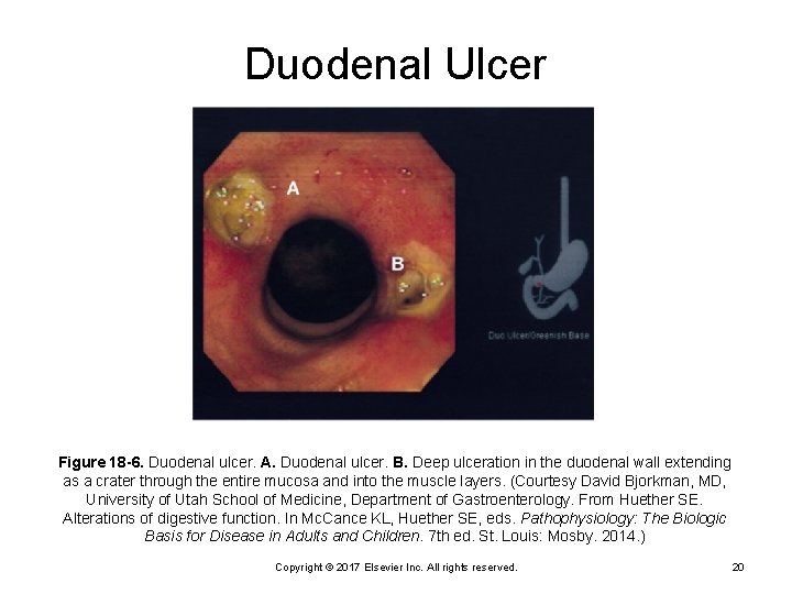
Duodenal Ulcer Figure 18 -6. Duodenal ulcer. A. Duodenal ulcer. B. Deep ulceration in the duodenal wall extending as a crater through the entire mucosa and into the muscle layers. (Courtesy David Bjorkman, MD, University of Utah School of Medicine, Department of Gastroenterology. From Huether SE. Alterations of digestive function. In Mc. Cance KL, Huether SE, eds. Pathophysiology: The Biologic Basis for Disease in Adults and Children. 7 th ed. St. Louis: Mosby. 2014. ) Copyright © 2017 Elsevier Inc. All rights reserved. 20
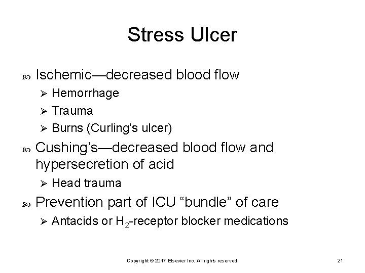
Stress Ulcer Ischemic—decreased blood flow Hemorrhage Ø Trauma Ø Burns (Curling’s ulcer) Ø Cushing’s—decreased blood flow and hypersecretion of acid Ø Head trauma Prevention part of ICU “bundle” of care Ø Antacids or H 2 -receptor blocker medications Copyright © 2017 Elsevier Inc. All rights reserved. 21
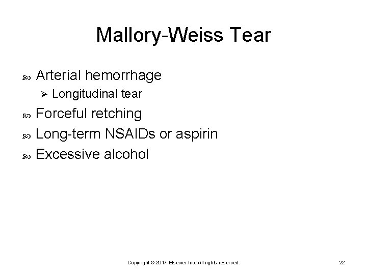
Mallory-Weiss Tear Arterial hemorrhage Ø Longitudinal tear Forceful retching Long-term NSAIDs or aspirin Excessive alcohol Copyright © 2017 Elsevier Inc. All rights reserved. 22
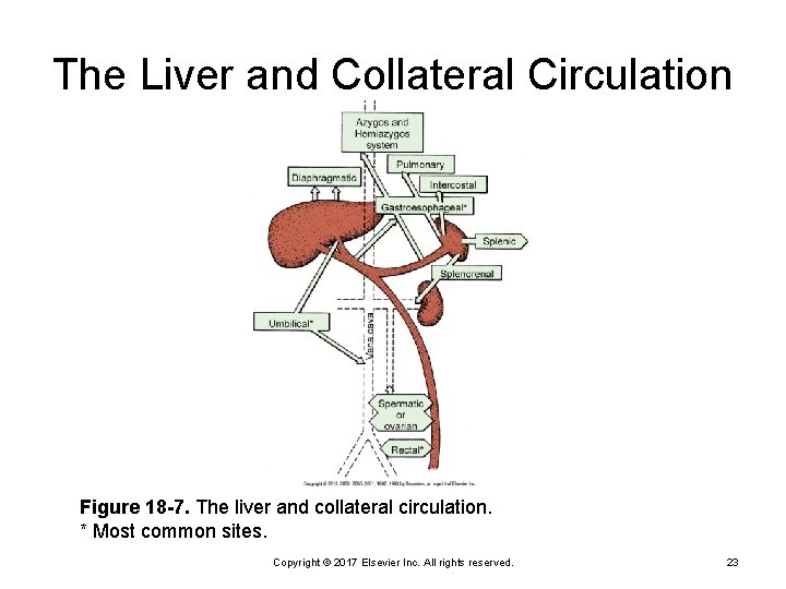
The Liver and Collateral Circulation Figure 18 -7. The liver and collateral circulation. * Most common sites. Copyright © 2017 Elsevier Inc. All rights reserved. 23
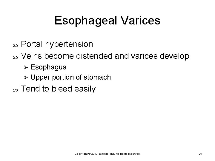
Esophageal Varices Portal hypertension Veins become distended and varices develop Esophagus Ø Upper portion of stomach Ø Tend to bleed easily Copyright © 2017 Elsevier Inc. All rights reserved. 24
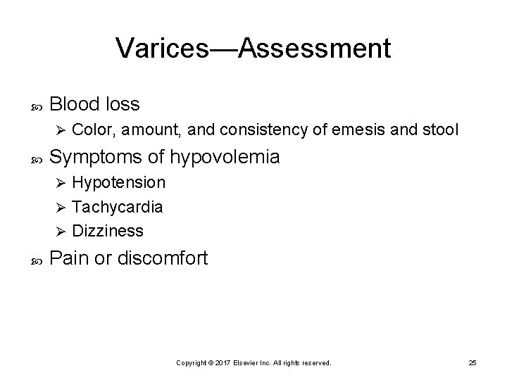
Varices—Assessment Blood loss Ø Color, amount, and consistency of emesis and stool Symptoms of hypovolemia Hypotension Ø Tachycardia Ø Dizziness Ø Pain or discomfort Copyright © 2017 Elsevier Inc. All rights reserved. 25
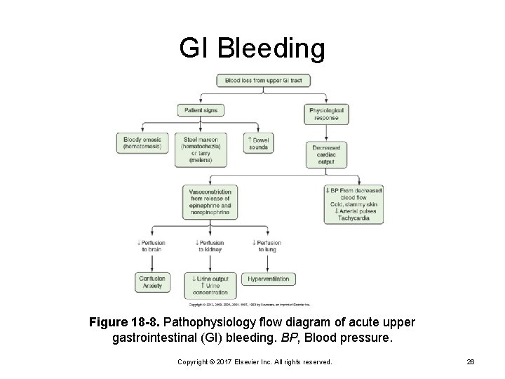
GI Bleeding Figure 18 -8. Pathophysiology flow diagram of acute upper gastrointestinal (GI) bleeding. BP, Blood pressure. Copyright © 2017 Elsevier Inc. All rights reserved. 26
Quick Quiz! The greatest risk of GI bleeding is: A. Anemia B. Hypovolemia C. Infection D. Increased cardiac output Copyright © 2017 Elsevier Inc. All rights reserved. 27
Critical Thinking Challenge When would hematochezia occur secondary to upper GI bleeding? Why are bowel sounds often hyperactive in GI bleeding? Copyright © 2017 Elsevier Inc. All rights reserved. 28
Nursing Assessment First priority is to assess blood loss Vital signs Ø Level of consciousness Ø Urine output Ø Hyperactive bowel sounds Copyright © 2017 Elsevier Inc. All rights reserved. 29
Laboratory Studies Complete blood count (CBC) Hemoglobin and hematocrit Ø White blood cell (WBC) count Ø Platelets Ø Electrolyte imbalance Increased blood urea nitrogen (BUN) and creatinine Copyright © 2017 Elsevier Inc. All rights reserved. 30
Laboratory Studies (Cont. ) Liver function Clotting profile Ammonia levels Copyright © 2017 Elsevier Inc. All rights reserved. 31
Diagnostic Tests Endoscopy Procedure of choice Ø Diagnosis and treatment of upper GI bleeding Ø Barium studies Ø Ulcers/bleeding, tumors Copyright © 2017 Elsevier Inc. All rights reserved. 32
Priority Treatment Hemodynamic stabilization Oxygen administration Ø Arterial pressure monitoring Ø Administration of fluids and/or blood products Ø Hematocrit may not reflect actual blood loss Ø Gastric lavage Upper GI bleeding Ø May be done prior to endoscopy to provide better visualization of gastric fundus Ø Copyright © 2017 Elsevier Inc. All rights reserved. 33
Pharmacological Therapy Proton pump inhibitors Antacids H 2 -receptor blockers Mucosal barrier enhancers Antibiotics (Helicobacter pylori) Copyright © 2017 Elsevier Inc. All rights reserved. 34
Endoscopic Treatment Sclerotherapy Thermal methods Band ligation (varices) Copyright © 2017 Elsevier Inc. All rights reserved. 35
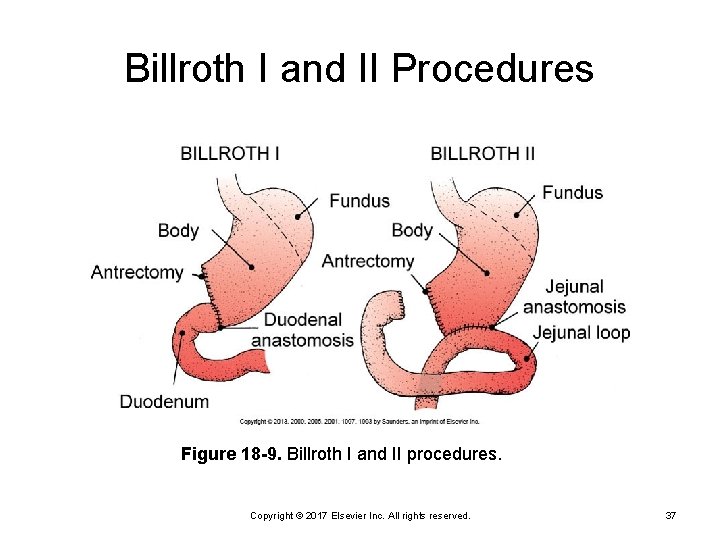
Surgical Treatment Types Gastric resection Ø Billroth I or II Ø Vagotomy Ø Pyloroplasty Ø Copyright © 2017 Elsevier Inc. All rights reserved. 36
Billroth I and II Procedures Figure 18 -9. Billroth I and II procedures. Copyright © 2017 Elsevier Inc. All rights reserved. 37
Nursing Diagnoses Risk for infection Altered nutrition Pain Fluid and electrolyte alterations Impaired gas exchange Copyright © 2017 Elsevier Inc. All rights reserved. 38
Critical Thinking Challenge What symptoms will you see if the patient is developing hypovolemic shock secondary to GI bleeding? What are your priorities, regardless of etiology? Copyright © 2017 Elsevier Inc. All rights reserved. 39
Variceal Bleeding Treatment Somatostatin or octreotide (long-acting) Decrease splanchnic blood flow Ø Reduce portal pressure Ø Vasopressin drip Vasoconstriction of splanchnic bed Ø Risk of fluid or electrolyte abnormalities Ø Continuous electrocardiogram (ECG) and blood pressure monitoring Ø Copyright © 2017 Elsevier Inc. All rights reserved. 40
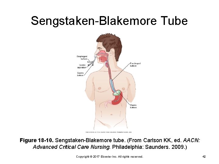
Variceal Bleeding Treatment (Cont. ) Esophagogastric (balloon) tamponade Sengstaken-Blakemore tube Ø Minnesota tube Ø Linton tube Ø Copyright © 2017 Elsevier Inc. All rights reserved. 41
Sengstaken-Blakemore Tube Figure 18 -10. Sengstaken-Blakemore tube. (From Carlson KK, ed. AACN: Advanced Critical Care Nursing. Philadelphia: Saunders. 2009. ) Copyright © 2017 Elsevier Inc. All rights reserved. 42
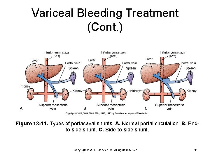
Variceal Bleeding Treatment (Cont. ) Sclerotherapy Transjugular intrahepatic portosystemic shunt (TIPS) Nonsurgical treatment for recurrent bleeding Ø Performed under fluoroscopy Ø Stent between hepatic and portal veins to reduce portal pressure Ø Surgery Copyright © 2017 Elsevier Inc. All rights reserved. 43
Variceal Bleeding Treatment (Cont. ) Figure 18 -11. Types of portacaval shunts. A. Normal portal circulation. B. Endto-side shunt. C. Side-to-side shunt. Copyright © 2017 Elsevier Inc. All rights reserved. 44
Quick Quiz! Portocaval shunt is done to prevent variceal bleeding. This surgery is associated with what negative outcome? A. Acute kidney injury B. Ascites C. Encephalopathy D. Gastric ulcer Copyright © 2017 Elsevier Inc. All rights reserved. 45
Critical Thinking Challenge What are the nursing priorities when these tubes are inserted? Copyright © 2017 Elsevier Inc. All rights reserved. 46
Acute Pancreatitis Inflammation of pancreas Mild to severe Ø Severe can lead to necrosis and hemorrhage Ø Severe has high mortality rate Ø Impaired exocrine and endocrine function Copyright © 2017 Elsevier Inc. All rights reserved. 47
Acute Pancreatitis (Cont. ) Etiology Alcoholism Ø Biliary tract disease Ø Medications Ø Trauma Ø Idiopathic Ø Copyright © 2017 Elsevier Inc. All rights reserved. 48
Pathophysiology Autodigestion leads to edema; interstitial hemorrhage; necrosis Release of histamine and bradykinin leads to increased vascular permeability; edema Inflammation may lead to obstruction of duct; ischemia Copyright © 2017 Elsevier Inc. All rights reserved. 49
Signs and Symptoms Severe pain Epigastric or midabdominal Ø Radiates to back Ø Nausea and vomiting Abdominal distension Ascites and jaundice (severe) Retroperitoneal hemorrhage Turner’s sign (discoloration of flank) Ø Cullen’s sign (discoloration around umbilicus) Ø Copyright © 2017 Elsevier Inc. All rights reserved. 50
Laboratory Data Elevated Serum amylase Ø Serum lipase Ø WBCs Ø Glucose Ø Liver function tests Ø Bilirubin Ø Triglycerides Ø Decreased Calcium Ø Albumin Ø Protein Ø Potassium Ø Copyright © 2017 Elsevier Inc. All rights reserved. 51
Diagnostic Tests Computed tomography (CT) of abdomen Magnetic resonance imaging (MRI) Ultrasound X-rays of abdomen and chest Upper gastrointestinal (UGI) IV cholangiogram Endoscopic retrograde cholangiopancreatography (ERCP) Copyright © 2017 Elsevier Inc. All rights reserved. 52
Treatment—Fluids Volume replacement Lactated Ringer’s (LR) Ø Colloids Ø Fresh frozen plasma (FFP) Ø Packed red blood cells (RBCs) (hemorrhagic type) Ø Monitor volume status Pulmonary artery (PA) catheter Ø Intake and output Ø Daily weights Ø Vasopressors may be indicated Copyright © 2017 Elsevier Inc. All rights reserved. 53
Treatment – New Modalities Surgical decompression of abdominal compartment syndrome (ACS) Copyright © 2013, 2009, 2005, 2001, 1997, 1993 by Saunders, an imprint of Elsevier Inc. 54
Fluids and Electrolytes Electrolyte replacements Calcium Ø Potassium Ø Treatment of hyperglycemia Copyright © 2017 Elsevier Inc. All rights reserved. 55
Rest the Pancreas Decrease secretions NPO controversial Ø Gastric suction Ø Issues related to early enteral versus parenteral nutrition Copyright © 2017 Elsevier Inc. All rights reserved. 56
Comfort Management Opiates PCA if able to use Ø Morphine may be acceptable as long as pancreatitis is not caused by gallbladder disease NG may help relieve pain Position patient Copyright © 2017 Elsevier Inc. All rights reserved. 57
Other Medical Therapy Pharmacological interventions Ø Antibiotics Drainage of pseudocysts Pancreatic resection Copyright © 2017 Elsevier Inc. All rights reserved. 58
Complications Hypovolemic shock Pulmonary complications Ø Atelectasis, acute respiratory distress syndrome (ARDS), and effusion Disseminated intravascular coagulation Pseudocysts and abscess Renal failure High glucose and triglycerides, low calcium Copyright © 2017 Elsevier Inc. All rights reserved. 59
Prognosis See Ranson’s criteria High severity of illness scores (APACHE III) Copyright © 2017 Elsevier Inc. All rights reserved. 60
Nursing Care Priorities Control pain; promote comfort Prevent and treat shock Maintain nutrition Prevent or minimize complications Copyright © 2017 Elsevier Inc. All rights reserved. 61
Hepatic Failure Hepatitis Decreased perfusion Cirrhosis Fatty liver disease Copyright © 2017 Elsevier Inc. All rights reserved. 62
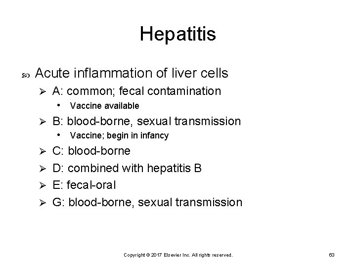
Hepatitis Acute inflammation of liver cells Ø A: common; fecal contamination • Vaccine available Ø B: blood-borne, sexual transmission • Vaccine; begin in infancy C: blood-borne Ø D: combined with hepatitis B Ø E: fecal-oral Ø G: blood-borne, sexual transmission Ø Copyright © 2017 Elsevier Inc. All rights reserved. 63
Symptoms of Hepatitis GI pain Fever, chills Jaundice Brown urine Right upper quadrant pain Clay-colored stools Copyright © 2017 Elsevier Inc. All rights reserved. 64
Management of Hepatitis No definitive treatment Rest Nutritional support Prevention of spread of the virus Copyright © 2017 Elsevier Inc. All rights reserved. 65
Cirrhosis Destruction of liver parenchyma and replacement by scar tissue Types Laënnec’s (alcoholic; portal) Ø Biliary Ø Cardiac Ø Postnecrotic Ø Copyright © 2017 Elsevier Inc. All rights reserved. 66
Assessment Liver enlarges due to increased fat accumulation Inflammation and necrosis of cells Yellow, orange, fatty, and scarred liver Liver shrinks; hobnail Ø Flow of blood is obstructed Ø Portal hypertension Ø Copyright © 2017 Elsevier Inc. All rights reserved. 67
Complications Portal hypertension Ø Varices Impaired metabolism Impaired clotting Ø Inability to detoxify drugs and toxins, including ammonia Ø Impaired bile formation and flow Copyright © 2017 Elsevier Inc. All rights reserved. 68
Signs and Symptoms Early: anorexia, dyspepsia, flatulence, nausea, vomiting, and changes in bowel patterns Related to altered metabolism Copyright © 2017 Elsevier Inc. All rights reserved. 69
Signs and Symptoms (Cont. ) Late Jaundice Ø Skin lesions Ø Hematological Ø Endocrine Ø Encephalopathy Ø Ascites Ø Copyright © 2017 Elsevier Inc. All rights reserved. 70
Diagnostic Tests Elevated liver enzymes Elevated bilirubin Elevated ammonia Coagulation studies Plasma proteins CT of abdomen Copyright © 2017 Elsevier Inc. All rights reserved. 71
Supportive Therapy Fluids Prevent injury and bleeding Treat hypoglycemia Copyright © 2017 Elsevier Inc. All rights reserved. 72
Aggressive Therapy Liver transplant Extracorporeal liver assist Experimental Ø Bridge to transplant or healing Ø Copyright © 2017 Elsevier Inc. All rights reserved. 73
Ascites Pathophysiology Low albumin plus obstruction to flow Ø Fluid in peritoneal cavity Ø Increased aldosterone • Sodium and water retention Ø Peripheral edema and further ascites Ø Copyright © 2017 Elsevier Inc. All rights reserved. 74
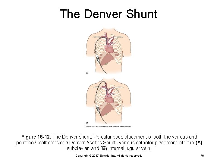
Medical Management: Ascites Bed rest Sodium and fluid restriction Diuretics Paracentesis Albumin Nutritional support Peritoneovenous shunt—long-term management Le. Veen shunt Ø Denver shunt Ø Copyright © 2017 Elsevier Inc. All rights reserved. 75
The Denver Shunt Figure 18 -12. The Denver shunt. Percutaneous placement of both the venous and peritoneal catheters of a Denver Ascites Shunt. Venous catheter placement into the (A) subclavian and (B) internal jugular vein. Copyright © 2017 Elsevier Inc. All rights reserved. 76
Nursing Management: Ascites Monitor fluid volume status Assess and facilitate respiratory status Assist with procedures to relieve pressure Copyright © 2017 Elsevier Inc. All rights reserved. 77
Quick Quiz! Hepatic encephalopathy is caused by an increase in: A. Ammonia B. Calcium C. Potassium D. Sodium Copyright © 2017 Elsevier Inc. All rights reserved. 78
Quick Quiz! The term for “flapping tremors” seen in hepatic encephalopathy is: A. Chvostek’s sign B. Cullen’s sign C. Asterixis D. Trousseau’s sign Copyright © 2017 Elsevier Inc. All rights reserved. 79
Encephalopathy Cerebral toxicity from elevated ammonia levels Asterixis Ø Impaired motor ability Ø Loss of consciousness (LOC) from confusion to coma Ø Copyright © 2017 Elsevier Inc. All rights reserved. 80
Medical Management: Encephalopathy Limit protein intake to 20 to 40 g/day Neomycin and lactulose Ø Reduce bacterial breakdown of protein in bowel Restrict toxic medications Sedation Copyright © 2017 Elsevier Inc. All rights reserved. 81
Hepatorenal Syndrome Acute renal failure End-stage cirrhosis and ascites Decreased albumin level Ø Portal hypertension Ø Poor outcome Copyright © 2017 Elsevier Inc. All rights reserved. 82
Trends Intraabdominal hypertension Leads to abdominal compartment syndrome Monitor bladder pressures Copyright © 2017 Elsevier Inc. All rights reserved. 83
Nursing Management: Hepatic Failure Priorities Improve nutritional status Ø Promote comfort Ø Observe for early signs of complications Ø Provide psychological support Ø Provide information Ø Copyright © 2017 Elsevier Inc. All rights reserved. 84