Chapter 18 Anatomy of the Cardiovascular System HEART

 Lies behind the body of the")
 Size and shape of the heart (Figures 18 -1 and 18")
 Coverings of the heart Structure of the heart coverings Pericardium Fibrous")
 Structure of the heart Wall of the heart: composed of three")


 Ventricles Two lower chambers known as pumping chambers because they")
 Valves of the heart: mechanical devices that permit the flow")
 Semilunar valves: halfmoon–shaped flaps growing out from the lining of")
 Skeleton of the heart Set of connected rings that serve")
 Blood supply of heart tissue Coronary arteries: myocardial cells receive")
 Veins of the coronary circulation As a rule, veins follow")
 Nerve supply of the heart Conduction system of the heart:")

 Types of blood vessels (cont. ) Capillaries Primary exchange vessels")
 Types of blood vessels (cont. ) Types of capillaries (Figure")
 Types of blood vessels (cont. ) Veins Carry blood toward")
")
 Systemic circulation: blood flows from the")
 Systemic circulation (cont. ) Hepatic portal circulation (Figures 18")
 Fetal circulation The basic plan of fetal circulation: additional")
 Fetal circulation Ductus venosus: continuation of the umbilical vein;")


- Slides: 38
Chapter 18: Anatomy of the Cardiovascular System
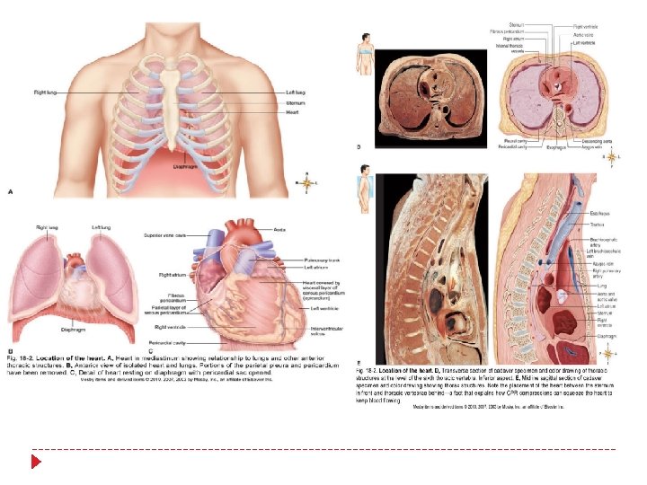
HEART Location of the heart (Figure 18 -2) Lies behind the body of the sternum between the points of attachment of ribs two through six Approximately two thirds of its mass is to the left of the midline of the body, and one third is to the right Apex lies on the diaphragm, pointing to the left Base lies just below the second rib Boundaries of the heart are clinically important as an aid in diagnosing heart disorders
HEART (cont. ) Size and shape of the heart (Figures 18 -1 and 18 -2) At birth, is transverse and appears large in proportion to the diameter of the chest cavity Between puberty and 25 years of age the heart attains its adult shape and weight In an adult, the shape of the heart tends to resemble that of the chest
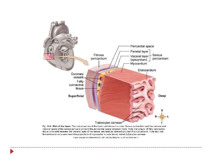
HEART (cont. ) Coverings of the heart Structure of the heart coverings Pericardium Fibrous pericardium: tough, loosefitting inextensible sac Serous pericardium: parietal layer lies inside the fibrous pericardium; visceral layer (epicardium) adheres to the outside of the heart; pericardial space with pericardial fluid separates the two layers Heart coverings protect against friction
HEART (cont. ) Structure of the heart Wall of the heart: composed of three distinct layers Epicardium: outer layer of heart wall Myocardium: thick, contractile middle layer of heart wall; compresses the heart cavities, and the blood within them, with great force Endocardium: delicate inner layer of endothelial tissue
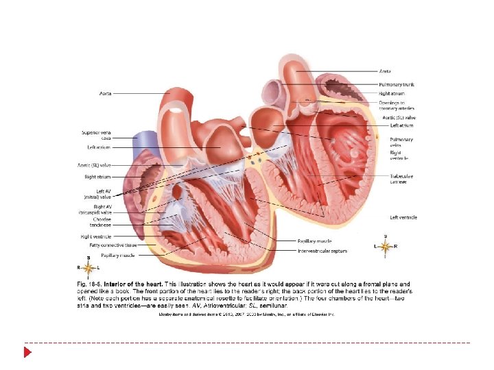
HEART: STRUCTURE Chambers: heart is divided into four cavities with the right and left chambers separated by the septum-sketch this
HEART: STRUCTURE Atria Two superior chambers known as receiving chambers because they receive blood from veins Atria alternately contract and relax to receive blood and then push it into ventricles Myocardial wall of each atrium is not very thick because little pressure is needed to move blood such a small distance Auricle: earlike flap protruding from each atrium
HEART: STRUCTURE (cont. ) Ventricles Two lower chambers known as pumping chambers because they push blood into the large network of vessels Ventricular myocardium is thicker than the myocardium of the atria because great force must be generated to pump the blood a large distance; myocardium of left ventricle is thicker than the right because it must push blood much further
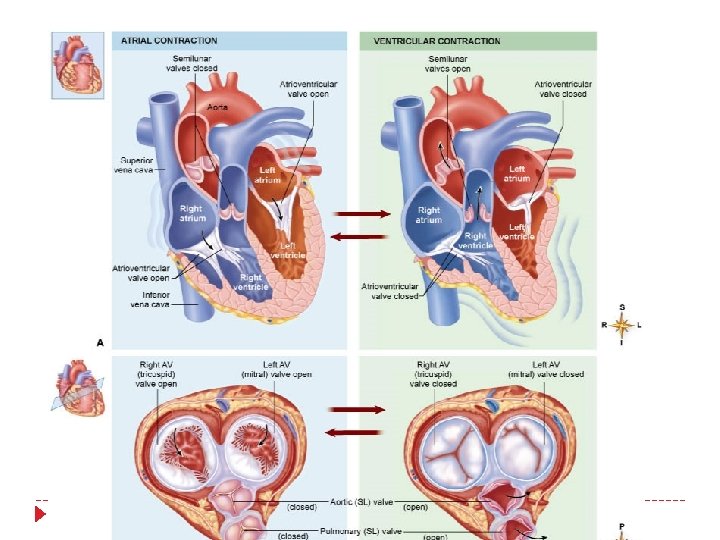
HEART: STRUCTURE (cont. ) Valves of the heart: mechanical devices that permit the flow of blood in one direction only Atrioventricular (AV) valves: prevent blood from flowing back into the atria from the ventricles when the ventricles contract Tricuspid valve (right AV valve): guards the right atrioventricular orifice; free edges of three flaps of endocardium are attached to papillary muscles by chordae tendineae Bicuspid, or mitral, valve (left AV valve): similar in structure to tricuspid valve except has only two flaps
HEART: STRUCTURE (cont. ) Semilunar valves: halfmoon–shaped flaps growing out from the lining of the pulmonary artery and aorta; prevent blood from flowing back into the ventricles from the aorta and pulmonary artery Pulmonary valve: valve at entrance of the pulmonary artery Aortic valve: valve at entrance of the aorta
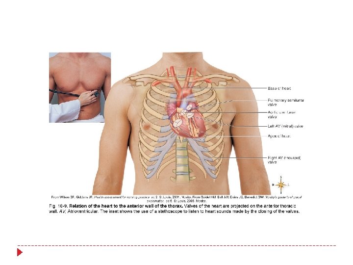
HEART: STRUCTURE (cont. ) Skeleton of the heart Set of connected rings that serve as a semirigid support for the heart valves and the attachment of cardiac muscle of the myocardium Serves as an electrical barrier between the myocardium of the atria and that of the ventricles Surface projection Flow of blood through heart
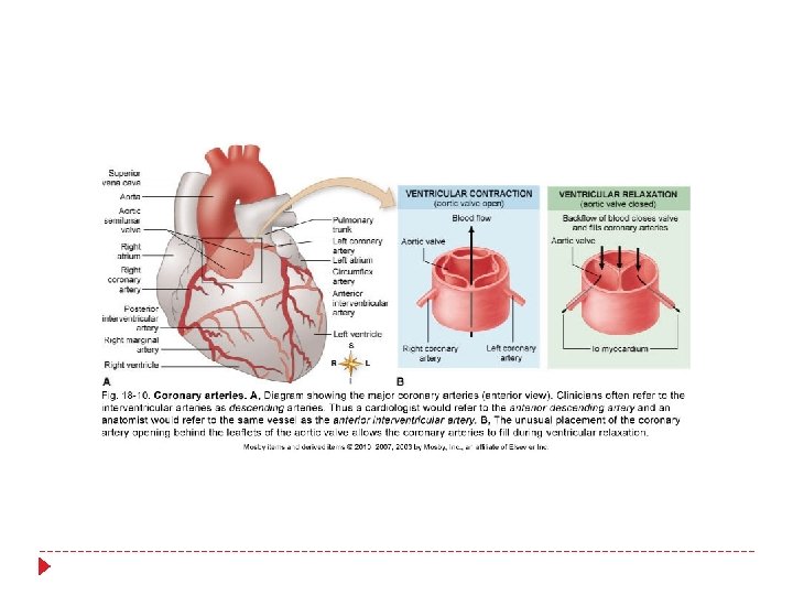
HEART: STRUCTURE (cont. ) Blood supply of heart tissue Coronary arteries: myocardial cells receive blood from the right and left coronary arteries First branches to come from the aorta Ventricles receive blood from branches of both right and left coronary arteries Each ventricle receives blood only from a small branch of the corresponding coronary artery
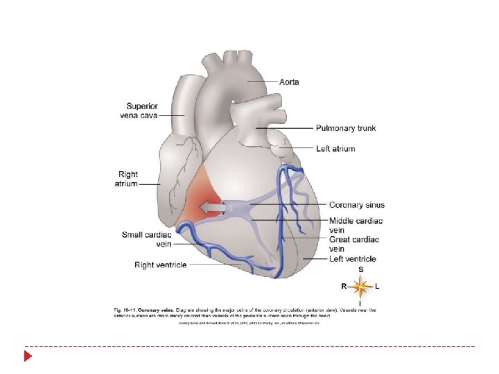
HEART: STRUCTURE (cont. ) Veins of the coronary circulation As a rule, veins follow a course that closely parallels that of coronary arteries After going through cardiac veins, blood enters the coronary sinus to drain into the right atrium Several veins drain directly into the right atrium
HEART: STRUCTURE (cont. ) Nerve supply of the heart Conduction system of the heart: composed of modified cardiac muscle, it generates and distributes the heart’s own rhythmic contractions; can be regulated by afferent(sensory) nerves Cardiac plexuses: located near the arch of the aorta; composed of sympathetic and parasympathetic fibers Sympathetic nerves: accelerator nerves Vagus fibers: inhibitory, or depressor, nerves
BLOOD VESSELS Types of blood vessels Arteries Carry blood away from heart; all arteries except pulmonary artery carry oxygenated blood Muscular (distributing) arteries Arterioles (resistance vessels) Smaller in diameter than elastic arteries Muscular layer is thick Smallest arteries Important in regulating blood flow to end organs Metarterioles Short connecting vessel between true arteriole and 20 to 100 capillaries Encircled by precapillary sphincters
BLOOD VESSELS (cont. ) Types of blood vessels (cont. ) Capillaries Primary exchange vessels Microscopic vessels Carry blood from arterioles to venules; together, arterioles, capillaries, and venules constitute the microcirculation
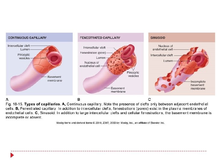
BLOOD VESSELS (cont. ) Types of blood vessels (cont. ) Types of capillaries (Figure 18 -15) True capillaries: receive blood flowing from metarteriole with input regulated by precapillary sphincters Continuous capillaries Continuous lining of endothelial cells Fenestrated capillaries Have both intercellular clefts and “holes, ” or fenestrations, through plasma membrane to facilitate exchange functions Sinusoids Very porous; permit migration of cells into or out of vessel lumen
BLOOD VESSELS (cont. ) Types of blood vessels (cont. ) Veins Carry blood toward the heart Act as collectors and reservoir vessels; called capacitance vessels
BLOOD VESSELS (cont. )
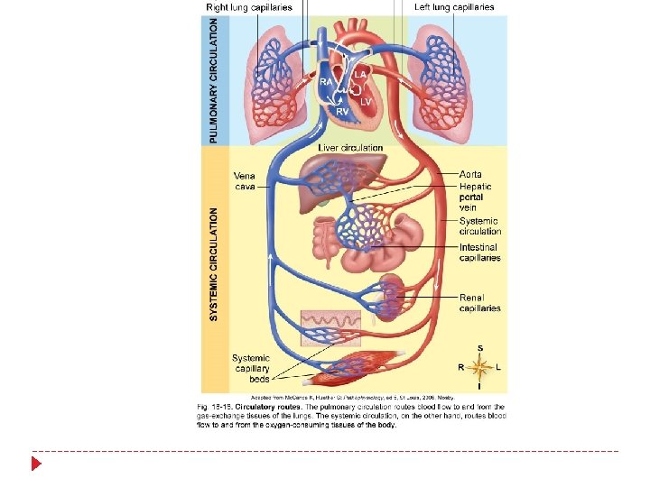
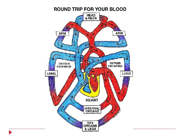
MAJOR BLOOD VESSELS Circulatory routes (Figure 18 -16) Systemic circulation: blood flows from the left ventricle of the heart through blood vessels to all parts of the body (except gas exchange tissues of lungs) and back to the right atrium Pulmonary circulation: venous blood moves from right atrium to right ventricle to pulmonary artery to lung arterioles and capillaries, where gases are exchanged; oxygenated blood returns to left atrium by pulmonary veins; from left atrium, blood enters the left ventricle
MAJOR BLOOD VESSELS (cont. ) Systemic circulation (cont. ) Hepatic portal circulation (Figures 18 -16 and 18 -28) Veins from the spleen, stomach, pancreas, gallbladder, and intestines send blood to the liver by the hepatic portal vein In the liver the venous blood mingles with arterial blood in the capillaries and is eventually drained from the liver by hepatic veins that join the inferior vena cava Venous blood from the lower extremities and abdomen drains into the inferior vena cava
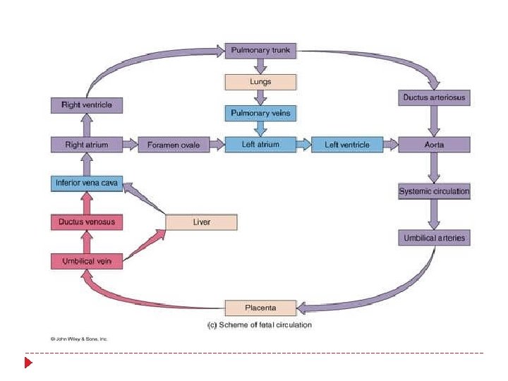
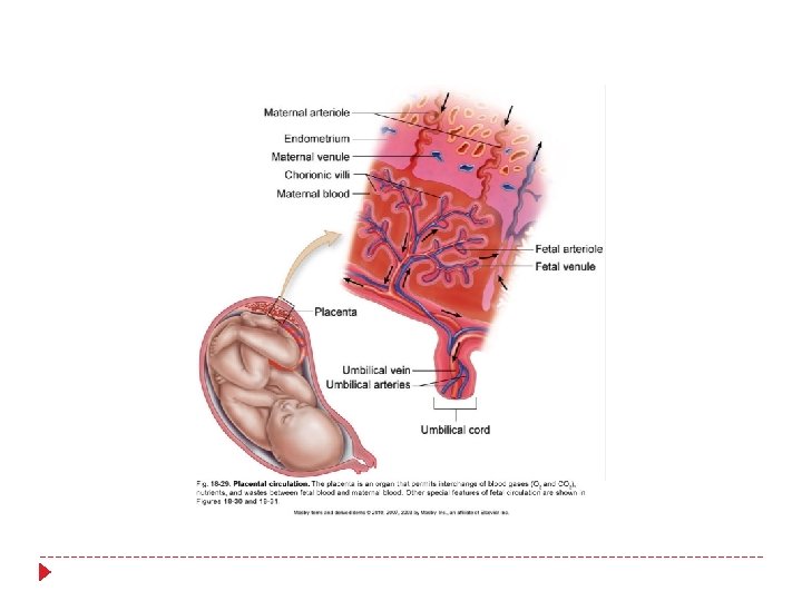
MAJOR BLOOD VESSELS (cont. ) Fetal circulation The basic plan of fetal circulation: additional vessels needed to allow fetal blood to secure oxygen and nutrients from maternal blood at the placenta (Figure 18 -31) Two umbilical arteries: extensions of the internal iliac arteries that carry fetal blood to the placenta Placenta: where exchange of oxygen and other substances between the separate maternal and fetal blood occurs; attached to uterine wall (Figure 18 -30) Umbilical vein: returns oxygenated blood from the placenta to the fetus; enters body through the umbilicus and goes to the undersurface of the liver, where it gives off two or three branches and then continues as the ductus venosus
MAJOR BLOOD VESSELS (cont. ) Fetal circulation Ductus venosus: continuation of the umbilical vein; drains into inferior vena cava Foramen ovale: opening in septum between the right and left atria Ductus arteriosus: small vessel connecting the pulmonary artery with the descending thoracic aorta
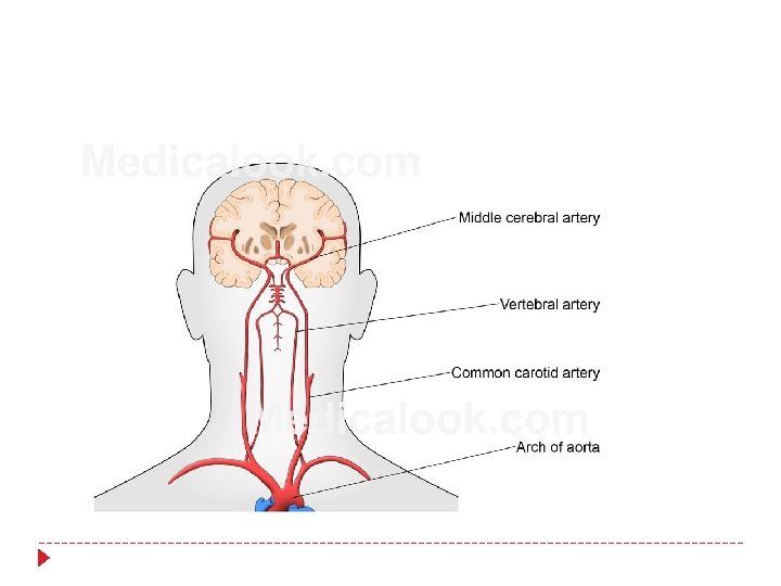
Cerebral Circulation
CYCLE OF LIFE: CARDIOVASCULAR ANATOMY Birth: change from placenta-dependent system Heart and blood vessels maintain basic structure and function from childhood through adulthood Exercise thickens myocardium and increases the supply of blood vessels in skeletal muscle tissue Adulthood through later adulthood: degenerative changes Atherosclerosis: blockage or weakening of critical arteries Heart valves and myocardial tissue degenerate, reducing pumping efficiency