
Chapter 17 The Special Senses 2012 Pearson Education
- Slides: 25
Chapter 17 The Special Senses © 2012 Pearson Education, Inc.
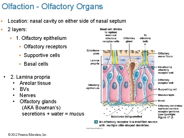
Olfaction - Olfactory Organs • Location: nasal cavity on either side of nasal septum • 2 layers: • 1. Olfactory epithelium • Olfactory receptors • Supportive cells • Basal cells • 2. Lamina propria • Areolar tissue • BVs • Nerves • Olfactory glands (AKA Bowman’s) secretions + water = mucus © 2012 Pearson Education, Inc.
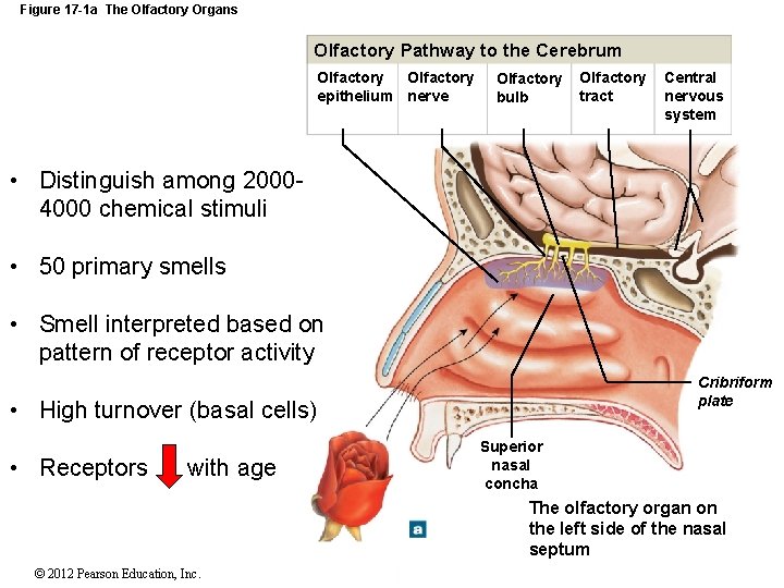
Figure 17 -1 a The Olfactory Organs Olfactory Pathway to the Cerebrum Olfactory epithelium nerve Olfactory bulb Olfactory tract Central nervous system • Distinguish among 20004000 chemical stimuli • 50 primary smells • Smell interpreted based on pattern of receptor activity Cribriform plate • High turnover (basal cells) • Receptors with age Superior nasal concha The olfactory organ on the left side of the nasal septum © 2012 Pearson Education, Inc.
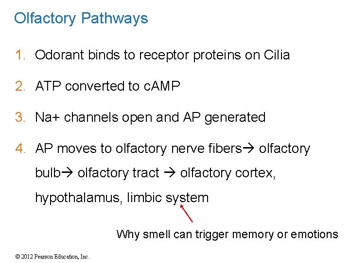
Olfactory Pathways 1. Odorant binds to receptor proteins on Cilia 2. ATP converted to c. AMP 3. Na+ channels open and AP generated 4. AP moves to olfactory nerve fibers olfactory bulb olfactory tract olfactory cortex, hypothalamus, limbic system Why smell can trigger memory or emotions © 2012 Pearson Education, Inc.
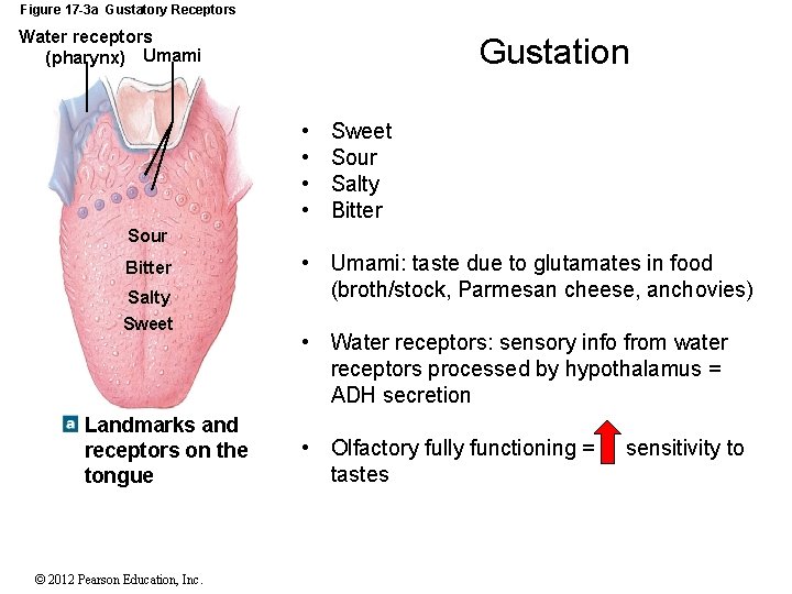
Figure 17 -3 a Gustatory Receptors Water receptors (pharynx) Umami Gustation • • Sweet Sour Salty Bitter Sour Bitter Salty Sweet Landmarks and receptors on the tongue © 2012 Pearson Education, Inc. • Umami: taste due to glutamates in food (broth/stock, Parmesan cheese, anchovies) • Water receptors: sensory info from water receptors processed by hypothalamus = ADH secretion • Olfactory fully functioning = tastes sensitivity to
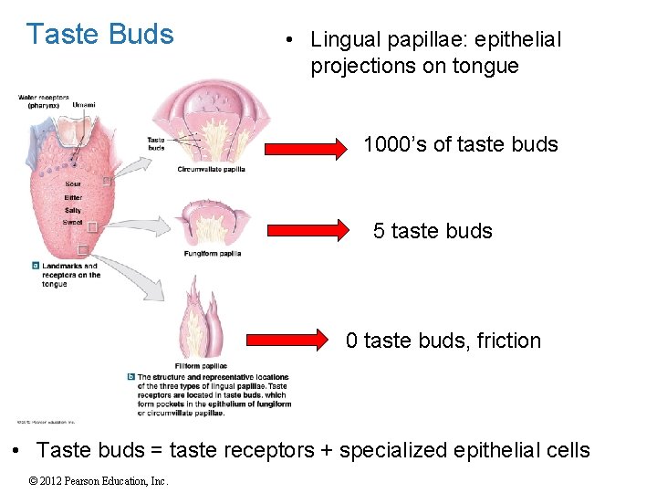
Taste Buds • Lingual papillae: epithelial projections on tongue 1000’s of taste buds 5 taste buds 0 taste buds, friction • Taste buds = taste receptors + specialized epithelial cells © 2012 Pearson Education, Inc.
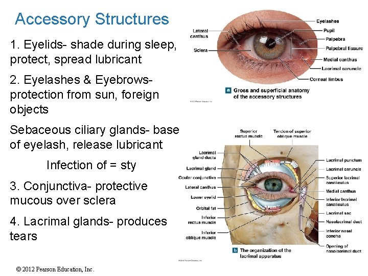
Accessory Structures 1. Eyelids- shade during sleep, protect, spread lubricant 2. Eyelashes & Eyebrowsprotection from sun, foreign objects Sebaceous ciliary glands- base of eyelash, release lubricant Infection of = sty 3. Conjunctiva- protective mucous over sclera 4. Lacrimal glands- produces tears © 2012 Pearson Education, Inc.
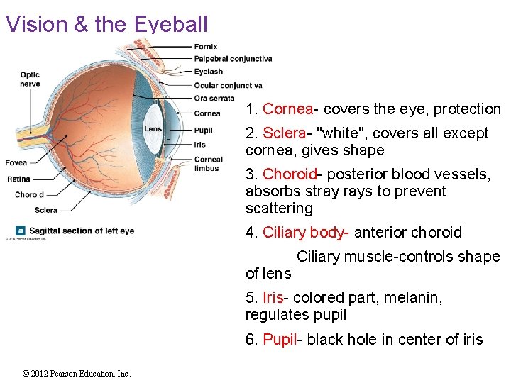
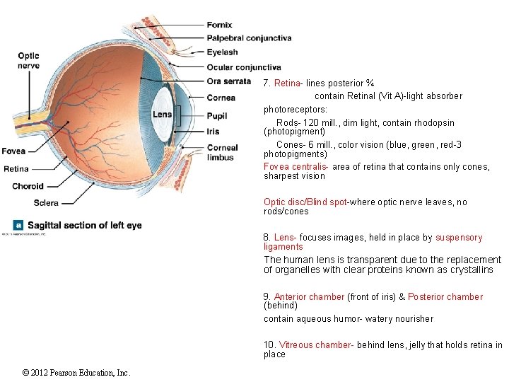
Vision & the Eyeball 1. Cornea- covers the eye, protection 2. Sclera- "white", covers all except cornea, gives shape 3. Choroid- posterior blood vessels, absorbs stray rays to prevent scattering 4. Ciliary body- anterior choroid of lens Ciliary muscle-controls shape 5. Iris- colored part, melanin, regulates pupil 6. Pupil- black hole in center of iris © 2012 Pearson Education, Inc.
7. Retina- lines posterior ¾ contain Retinal (Vit A)-light absorber photoreceptors: Rods- 120 mill. , dim light, contain rhodopsin (photopigment) Cones- 6 mill. , color vision (blue, green, red-3 photopigments) Fovea centralis- area of retina that contains only cones, sharpest vision Optic disc/Blind spot-where optic nerve leaves, no rods/cones 8. Lens- focuses images, held in place by suspensory ligaments The human lens is transparent due to the replacement of organelles with clear proteins known as crystallins 9. Anterior chamber (front of iris) & Posterior chamber (behind) contain aqueous humor- watery nourisher 10. Vitreous chamber- behind lens, jelly that holds retina in place © 2012 Pearson Education, Inc.
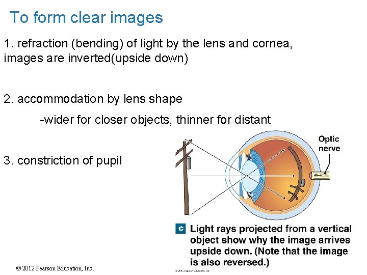
To form clear images 1. refraction (bending) of light by the lens and cornea, images are inverted(upside down) 2. accommodation by lens shape -wider for closer objects, thinner for distant 3. constriction of pupil © 2012 Pearson Education, Inc.
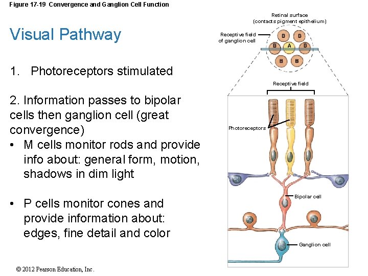
Figure 17 -19 Convergence and Ganglion Cell Function Retinal surface (contacts pigment epithelium) Visual Pathway Receptive field of ganglion cell 1. Photoreceptors stimulated Receptive field 2. Information passes to bipolar cells then ganglion cell (great convergence) • M cells monitor rods and provide info about: general form, motion, shadows in dim light • P cells monitor cones and provide information about: edges, fine detail and color Photoreceptors Bipolar cell Amacrine cell Ganglion cell © 2012 Pearson Education, Inc.
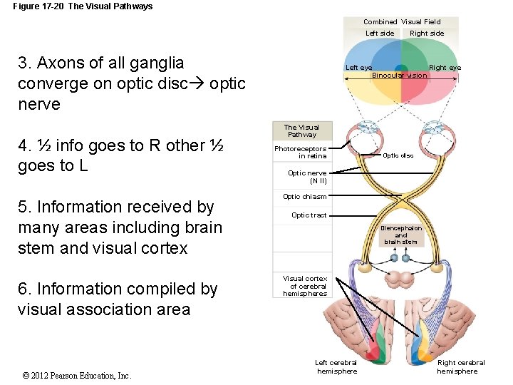
Figure 17 -20 The Visual Pathways Combined Visual Field Left side 3. Axons of all ganglia converge on optic disc optic nerve 4. ½ info goes to R other ½ goes to L 5. Information received by many areas including brain stem and visual cortex 6. Information compiled by visual association area © 2012 Pearson Education, Inc. only Right side Left eye Right eye only Binocular vision The Visual Pathway Photoreceptors in retina Optic disc Optic nerve (N II) Optic chiasm Optic tract Diencephalon and brain stem Visual cortex of cerebral hemispheres Left cerebral hemisphere Right cerebral hemisphere
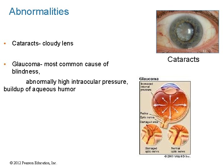
Abnormalities • Cataracts- cloudy lens • Glaucoma- most common cause of blindness, abnormally high intraocular pressure, buildup of aqueous humor © 2012 Pearson Education, Inc. Cataracts
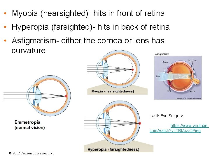
• Myopia (nearsighted)- hits in front of retina • Hyperopia (farsighted)- hits in back of retina • Astigmatism- either the cornea or lens has curvature Lasik Eye Surgery: https: //www. youtube. com/watch? v=TBf. Auv. OPjeg © 2012 Pearson Education, Inc.
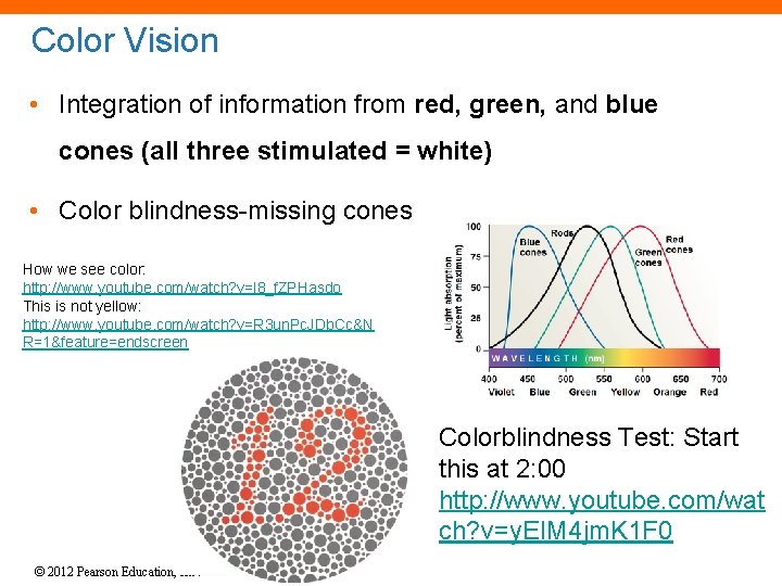
Color Vision • Integration of information from red, green, and blue cones (all three stimulated = white) • Color blindness-missing cones How we see color: http: //www. youtube. com/watch? v=l 8_f. ZPHasdo This is not yellow: http: //www. youtube. com/watch? v=R 3 un. Pc. JDb. Cc&N R=1&feature=endscreen Colorblindness Test: Start this at 2: 00 http: //www. youtube. com/wat ch? v=y. EIM 4 jm. K 1 F 0 © 2012 Pearson Education, Inc.
Vision and Pigments • Pigment = light absorbing molecule • Pigments of Rods • Rhodopsin (with protein opsin) • Retinal = synthesized from vitamin A • Pigments of Cones • Retinal • Other forms of opsin • Type of opsin stimulated = absorption of specific wavelength of light = color vision © 2012 Pearson Education, Inc.
Why should you eat Carrots to have good eyes? • Carrots do not contain Vitamin A- Instead they contain CAROTENE, an orange pigment, which is converted to retinol (Vit A) in the body. • Retinal, the pigment in the rhodopsin molecule, is synthesized from Vit A • Lack of Vit A can cause night blindness and even permanent blindness! © 2012 Pearson Education, Inc.
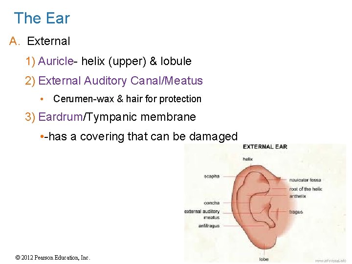
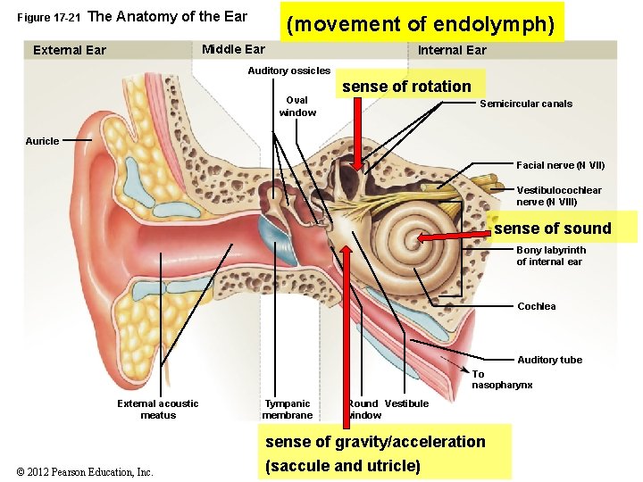
The Ear A. External 1) Auricle- helix (upper) & lobule 2) External Auditory Canal/Meatus • Cerumen-wax & hair for protection 3) Eardrum/Tympanic membrane • -has a covering that can be damaged © 2012 Pearson Education, Inc.
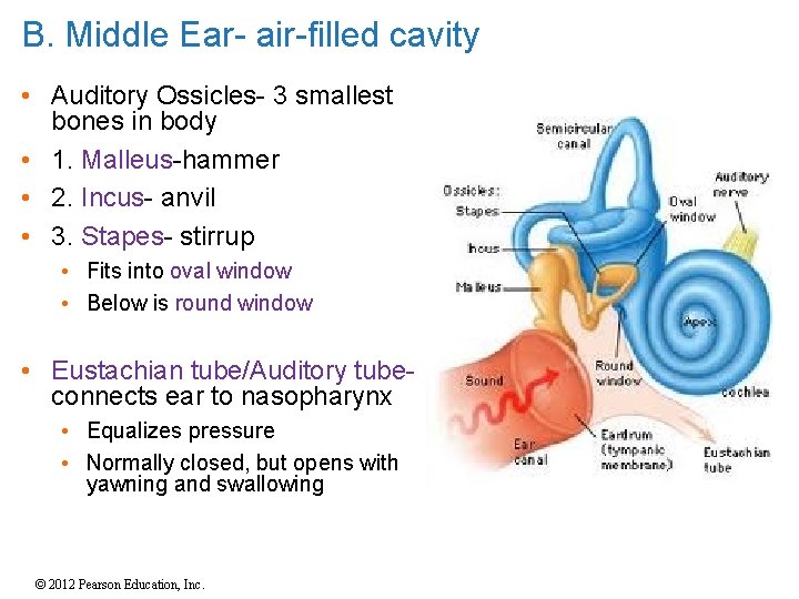
B. Middle Ear- air-filled cavity • Auditory Ossicles- 3 smallest bones in body • 1. Malleus-hammer • 2. Incus- anvil • 3. Stapes- stirrup • Fits into oval window • Below is round window • Eustachian tube/Auditory tubeconnects ear to nasopharynx • Equalizes pressure • Normally closed, but opens with yawning and swallowing © 2012 Pearson Education, Inc.
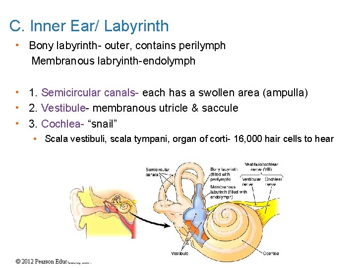
C. Inner Ear/ Labyrinth • Bony labyrinth- outer, contains perilymph Membranous labryinth-endolymph • 1. Semicircular canals- each has a swollen area (ampulla) • 2. Vestibule- membranous utricle & saccule • 3. Cochlea- “snail” • Scala vestibuli, scala tympani, organ of corti- 16, 000 hair cells to hear © 2012 Pearson Education, Inc.
Figure 17 -21 The Anatomy of the Ear (movement of endolymph) Middle Ear External Ear Internal Ear Auditory ossicles Oval window sense of rotation Semicircular canals Auricle Facial nerve (N VII) Vestibulocochlear nerve (N VIII) sense of sound Bony labyrinth of internal ear Cochlea Auditory tube To nasopharynx External acoustic meatus Tympanic membrane Round Vestibule window sense of gravity/acceleration © 2012 Pearson Education, Inc. (saccule and utricle)
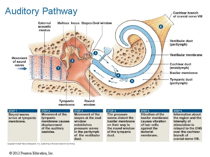
Auditory Pathway © 2012 Pearson Education, Inc.
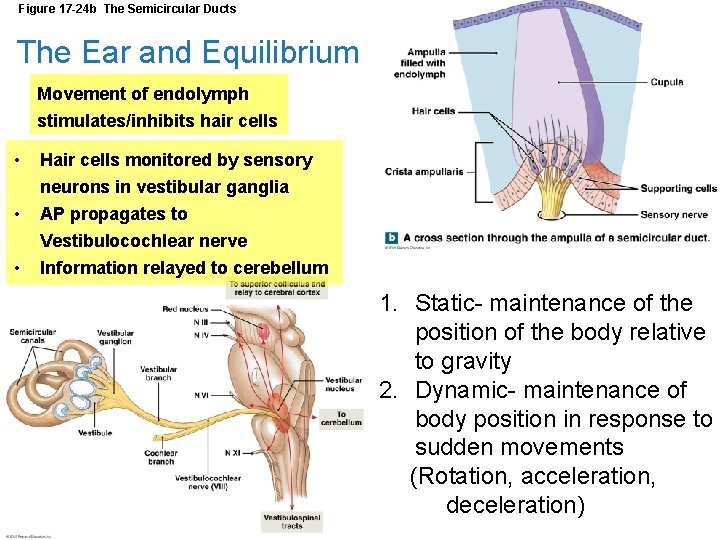
Figure 17 -24 b The Semicircular Ducts The Ear and Equilibrium Movement of endolymph stimulates/inhibits hair cells • Hair cells monitored by sensory • neurons in vestibular ganglia AP propagates to • Vestibulocochlear nerve Information relayed to cerebellum 1. Static- maintenance of the position of the body relative to gravity 2. Dynamic- maintenance of body position in response to sudden movements (Rotation, acceleration, deceleration) © 2012 Pearson Education, Inc.
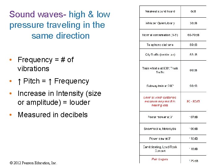
Sound waves- high & low pressure traveling in the same direction • Frequency = # of vibrations • ↑ Pitch = ↑ Frequency • Increase in Intensity (size or amplitude) = louder • Measured in decibels © 2012 Pearson Education, Inc.
Hearing and Age • Young children have greatest hearing range • Injuries to ear accumulate with age • Tympanic membrane loses flexibility • Articulations between ossicles stiffen • Round windows ossify © 2012 Pearson Education, Inc.