CHAPTER 16 Physiology and care during the first


 defines normal labour as one that is low risk")


 of the cervical canal into")


. v her choice of clothes for labour.")


 have become a routine procedure in labour there")
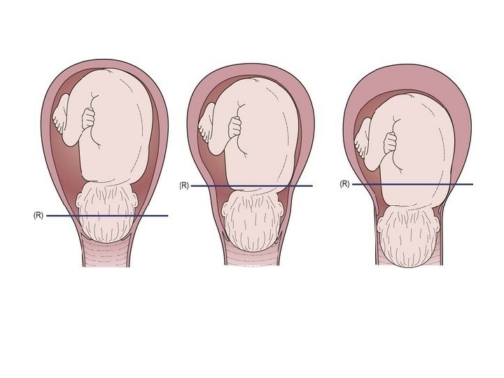
 Diagrams showing descent of the fetal head through the pelvic brim. (B) Diagrams")

. If")


 is not recommended in low-risk labour")


 is a widely used and well- appreciated method of")


")


 analgesia Epidurals are e�ective in relieving pain in labour and may be")
 analgesia An intravenous infusion of crystalloid fluids is commenced prior to siting")


 Prelabour rupture of membranes (PROM) at")


 occurs before 37 completed weeks' gestation, where")


- Slides: 114
CHAPTER 16 Physiology and care during the first stage of labour
Defining labour A human pregnancy is considered to last approximately 40 weeks, with labour usually occurring between 37 and 42 weeks' gestation (National Institute for Health and Clinical Excellence [NICE] 2007). Labour, purely in the physical sense, may be described as the process by which the fetus, placenta and membranes are expelled through the birth canal;
The World Health Organization (WHO) defines normal labour as one that is low risk throughout, spontaneous in onset with the fetus presenting by the vertex, culminating in the mother and infant being in good condition following birth (WHO 1999). labour where the fetus is presenting by the breech with no other risk factors should also be considered normal (Burvill 2005). Furthermore, all definitions of labour appear to be purely physiological and do not encompass the psychological wellbeing of the woman.
stages of labour The first stage of labour is usually recognized by the onset of regular uterine contractions, an accompanying e�acement and at least 4 cm dilatation of the cervix and finally culminates in full dilatation of the cervix. First stage consist of : the latent, is prior to the active phase stage of labour and may last 6– 8 hours in primigravidae when the cervix dilates from 0 cm to 4 cm dilated active This begins when the cervix is at least 4 cm dilated and, in the presence of rhythmic contractions, progressively dilates to 10 cm or full dilatation. transitional phases, the cervix is around 8 cm dilated until it is fully dilated or until expulsive contractions associated with the second stage of labour are felt by the woman.
The onset of spontaneous physiological labour The onset of labour is determined by a complex interaction of maternal and fetal hormones and is not fully understood. It would appear to be multifactorial in origin, being a combination of hormonal and mechanical factors. Increase level of oestrogen during the last weeks of pregnancy, resulting in changes cause uterine muscle fibres to display oxytocic receptors and form gap junctions with each other. Oestrogen also stimulates the placenta to release prostaglandins. Uterine activity may also result from mechanical stimulation of the uterus and cervix as q Overstretching q pressure from a presenting part on the cervix.
Recognition of the onset of labour The onset of labour is a process, not an event; therefore it is very di�cult to identify exactly when the painless (sometimes painful) contractions of prelabour develop into the progressive rhythmic contractions of established labour. It is part of the role of the midwife to ensure that women have su�cient information to assist them in recognizing the onset of established labour. This information is also needed to enable women to make informed choices based on current and unbiased evidence. The complex physical, psychological and emotional experience of labour a�ects every woman di�erently and midwives must have sound knowledge and experience to enable the woman to maintain control over the birth of her baby.
Ways to enable the woman to maintain birth control v Women in labour should be encouraged to trust their own instincts. v Women should listen to their own body and verbalize their feelings. v Support & reassurance should be given and discussion of this potential situation earlier in the pregnancy can enable the woman and her partner to prepare for labour more e�ectively. v Contact with the midwife should be made when regular, rhythmic, uterine contractions are experienced, and these are perceived by the woman as uncomfortable or painful.
Physiology of the first stage of labour Duration The length of labour varies widely and is influenced by parity, birth interval, psychological state, presentation and position of the fetus. Maternal pelvic shape and size and the character of uterine contractions also affect timescale. Over the years there has been much debate surrounding the length of physiological active labour in low-risk populations of childbearing women. cervical dilatation occurred at the rate of 1 cm per hour (Albers 1999; Lavender et al 2006; Zhang et al 2010). A cervical dilatation rate of 0. 5 cm per hour, however, has now been incorporated into the NICE (2007) intrapartum guidelines as being within the parameters of normal labour.
Cervical effacement Effacement refers to the inclusion (taking up) of the cervical canal into the lower uterine segment. the segment and the cervix merges into the lower uterine segment. The cervical canal widens at the level of the internal os, whereas the state of the external os remains unchanged (Cunningham et al 2010) (Fig. 16. 1).
E�acement may occur late in pregnancy, or it may not take place until labour begins. In the nulliparous woman the cervix will not usually dilate until e�acement is complete, whereas in the multiparous woman, e�acement and dilatation usually occurs simultaneously and a small canal may be felt in early labour.
Cervical dilatation Dilatation of the cervix is the process of enlargement of the os from a tightly closed to an opening large enough to permit passage of the fetus. Dilatation is assessed in centimeters and full dilatation at term equates to about 10 cm. However, acknowledging that all women are di�erent sizes and shapes means that full cervical dilatation may be between 9 and 11 cm in individual women (Walsh 2012). Dilatation occurs as a result of uterine action and the counter-pressure applied by either the intact bag of membranes or the presenting part, or both. A well-flexed fetal head closely applied to the cervix favors e�cient dilatation. Pressure applied evenly to the cervix causes the uterine fundus to respond by contraction and retraction, referred to as the Ferguson reflex
Ut er in e ac tio n Fundal dominance Each uterine contraction commences in the fundus near one of the cornua and spreads across and downwards.
Polarity is the term used to describe the neuromuscular harmony that prevails between the two poles or segments of the uterus throughout labour. During each uterine contraction, these two poles act harmoniously. The upper pole contracts strongly and retracts to expel the fetus; the lower pole contracts slightly and dilates to allow expulsion to take place. If polarity is disorganized then the progress of labour is inhibited.
Contraction and retraction Uterine muscle has a unique property. During labour the contraction does not pass o� entirely, as muscle fibres retain some of the shortening of contraction instead of becoming completely relaxed This is termed retraction. This process assists in the progressive expulsion of the fetus, such that the upper segment of the uterus becomes gradually shorter and thicker and its cavity diminishes.
Intensity and resting tone Each labour is individual and does not always conform to expectations, but generally before labour becomes established, uterine contractions may occur every 15– 20 minutes, lasting for about 30 seconds. They are often fairly weak and may even be imperceptible to the woman. The contractions usually occur with rhythmic regularity and the intervals between them where the muscle relaxes (resting tone) gradually lessen while the length and strength gradually intensifies through the latent phase and into the active phase of the first stage of labour. By the end of the first stage, the contractions may occur at 2– 3 minute intervals, last for 50– 60 seconds and are very powerful
Formation of upper and lower uterine segments By the end of pregnancy, the body of the uterus is described as having divided into two segments, which are anatomically distinct (Fig. 16. 4). The upper uterine segment, having been formed from the body of the fundus, is mainly concerned with contraction and retraction, and is thick and muscular. The lower uterine segment is formed of the isthmus and the cervix, and is about 8– 10 cm in length. The lower segment is prepared for distension and dilatation.
The retraction ring A ridge develops between the upper and lower uterine segments, known as the retraction ring (Fig. 16. 5). The physiological retraction ring gradually rises as the upper uterine segment contracts and retracts and the lower uterine segment thins out to accommodate the descending fetus. Once the cervix is fully dilated and the fetus can leave the uterus, the retraction ring rises no further. However, in extreme cases of mechanically obstructed labour, this physiological retraction ring becomes visible above the symphysis pubis and is described as Bandl's ring. A Bandl's ring may consequently be associated with fetal compromise
Show As a result of the dilatation of the cervix, the operculum, which formed the cervical plug during pregnancy, is released. The woman may observe a bloodstained mucoid discharge a few hours before, or within a few hours after, labour commences. The blood comes from ruptured capillaries in the parietal decidua where the chorion has become detached from the dilating cervix and should only be a staining (Impey and Child 2012 , a small loss of bright red blood referred to as a show.
Mechanical factors Formation of the forewaters and hindwaters As the lower uterine segment forms and stretches, the chorion becomes detached from it and the increased intrauterine pressure causes this loosened part of the sac of fluid to bulge downwards into the internal os, to the depth of 6– 12 mm. The well-flexed fetal head fits snugly into the cervix and cuts o� the amniotic fluid in front of the head from that which surrounds the body, forming two separate pools of fluid. The former is known as the forewaters and the lafer, the hindwaters. In early labour it is often possible to feel intact forewaters bulging even when the hindwaters have ruptured, making ruptured membranes a difficult diagnosis at times.
General fluid pressure While the membranes remain intact, the pressure of the uterine contractions is exerted on the amniotic fluid and, as fluid is not compressible, the pressure is equalized throughout the uterus and over the fetal body, known as general fluid pressure (Fig. 16. 6). When the membranes rupture and a quantity of fluid emerges, the fetal head, the placenta and umbilical cord are compressed between the uterine wall and the fetus during contractions with a consequential reduction in the oxygen supply to the fetus. Preserving the integrity of the membranes, therefore, optimizes the oxygen supply to the fetus and also helps to prevent intrauterine and fetal infection (Howie and Rankin 2010).
Rupture of the membranes The optimum physiological time for the membranes to rupture spontaneously is at the end of the first stage of labour, after the cervix becomes fully dilated and no longer supports the bag of forewaters. The membranes may sometimes rupture days before labour, in most cases there is no apparent reason for early spontaneous membrane rupture. Early rupture of membranes may lead to an increased incidence of variable decelerations on (CTG), resulting in an increase in caesarean sections if fetal blood sampling is not available. All women are required to give consent for this intervention and the midwife should have a clear indication for performing an ARM: details of which should be recorded in the woman's labour records (Nursing and Midwifery Council [NMC] 2009, 2012).
Fetal axis pressure During each contraction, the uterus rises forward and the force of the fundal contraction is transmitted to the upper pole of the fetus, down the long axis of the fetus and applied by the presenting part to the cervix. This is known as fetal axis pressure (Fig. 16. 7) and becomes much more significant after rupture of the membranes and during the second stage of labour.
Recognition of the first stage of labour Education during pregnancy is important to enable the woman to recognize the beginning of labour and understand the latent phase in order to consider possible strategies she may use for labour and birth. Women should appreciate that in late pregnancy vaginal secretions without any bloodstaining increase. In addition, they should be aware that a show, which is usually a pink or bloodstained jelly-like loss, prior to the onset of labour or in early labour, is quitecommon. If a woman is examined vaginally in late pregnancy they should also be informed that there may be some slight blood loss after the procedure. Braxton Hicks contractions are more noticeable in late pregnancy and some women experience them as painful. In active labour, contractions exhibit a pattern of rhythm and regularity, usually increasing in length, strength and frequency as time goes on.
When the woman first feels contractions she may be aware only of backache, but if she places a hand on her abdomen she may perceive simultaneous hardening of the uterus. If the pregnancy has no problem, with a normal birth anticipated, the midwife should advise the woman to stay in her own surroundings, continue with her normal activities, to eat, be active and upright. It is sometimes di�cult to be certain whether or not the membranes have ruptured spontaneously prior to labour or in early labour. The woman may be experiencing some degree of stress incontinence, so she may be unsure if it is liquor or urine that she is passing. If there is any doubt, the woman should contact her midwife who may decide to insert a speculum into the vagina to observe for any amniotic fluid. Digital examination should be avoided if the woman is not in labour as it can increase the risk of ascending infection (Shepherd et al 2010).
Initial meeting with the midwife Ideally, the woman should know her own midwife and be able to contact her when labour starts. Where this is not possible, it is crucial that the first meeting between the midwife, the labouring woman and her partner is very important. If the woman is planning to birth in hospital, she may worry about the reception she and her companion will receive and the atitude of the people there. In addition, an unfamiliar environment may provoke feelings of vulnerability and undermine her confidence. Comfortable surroundings, a welcoming manner and a midwife who greets the woman as an equal in a partnership will engender feelings of mutual respect, thus enabling the woman to relax and respond positively to the amazing forces of labour and to her baby aher it is born (Berry 2006; Fisher et al 2006; Raynor and England 2010).
The language of childbirth It has been recognized that some of the childbirth terminology used when communicating with women appears medical not clear to women. The terms pain and labour are suggestive of difficulty and trouble. It is therefore vital the midwife observes what she says to women during childbirth and uses appropriate and adapted language which is woman-friendly. The word delivery has been replaced by the term birthing or birth as these appear more suitable when discussing the concept and practice of normality within midwifery.
Communication The key issues for women relate to achieving a safe birth, feeling in control within the birth environment, developing supportive relationships with their carers, and being treated with kindness, respect, dignity and cultural sensitivity if they are to realize a positive experience of birth. E�ective communication between the midwife and the woman and her partner, and with other clinicians in the multidisciplinary team, is essential to providing e�ective safe supportive care in labour and achieving the woman's objectives. Communication does not consist only of the content of what is said, but also includes non-verbal communication and written records, such as the woman's birth plan. Poor communication is the commonest cause of preventable adverse outcomes in hospitals and remains a significant cause of writen complaints.
Interpreting services If the woman and midwife are unable to understand each other, communication will be ine�ective and it is essential that adequate interpretation services are available when necessary. Although there is a tendency to rely on family members or friends to provide interpreting services, the use of such interpreters is deemed inappropriate when the midwife wishes to discuss sensitive issues such as past history, domestic abuse or the need for interventions. Wherever possible, professional interpretation services (axcess) should be provided for all non-English-speaking women. If a face-to-face interpreter cannot be obtained, then the use of a telephone or Internet interpretation service should be considered.
Birth plan Regardless of where the woman plans to give birth, a birth plan is a valuable tool for midwives to observe and use to facilitate the provision of holistic, individualized care. The birth plan therefore provides the opportunity for the midwife to discuss with each woman and her partner any plans about the type of birth they would like that they may have already prepared with support from their community midwife. An outline may be present in the case notes, or the couple may bring a birth plan with them. Frequently, the partner is involved in this forward planning, which should be a flexible proposal that can be reviewed and revised as labour progresses (Department of Health [DH] 2007). Some women, however, may not have prepared a birth plan and so the midwife should encourage them to consider any preferences that they may have, for example:
example: v her choice of birth companion(s). v her choice of clothes for labour. v ambulation and fetal monitoring (intermittent, electronic or a combination). v strategies for labour (water immersion, massage, pharmacological pain relief). v position for labour and birth. v cutting of the umbilical cord. v skin-to-skin contact and feeding the baby after birth. Having the opportunity to discuss such issues in early labour enables the establishment of a trusting relationship between the woman and the midwife to develop where the woman feels valued and involved in intrapartum decision-making: all details of which should be clearly documented in the intrapartum records
Emotional and psychological care When a woman begins to labour, she may have a mixture of emotions. Most women anticipate labour with a degree of excitement, anxiety, fear and hope. Many other emotions are influenced by cultural expectations and previous life experiences. The state of the woman's knowledge, her fears and expectations are also influenced by her companions during labour, including the attitude and behavior of the caregiver. By the time labour starts, a decision will already have been reached about where the woman plans to give birth. Some women may choose to give birth at home, some in a midwife-led unit/birthing center and others in hospital. Some women may also wish to labour as long as possible at home but give birth in hospital. Whatever choice the woman makes, she must able to feel she is in control of what is happening and contributing to the decisions made about her care.
The concept of continuous support in labour There is evidence that the presence of the midwife and one-to-one personal affection is positively associated with a woman's satisfaction with her care. Kennedy et al (2010) describe that the presence of the midwife can enhance the woman's trust in her own ability to cope. In a systematic review by Hodnef et al (2011), the value of continuous support during labour and birth is clearly evident. The review, consisting of 21 randomized clinical trials, involving over 15 000 women, showed evidence that women who laboured with continuous support had shorter labours and were less likely to experience intrapartum interventions. These women were also less likely to have an epidural or other forms of pain medication, give birth by caesarean section, ventouse or forceps and consequently appeared more satisfied with their overall experience of childbirth (Hodnef et al 2011).
Reducing the risk of infection In the latest Confidential Enquiries into Maternal Deaths report, the leading cause of direct deaths during the triennium 2006– 2008, was sepsis, accounting for 26 deaths (Harper 2011). Hand hygiene, the combination of processes including hand washing, the use of alcohol hand rub and carefully drying and caring for the skin and nails, is considered to be the single most important measure in preventing the spread of infection. A clean environment is essential if infection rates are to be kept to a minimum and the midwife has an important role to play in ensuring that all equipment is cleaned according to local Trust guidelines and that there is adherence to all infection control measures. Rooms, birthing pools, beds and any equipment used by the midwife should be effectively cleaned before use. When a woman is admitted to hospital, invasive procedures should be kept to a minimum as, such as the performance of vaginal examinations , intravenous fluids, repeated vaginal examinations, epidural analgesia and fetal blood sampling, all of which will increase the risk of infection.
The midwife's initial physical examination of the woman The initial examination should include a discussion with the woman about when labour commenced, whether the membranes have ruptured and the frequency and strength of the contractions. The midwife should be aware that the woman will be very conscious of her body and may be unable to concentrate on the conversation or respond while experiencing a contraction. Since the woman has embarked on an intensely energy- demanding process, enquiry should be made about her ability to sleep and her most recent intake of food. If she is in early labour and there are no concerns about the pregnancy, the woman should be advised she can eat and drink as she wishes, remain mobile and maybe bathe if she would find this relaxing. Consideration should be given to the woman's social circumstances, including the care of other children and whether a birthing partner is available and has been contacted.
Past history Of particular relevance at the onset of a woman's labour are: qthe contents of the birth plan. qher parity and age. qthe gestational age and outcomes of previous labours. qthe weights and condition of previous babies. qher blood results including grouping, Rhesus factor and haemoglobin qher attendance at any specialist clinics. qevidence of any known problems: social or physical.
Consent Prior to touching the woman, a sound explanation of the proposed examination and their significance should be given. Verbal consent should be obtained and recorded in the notes (NMC 2008, 2009). The midwife must be aware that a competent woman, with a capacity to make decisions, is within her rights to refuse any treatment regardless of the consequences to her and her unborn baby and does not have to give a reason (DH 2009). Should the midwife be providing care to a pregnant teenager under the age of 16 years, it is important to carefully assess whethere is evidence that she has su�cient understanding in order to give valid consent, i. e. complies with the Fraser guidelines, previously referred to as being Gillick competent (Gillick v West Norfolk and Wisbech AHA 1986; GMC 2013).
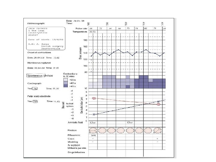
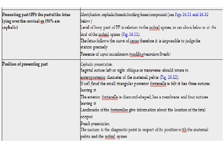
General assessment Basic observations, including pulse rate, temperature and blood pressure, are assessed and recorded. The woman's hands and feet are usually examined for signs of oedema. Slight swelling of the feet and ankles is physiological, but pre-tibial oedema or puffiness of the fingers or face is not. A detailed abdominal examination , partogram, fetal heart rate should be auscultated for a minimum of 1 minute immediately after a contraction using a Pinard stethoscope and the rate should be recorded as an average, in a single figure. A vaginal examination (VE) may also be undertaken to help confirm the onset of labour and determine the extent of cervical e�acement and dilatation (Fig. 16. 8), with some women requesting it when seeking reassurance about the status of their labour.
Records The midwife's record of labour is a legal document. The records may be examined by any court for up to 25 years, Records should be as contemporaneous as possible. Each entry should be authenticated with the midwife's full signature with the name printed underneath. Records should be comprehensive but concise and consist of the woman's observations, her physical, psychological and sociological state, and any problem that arises as well as the midwife's response and any subsequent interventions. The records should be kept in chronological order as their accuracy provides the basis from which clinical improvements, progress or deterioration of the woman or fetus can be judged. The record is shared between the midwife and obstetrician The obstetrician is also responsible to record their findings. The midwife usually enters in the records the summary of labour and initial details about the health of the baby. A midwife must ensure all records are stored securely and should not destroy or arrange for their destruction.
The charts are usually designed to allow for recordings at 15 minute intervals and include: fetal heart rate maternal temperature, pulse and blood pressure frequency and strength of contractions every 10 minutes descent of the presenting part cervical effacement and dilatation colour of amniotic fluid degree of caput succedaneum/moulding fluid balance urine analysis drugs administered.
Subsequent care in the first stage of labour Assessing progress Physical examination of the cervix is not the only way to assess labour. Midwives can use a range of skills, including visualization of the purple line, appearing from the woman's anal margin gradually extending to the nape of the bufocks (Hobbs 1998; Shepherd et al 2010), and observing the Rhombus of Michaelis, a kite-shaped area between the sacrum and ilea which becomes increasing visible as the fetal head descends in the pelvis (Shepherd et al 2010). In addition, the midwife should be alert in observing for changes in the woman's breathing, behaviour, noises, movements and posture alongside changes in the nature of contractions.
Abdominal examination An abdominal examination should be repeated by the midwife at intervals throughout labour in order to assess the length, strength and frequency of contractions and the descent of the presenting part. Palpation is of benefit prior to undertaking a vaginal examination, as the findings will assist the midwife to be accurate when defining the position and station of the head/breech. It is also useful to record the position of the fetus contemporaneously during the labour, as this can assist with the analysis of events should a shoulder dystocia occur.
Contractions The frequency, length and strength of the contractions should be noted and recorded on the partogram, usually at 30 minute intervals. The uterus should always feel softer between contractions and failure to relax is evidence of hyper tonicity that is usually defined as a contraction lasting more than 2 minutes (NICE 2007). The contraction rate is usually assessed by counting the number of contractions in 10 minutes, over a 20 -minute period. Evidence of 5 contractions or more in 10 minutes is evidence of tachy-systole in spontaneous labour, or hyper stimulation in induced labour (Chapter 19). An excessive number of contractions can result in fetal compromise as a result of prolonged cord compression or reduction in placental perfusion with consequent reduction in blood supply to the fetus.
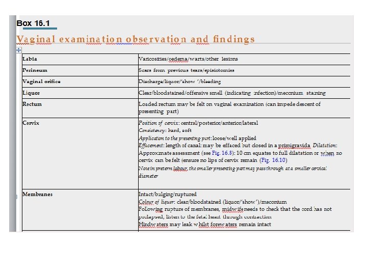
Vaginal examination Although vaginal examinations (VE) have become a routine procedure in labour there is very lifle evidence to support their e�cacy. Dixon and Foureur (2010) state that vaginal examinations are arguably considered to be both an intervention and an essential clinical assessment tool in labour. Midwives should remember that to women who have survived sexual abuse, experienced female genital mutilation (FGM), or are extremely anxious, a vaginal examination can be very distressing and sometimes impossible. examinations are undertaken using aseptic principles. Ideally the same person should perform the vaginal examinations to be in a better position to judge any changes. Observations and findings as detailed in Box 16. 1 should be noted and recorded accordingly by the midwife.
(A) Diagrams showing descent of the fetal head through the pelvic brim. (B) Diagrams showing dilatation of the cervix and rotation of the fetal head as felt on vaginal examination.
Indications for vaginal examination There should be valid reasons to undertake a VE in labour, which are to: make a positive identification of the presentation determine whether the head is engaged in case of doubt ascertain whether the forewaters have ruptured, or to rupture them artificially exclude cord prolapse after rupture of the forewaters, especially if there is an ill-fitting presenting part or there are fetal heart rate changes assess progress or slow labour confirm full dilatation of the cervix
Maternal observations Pulse rate The pulse rate should be recorded hourly (NICE 2007). If the rate increases to >100 bpm it may be indicative of anxiety, pain, infection, ketosis or haemorrhage. Temperature A rise in temperature can be indicative of infection or dehydration. The temperature should be recorded 4 -hourly (NICE 2007) and additionally when there is a clinical indication. Blood pressure should be measured every 4 hours Hypotension may be caused by the woman being in the supine position, by shock or as a result of vasodilation associated with epidural anaesthesia. Hypertension is an indicator of pre-eclampsia. It is usual practice to monitor the blood pressure at 5 - minute intervals for 20 minutes following the administration of an epidural anaesthetic and following the administration of any bolus dose.
Bath or shower Immersion in a warm bath or birthing pool can be an e�ective form of pain relief for labouring women that facilitates increased mobility with no increased incidence of adverse outcome for the woman or fetus (Da Silva et al 2009). The midwife should invite the woman who is mobile to have a bath or shower whenever she wishes during labour. Clothing It is entirely up to the individual woman what she wears in labour. If in hospital she may prefer to wear the loose gown o�ered or she may feel more comfortable wearing her own choice of clothing.
Position and mobility There are physical benefits if the woman maintains an upright position, including a shorter labour and a reduction in the need for analgesia , fewer episiotomies and fewer abnormal fetal heart rate paferns (Gupta et al 2012). Other benefits include increasing the woman's sense of control (Coppen 2005), thus contributing to a positive birth experience. . Despite this, the majority of women, including some obese women, will be able to maintain an upright position, retain a degree of mobility and achieve birth away from a birthing bed if supported in doing so. A key factor in encouraging di�erent labour positions and mobility is the environment. The birthing room should contain equipment such as beanbags, birthing balls and chairs (Albers 2007) and the midwife should be proactive in encouraging the woman to remain active and to change her position.
Nutrition in labour It has been estimated that in established labour a woman requires a calorie intake to prevent. Most women will be able to draw on glucose stores to provide energy but if insu�cient carbohydrate is available energy will be obtained from body fat and this will release ketones, resulting in ketoacidosis. Nutrition is a controversial issue, Hospital policies are usually based on the need to restrict food intake in order to prevent gastric aspiration. However, the number of women experiencing gastric aspiration is extremely low and there is no evidence to support this. As there is evidence that the desire to eat is more common in women in early labour (Singata et al 2010) and most women do not want to eat in active labour, it is important that the woman is not encouraged to eat if she has no desire to do so.
Bladder care Although frequent bladder emptying during labour is recommended, there is no evidence to support any particular regime of intervention. It is therefore reasonable to consider that the bladder should be emptied at least 4 -hourly or more frequently if it is palpable abdominally. There is some evidence that infrequent bladder emptying in labour is associated with an increased risk of urinary incontinence in the postpartum period (Birch et al 2009). A full bladder may increase pain, reduce e�ciency of uterine contractions and delay descent of the presenting part. The woman's sensation to micturate during labour may be reduced by pressure of the presenting part during its descent through the pelvis, or by an e�ective epidural block. In all cases of delay in labour, the midwife should ascertain whether the bladder is full or not.
it may become necessary to introduce a catheter into the bladder. As the risk of infection is increased with the use of retaining catheters, it is generally recommended that an ‘in-out’ catheter is used.
Assessing the wellbeing of the fetus The aim of fetal monitoring in labour is to maintain wellbeing, detect fetal hypoxia and initiate appropriate intervention before the fetus becomes asphyxiated and neurological damage occurs. Despite the widespread practice of both interment and continuous fetal monitoring, there are no statistical di�erences in the rates of cerebral palsy, infant mortality or other standard measures of neonatal wellbeing with either form of monitoring. The midwife should discuss the recommendations for fetal monitoring and the available evidence with the woman in order that she can make an informed decision about how her baby's wellbeing will be monitored during labour.
Intermittent auscultation Intermifent auscultation involves listening to the fetal heart rate at intervals using a Pinard stethoscope or a hand-held Doppler. The hand-held Doppler uses ultrasound to detect movement, either of the fetal heart muscle or valves, which are converted into a sound that can be heard and counted, such that the woman and her partner can also hear the fetal heart. It should be recognized that as the Doppler converts movement into sound it is possible for the maternal pulse to be detected and be mistaken for the fetal pulse. The normal fetal heart will have a rate of 110– 160 bpm and there should be no audible decelerations (NICE 2007). If at any time a fetal heart rate abnormality is suspected or there is any concern about fetal wellbeing, such as following the detection of meconium staining of the liquor, electronic fetal monitoring (EFM) should be commenced following discussion with the woman and her companion (NICE 2007).
The current guidance is for intermittent auscultations to be performed in the active first stage of labour at least every 15 minutes for a full minute immediately following a contraction (NICE 2007). As with any observation, every recording should be documented in the labour records. In order to demonstrate compliance with the recommendation regarding the timing of auscultations, it is helpful at least once to provide a description in the intrapartum records of how and when auscultation is being performed.
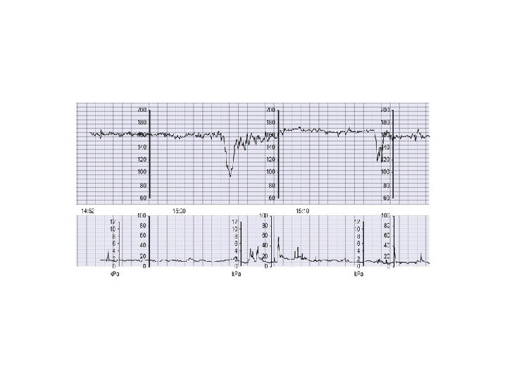
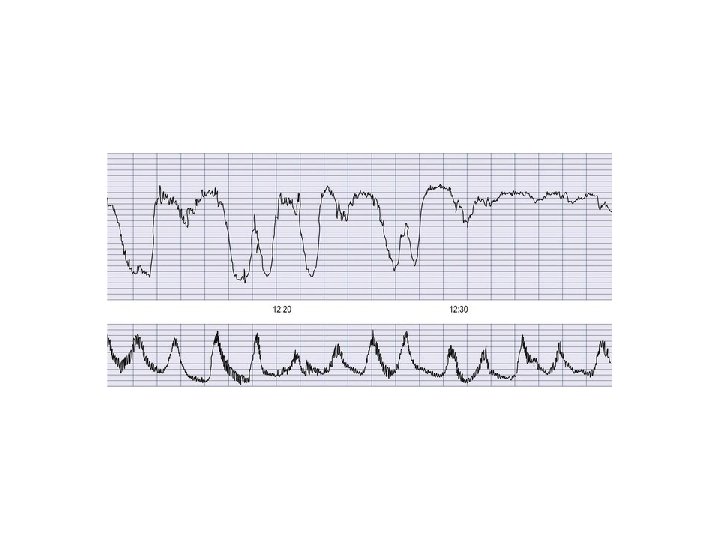
Continuous electronic fetal monitoring An admission cardiotocograph (CTG) is not recommended in low-risk labour it was confirm that such traces do not provide any benefit in terms of reducing perinatal morbidity or mortality but can increase the rate of caesarean section by approximately 20%. Continuous electronic fetal monitoring (EFM) is recommended for any labour where there are risks to fetal wellbeing, including the use of oxytocin and epidural analgesia. EFM will detect suspect pathology, & can detect changes in the pattern of the fetal heart that could indicate hypoxia but cannot provide a diagnosis as the CTG is a highly sensitive tool but the condition it is designed to detect is of low prevalence,
Baseline rate This is the mean level of the fetal heart rate between contractions, excluding accelerations and decelerations. A rising baseline rate may be of concern even if it remains within the normal range, if other nonreassuring or abnormal features are present. Baseline variability This is the degree to which the baseline varies over the period of a minute. The baseline variability is under the influence of the autonomic nervous system and this produces an irregular jagged appearance on the CTG trace. However, if repeated accelerations are seen on a trace with reduced variability, the variability should not be assessed as non- reassuring.
Decelerations These are reflected as a fall in the baseline rate of at least 15 bpm for at least 15 seconds. True uniform decelerations, usually referred to as early, are rare and benign, and are therefore not significant. Most decelerations in labour are variable, which means they are variable in presentation and appearance. There are two types of variable decelerations, typical and atypical, and it is important to distinguish between them.
Typical variable decelerations occur in response to interment cord compression and are commonly seen during the second stage of labour. They are quick to recover to the normal baseline, have normal variability, last less than 2 minutes and have evidence of shouldering, which is a normal physiological response to intermittent cord compression.
Atypical variable decelerations These decelerations can be an indicator of hypoxia and have some or all of the following features: Loss of acceleration (shouldering) before and after deceleration Delayed recovery back to baseline Late component/biphasic deceleration Rebound tachycardia – caused by catecholamine release in response to stress Loss of variability/change in baseline rate.
Late decelerations These decelerations are uncommon and are usually considered to be indicative of fetal hypoxia. The main features being: Reduced variability Repetitive and uniform shape Begin at or after the peak of the contraction Their lowest point is 20 seconds or more after the peak of contractions.
Accelerations These particular features reflect an elevation in the baseline rate of 15 bpm for 15 seconds or more. The significance of the absence of accelerations in an otherwise normal trace is uncertain.
Normal If no other indication for CTG, discontinue and commence intermifent auscultation every 15 minutes
Suspicious The trace should continue and the possible causes be considered. The woman's position should also be changed to the leh lateral, her pulse and blood pressure assessed and fluids administered if appropriate. If the trace reverts to normal and there is no other indication for the CTG it could be discontinued; however review by an obstetrician may be required. Should the trace remain suspicious, the appropriate level obstetrician should be consulted. Fetal blood sampling may be required
Pathological The woman's position should be changed to the leh lateral, her pulse and blood pressure assessed and fluids administered if appropriate. If there is a bradycardia in a hospital sefing the emergency call bell should be used to summon assistance and referral to a Registrar or Consultant should be made immediately. Fetal blood sampling or the need to expedite the birth may be necessary. If fetal blood sampling is performed this should be prior to the administration of pethidine, epidural or syntocinon, should the trace remain pathological
Fetal blood sampling Maternity units that use electronic fetal monitoring should have 24 hours access to fetal blood sampling (FBS) facilities. When the fetal heart rate pattern is suspicious or pathological and fetal acidosis is suspected, then FBS should always be undertaken (NICE 2007) (Fig. 16). The procedure involves a small sample of blood being taken from the scalp of the fetal head with the woman in the left lateral position as the lithotomy position is more distressing for both the woman and fetus, causing supine hypotension which can increase fetal hypoxia. If the CTG trace is bradycardic or significantly pathological with no prospect of spontaneous birth, time should not be wasted performing a FBS. This would be the clinical decision of a senior obstetrician.
A normal fetal blood sample result should be repeated no more than an hour later if the trace remains pathological, or earlier if the heart rate pafern deteriorates. A fetal blood sample result of <7. 25 should be repeated no more than 30 minutes later, whereas one that is <7. 20 indicates that the baby requires immediate birth (NICE 2007).
Women's control of pain during labour The issue of pain during labour should be woman-centred, not medically oriented. A clear di�erentiation must be made between the traditional goal of pain relief and the control of pain in labour. midwives adopt one of two paradigms: the pain relief model and the working with pain model. In the pain relief model, midwives present a menu of pain-relieving options to the woman, but while this approach has the best of intentions, it inevitably undermines the woman's confidence in herself and her body to give birth without the aid of medication. However, in the working with pain model it is expected that most women in labour will experience some degree of pain that is fundamental to a physiological labour. Pain of normal labour is positive and has a purpose, and if this philosophy is embraced, midwives and women can work together to ensure labour is an enabling and ultimately uplifting experience. pain can be modified by past experience, anxiety, emotion and suggestion. Lack of food, rest and sleep also impact on the woman's perception of pain.
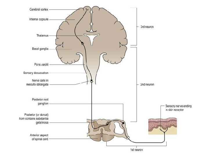
The physiology of pain Pain stimulus and pain sensation The discomfort or pain of labour is caused by the descent of the fetal head further into the pelvis (pressure). It is also caused by pressure on the cervix and the stretching of the vaginal walls and pelvic floor muscles, as descent of the presenting part occurs. Pain is caused by a stimulus that may cause, tissue damage. Pain sensation may therefore be distinguished from other sensations, although emotions such as fear and anxiety are also experienced at the same time, thereby a�ecting the person's perception of pain. It must also be remembered that a painful stimulus may also induce such changes by the sympathetic nervous system as increased heart rate, a rise in blood pressure, release of adrenaline (epinephrine) into the bloodstream and an increase in blood glucose levels. There is also a decrease in gastric motility and a reduction in the blood supply to the skin, causing sweating. Thus, stimuli that cause pain result in a sensory incident or occurrence.
Pain transmission The pain pathway or ascending sensory tract originates in the sensory nerve endings at the site of trauma. The impulse travels along the sensory nerves to the dorsal root ganglion of the relevant spinal nerve and into the posterior horn of the spinal cord. This is known as the first neuron. The second neuron arises in the posterior horn, crosses over within the spinal cord (the sensory decussation) and transmits the impulse via the medulla oblongata, pons varolii and the mid-brain to the thalamus. From here, it travels along the third neuron to the sensory cortex (Fig. 16. 17).
In cases of acute pain, sensations are transmitted along fibers, which are large diameter nerve fibers. This type of pain is perceived as being a pricking pain that is readily localized by the su�erer. The pathway for chronic pain is slightly di�erent as the nerve fibers involved are of smaller diameter and are called C fibers. Chronic pain is often described as a burning pain that is difficult to localize.
Somatosensory function Somatic sensation refers to the sensory function of the skin and body walls. This is moderated by a variety of somatic receptors of which there are particular receptors for each sensation, such as heat, cold, touch, pressure, etc. On entering the central nervous system, the a�erent nerve fibres from somatic receptors form synapses with interneurons that comprise the specific ascending pathways going to the somatosensory cortex via the brain stem and the thalamus. A n a�erent neuron, with its receptor, makes up a sensory unit. Usually the peripheral end of an a�erent neuron branches into many receptors. The receptors whose stimulation gives rise to pain are situated in the peripheries of small unmyelinated or slightly myelinated a�erent neurons. These receptors are known as nociceptors because they detect injury (noci, being the Latin word for ‘harm’, ‘injury’). The primary a�erents coming from nociceptors form synapses with interneurons after entering the central nervous system. .
Endorphins and enkephalins Endorphins are described as being opiate-like peptides, or neuropeptides, which are produced naturally by the body at neural synapses at various points in the central nervous system pathways. They control the transmission of pain perception in these areas. Endorphins are found in the limbic system, hypothalamus and reticular formation (Martini et al 2011). They bind to the presynaptic membrane, inhibiting the release of substance P, and therefore inhibit the transmission of pain. Enkephalins are also neuropeptides that have the ability to inhibit neurotransmitters along the pathway of pain transmission, thereby reducing it. These act like a natural pain relieving substance.
Non-pharmacological methods for pain control in labour Increasingly, non-pharmacological methods are being used by women during labour. there is some evidence that hypnosis, massage, acupuncture and acupressure reduce pain or decrease the need for pharmacological analgesia during labour (Tiran 2010 a) The midwife needs to be mindful that to administer any complementary alternative therapy to a woman in labour it is both essential and a professional requirement that an approved course of training is undertaken to be deemed knowledgeable and competent to practise such a skill. (NMC 2010, 2012)
Aromatherapy is the use of essential oils for a range of purposes, for example to induce relaxation, reduction of pain or nausea and vomiting. These oils may be massaged into the skin, inhaled through di�users or oil burners, or used in conjunction with hydrotherapy. This particular complementary therapy has become popular among childbearing women and midwives, with many maternity units in the UK providing this service for women during labour and birth.
Homeopathy uses small doses of natural medicines to stimulate the body's own physiological response to heal itself (Idarius 2010). Homeopathic remedies are prepared from plant extracts and from minerals. Professional advice is recommended during pregnancy as the holistic approach of this method entails a consideration of all the facets and the requirements of the individual. Aconite may be used to relieve fear and anxiety and Kali Carbonate to alleviate back pain during labour (Steen and Calvert 2006).
Hydrotherapy Immersion in water during labour as a means of analgesia has been used for many years. cite that the e�ectiveness of hydrotherapy is due to heat- relieving muscle spasm, and therefore pain, and hydrokinesis eliminates the e�ects of gravity and also the discomfort and strain on the pelvis. Using water in the first stage of labour reduces the use of epidural/spinal analgesia and no evidence of an increase in adverse e�ects on fetus/neonate or the woman. Water immersion is usually highly rated by both women and midwives, and the calming atmosphere of a pool room can benefit everyone as the woman appears less anxious and therefore feels less pain (Benjoya Miller 2006).
Music therapy As well as at home, many birthing rooms are equipped with radio or CD apparatus and this is often a useful means to help women relax, be entertained and find some distraction during the early stages of labour. Many types of music are available for relaxation, some of which are specifically for childbirth. Henley-Einion (2007) recounts that music can have a positive e�ect on the woman's body, mind and spirit by providing empowerment and creating an enabling effect.
Transcutaneous electrical nerve stimulation (TENS) is a widely used and well- appreciated method of pain relief. It consists of a small device that distributes low intensity electrical charges across the skin which is thought to prevent pain signals from the uterus, vagina and cervix arriving at the brain
TENS works by stimulating low threshold a�erent fibers, such as the fibers of touch receptors which inhibit neurons in the pain pathways. As pathways activated by the touch receptors add a synaptic input into the pain pathways, the individual may massage a painful area to relieve the pain which is how TENS functions. The electrodes are positioned at the level of T 10 and L 1 on the woman's back and have been found to be e�ective in reducing pain during the first stage of labour. The remaining electrodes are placed between S 2 and S 4 and provide control of pain during the second stage of labour.
Pharmacological methods for pain control
Inhalation analgesia A premixed gas made up of 50% nitrous oxide (N 2 O) and 50% oxygen (O 2) administered via the Entonox apparatus is the most commonly used inhalation analgesia in labour. Nitrous oxide (also known as laughing gas), like many other forms of analgesia, acts by limiting the neuronal and synaptic transmission within the central nervous system. Evidence shows that N 2 O induces opioid peptide release in the periaqueductal grey area of the mid-brain leading to the activation of the descending inhibitory pathways, resulting in modulation of the pain/nociceptive processing in the spinal cord (Fujinaga and Maze 2002). The mixture of gases is stable at normal temperature, but separates below − 7 °C. In many large obstetric units the gas is stored in a bank and piped to each birthing room or is available in cylinders. . The cylinders must be brought into a warm room if they have been exposed to cold temperatures, and the gases remixed by inverting the cylinder at least three times before use.
The gases take e�ect within 20 seconds and it is important that the woman uses it before a contraction commences. The maximum e�cacy of the gases occurs aher about 45– 50 seconds which should coincide with the height of the contraction, providing maximum relief for the woman. This method of pain control is useful in that the woman is able to administer it herself, but its e�ectiveness is determined by the woman's ability to use the equipment as advised. nitrous oxide and oxygen provided befer analgesia and had more beneficial effects when compared to pethidine. Exposure to high levels of nitrous oxide can cause teratogenic and other side-e�ects such as infertility among midwives and other sta�. It is important that scavenging equipment to extract expired gases is installed in all birthing rooms to reduce such effects on staff (RCM 2012 c).
Opiate drugs are frequently used during childbirth because of their powerful analgesic properties. The action of these drugs lies in their ability to bind with receptor sites which are mainly found in the substantia gelatinosa of the dorsal horn of the spinal cord (Anderson 2011). Others are located in the midbrain, thalamus and hypothalamus. In the UK three systemic opioids are commonly used for pain relief in labour: vpethidine (meperidine in the USA) vdiamorphine vmeptazinol (Meptid). All have similar pain-relieving properties, but lifle evidence exists in relation to their e�ectiveness, maternal satisfaction of their use or their e�ect on the fetus/neonate in labour (Ullman et al 2010). There are numerous side-e�ects of opiate drugs and the extent to which they are experienced is influenced by the woman's metabolism of the drug, the degree and speed of transfer of the drug and metabolites from maternal to fetal circulation and the ability of the fetus to process and excrete both.
Common side-e�ects of opiate drugs include: nausea and vomiting delayed emptying of the stomach drowsiness reduction in fetal heart rate variability depression of the baby's respiratory center at birth An anti-emetic agent is sometimes given to the woman at the same time to reduce the feeling of nausea. It is therefore important to ensure that the woman is fully informed during pregnancy of the e�ects of these drugs so that she can make informed decisions about methods of pain control.
Pethidine is a synthetic compound acting on the receptors in the body and is the most frequently used systemic narcotic analgesic in the UK. It is usually administered intramuscularly in doses of 50– 150 mg, depending on the woman's size, and takes about 20 minutes to have an e�ect. Pethidine can be administered intravenously for a faster e�ect and some maternity units use a machine to enable the woman to control the administration: known as patient-controlled analgesia (PCA). Some reports show that opiates, especially pethidine, slow down the process of labour and are not significantly e�ective in relieving labour pain, as ohen sedation is confused with analgesia ( Anderson 2011).
Diamorphine has been found to provide e�ective analgesia for up to 4 hours in labour with the usual dose being 5 mg. It is more rapidly metabolized, accounting for its greater speed of e�ectiveness and consequently it is eliminated more readily from maternal and neonatal plasma (Fernando and Jones 2009). Diamorphine is used far less commonly than other opiates in labour, even though some claim it gives befer pain relief and hence more comparative studies are needed (Jones et al 2012). It is possible that the lack of use of diamorphine in labour might be due to fears of its potentially addictive nature.
Meptazinol is usually given in doses of 100– 150 mg intramuscularly. It is fast-acting and is e�ective for about 4 hours. This opiate provides similar pain relief to pethidine and like other opiates, meptazinol is also associated with an increased incidence of nausea and vomiting (Ullman et al 2010).
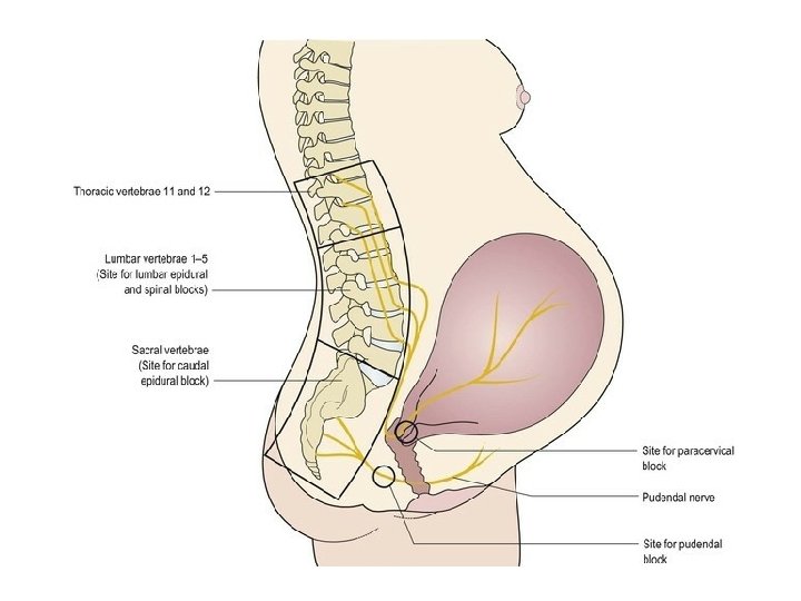
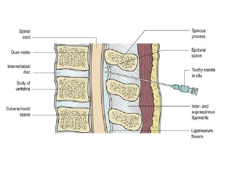
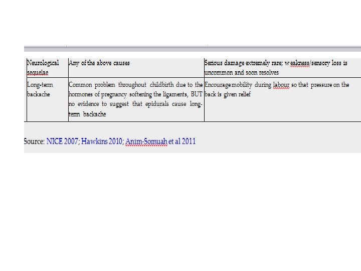
Regional (epidural) analgesia Epidurals are e�ective in relieving pain in labour and may be requested by women at any point during the first stage of labour. , usually between L 2 and L 3 Women who have used other methods of pain relief on experiencing strong contractions may decide to request an epidural when labour is well advanced. This makes explanation of the benefits and risks of epidural analgesia during pregnancy even more important. The pain relief from an epidural is obtained by blocking the conduction of impulses along sensory nerves as they enter the spinal cord. It is an invasive procedure that requires informed consent from the woman and an experienced (obstetric) anaesthetist to initiate under strict aseptic conditions.
Regional (epidural) analgesia An intravenous infusion of crystalloid fluids is commenced prior to siting the epidural. The need for preloading has reduced since lowdose epidural blockades have been used, reducing the risk of hypotension (NICE 2007). The woman is either positioned in the leh lateral position to reduce the risk of supine hypotension or in a sifing position to flex the spine, in an e�ort to separate the vertebrae, thus facilitating the management of the procedure. The fetal heart rate and the woman's blood pressure must be recorded throughout the procedure.
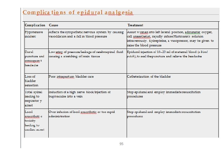
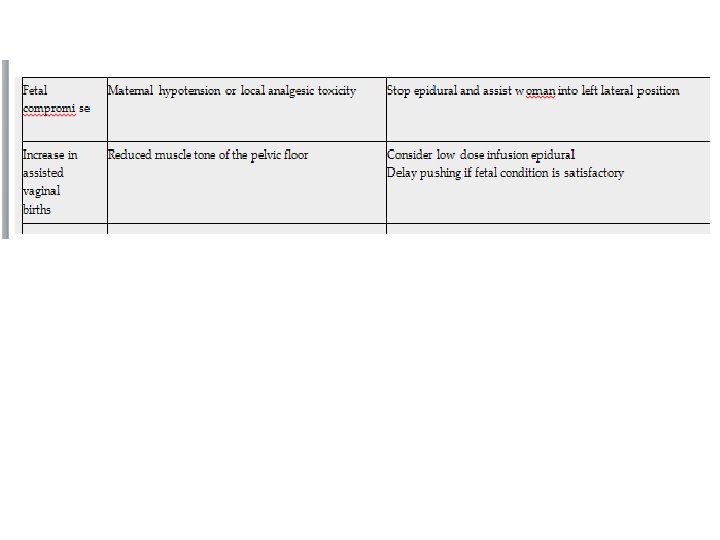
Observations and care by the midwife Each midwife is personally responsible for ensuring that they are competent to care for women who have epidural analgesia, including topping up the epidural block as specifically prescribed by the anaesthetist, being aware of the possible complications and their immediate treatment. After the administration of the first dose of bupivacaine and subsequent top-up doses of local anaesthetic the blood pressure and pulse should be measured and recorded every 5 minutes for 15 minutes and then every 30 minutes (NICE 2007). Temperature should also be recorded regularly. The woman may adopt any position she finds comfortable avoiding aorto-caval compression and encouraged to change position regularly to avoid soft tissue damage. The fetal heart is usually monitored electronically.
The spread of the block should be assessed hourly by the midwife using a cold object or ethyl chloride spray (NICE 2007). The sensation to void urine may be reduced so the midwife must ensure that the woman is encouraged to empty her bladder regularly to avoid postnatal urinary retention. Similarly the woman may not be as sensitive to feeling uterine contractions or the desire to bear down in the second stage of labour. This is due to the pelvic floor muscles being relaxed and a�ecting rotation of the fetal presenting part. The midwife therefore needs to be observant of these physiological changes.
Prelabour rupture of fetal membranes at term (PROM) Prelabour rupture of membranes (PROM) at term (>37 weeks) complicates between 8 and 10% of all pregnancies and most women with PROM will labour spontaneously within 24 hours (NICE 2007). Following PROM with no signs of labour, regardless of whether or not liquor is draining, digital examination should be avoided owing to an increased risk of ascending infection (NICE 2007; NICE 2013). If there is doubt about whether the membranes have ruptured, a sterile speculum examination can be performed in order to observe whethere is pooling of liquor in the posterior fornix of the vagina (NICE 2013). If there are no facilities for this, the woman can be encouraged to wear a sanitary pad for an hour or two in order for the midwife to re-assess for signs of any liquor before a definite diagnosis can be made. The taking of low vaginal swabs is not recommended (NICE 2007). Initial assessment of the woman should include observation of her pulse, respiration rate, blood pressure, temperature, oxygen saturation and urinalysis.
Following PROM the risk of serious neonatal infection is increased from 0. 5% to 1%, compared with women whose membranes remain intact, and the woman should be advised of this (NICE 2007). In view of this, and in the absence of any clinical indication for immediate induction, such as Group B Streptococcus, maternal infection or meconium staining of the liquor, it is usual practice to advise the woman that if she does not go into spontaneous labour within 24 hours, labour should be induced aher PROM (NICE 2007) (see Chapter 19). Women should be given adequate information to decide between expectant management and active management of labour following PROM. Hospital admission, in the absence of any other concerns, is not required whilst waiting for induction to take place. .
Until the induction is commenced or if expectant management beyond 24 hours is chosen by the woman the following recommendations regarding advice to women and subsequent care should be followed (NICE 2007): Bathing or showering are not associated with an increase in infection, but having sexual intercourse may be. Body temperature should be recorded every 4 hours during waking hours and any change in the colour or smell of the vaginal loss should be reported to the midwife immediately. Fetal movements should be observed. In the absence of any risk indicators and with satisfactory evidence of fetal movements and a normal fetal heart rate, there is no reason for a CTG to be performed. Low vaginal swabs and blood samples to assess maternal C-reactive protein should not be taken
Preterm prelabour rupture of the membranes (PPROM) occurs before 37 completed weeks' gestation, where the fetal membranes rupture without the onset of spontaneous uterine activity and the consequential cervical dilatation.
The responsibilities of the midwife The midwife has an important enabling and facilitating role to support the woman during childbirth. It is vital that shared decision-making takes place between women and their caregivers at all times (Hodnef et al 2012). Accurate and detailed records of all care given during the first stage of labour, including the careful administration and monitoring of any medicines, is essential to the provision of quality care. These in turn will provide a good basis from which proper decisions may be made concerning the progress and the needs of the woman to optimize her labour experience and eventual birth outcome.