
Chapter 14 The Urinary System Lesson 14 1
- Slides: 30
Chapter 14: The Urinary System Lesson 14. 1 Anatomy of the Kidney
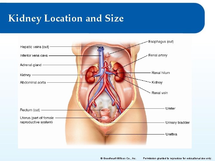
Kidney Location and Size © Goodheart-Willcox Co. , Inc. Permission granted to reproduce for educational use only.
Anatomy of the Kidney • Retroperitoneal and fat covered – 11 cm X 3 cm X 6 cm – 150 gm • renal cortex – Outer, light colored • renal medulla – Middle, darker – Renal pyramids • renal pelvis – Inner, leads to ureter – hollow © Goodheart-Willcox Co. , Inc. Permission granted to reproduce for educational use only.
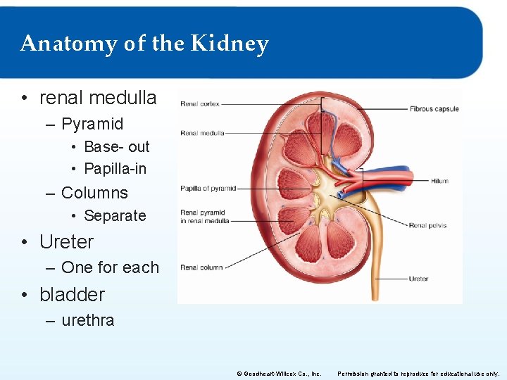
Anatomy of the Kidney • renal medulla – Pyramid • Base- out • Papilla-in – Columns • Separate • Ureter – One for each • bladder – urethra © Goodheart-Willcox Co. , Inc. Permission granted to reproduce for educational use only.
Nerve and Blood Supply • • 20– 25% of blood flows to kidneys renal artery renal vein sympathetic nerve system – renal nerve fibers – Mesh around renal artery • Everything connects at the Hilum © Goodheart-Willcox Co. , Inc. Permission granted to reproduce for educational use only.
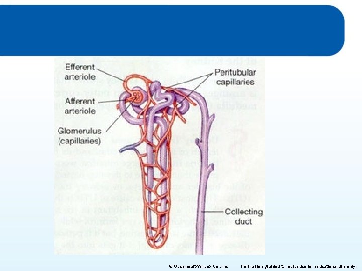
The Nephron • basic working unit of each kidney – Each kidney has 1 million nephrons – Each nephron has a blood supply and creates urine • two main parts – renal corpuscle • • Glomerulus (capillaries) Glomerular capsule ( collecting cup) Enters at afferent, exits at efferent (arterioles) Like a cul-de-sac – renal tubule • processing © Goodheart-Willcox Co. , Inc. Permission granted to reproduce for educational use only.
© Goodheart-Willcox Co. , Inc. Permission granted to reproduce for educational use only.
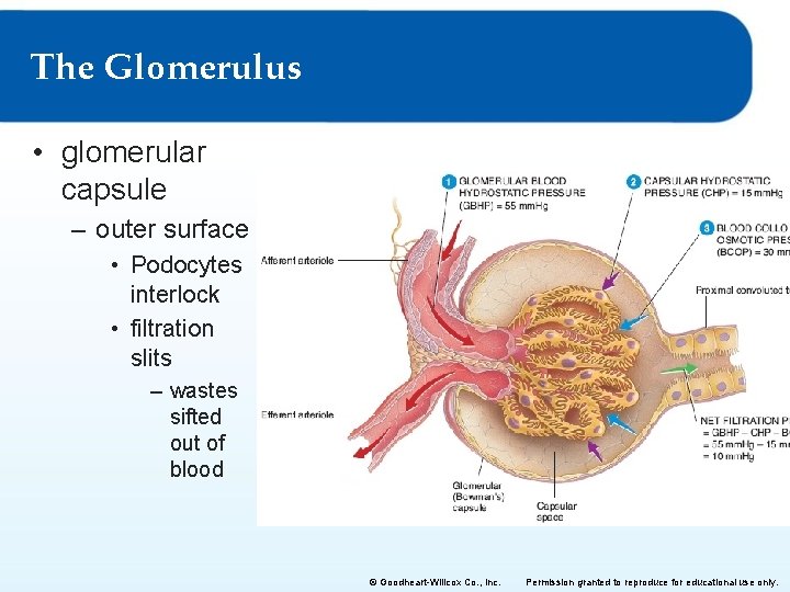
The Glomerulus • glomerular capsule – outer surface • Podocytes interlock • filtration slits – wastes sifted out of blood © Goodheart-Willcox Co. , Inc. Permission granted to reproduce for educational use only.
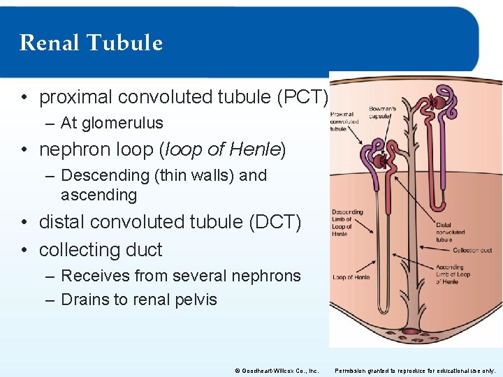
Renal Tubule • proximal convoluted tubule (PCT) – At glomerulus • nephron loop (loop of Henle) – Descending (thin walls) and ascending • distal convoluted tubule (DCT) • collecting duct – Receives from several nephrons – Drains to renal pelvis © Goodheart-Willcox Co. , Inc. Permission granted to reproduce for educational use only.
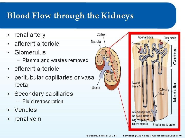
Blood Flow through the Kidneys • renal artery • afferent arteriole • Glomerulus – Plasma and wastes removed • efferent arteriole • peritubular capillaries or vasa recta • Secondary capillaries – Fluid reabsorption • Venules • renal vein © Goodheart-Willcox Co. , Inc. Permission granted to reproduce for educational use only.
Chapter 14: The Urinary System Lesson 14. 2 Urine Formation, Storage, and Excretion
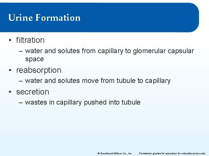
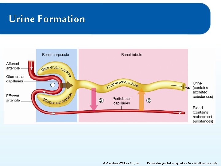
Urine Formation • filtration – water and solutes from capillary to glomerular capsular space • reabsorption – water and solutes move from tubule to capillary • secretion – wastes in capillary pushed into tubule © Goodheart-Willcox Co. , Inc. Permission granted to reproduce for educational use only.
Urine Formation © Goodheart-Willcox Co. , Inc. Permission granted to reproduce for educational use only.
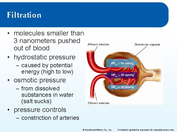
Filtration • molecules smaller than 3 nanometers pushed out of blood • hydrostatic pressure – caused by potential energy (high to low) • osmotic pressure – from dissolved substances in water (salt sucks) • pressure controls – constriction of arteries © Goodheart-Willcox Co. , Inc. Permission granted to reproduce for educational use only.
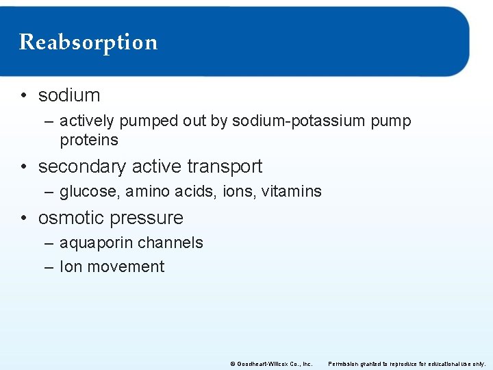
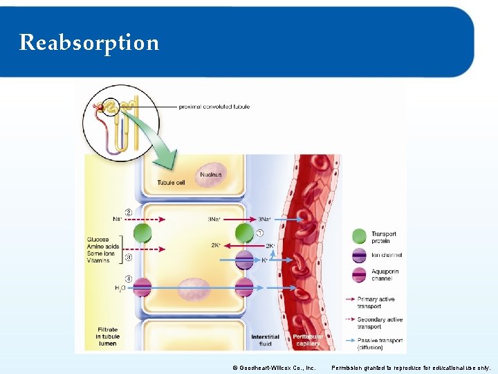
Reabsorption • sodium – actively pumped out by sodium-potassium pump proteins • secondary active transport – glucose, amino acids, ions, vitamins • osmotic pressure – aquaporin channels – Ion movement © Goodheart-Willcox Co. , Inc. Permission granted to reproduce for educational use only.
Reabsorption © Goodheart-Willcox Co. , Inc. Permission granted to reproduce for educational use only.
Secretion • maintain arterial blood p. H at 7. 4 (active) – hydrogen ions – bicarbonate ions • some drugs secreted – penicillin – aspirin © Goodheart-Willcox Co. , Inc. Permission granted to reproduce for educational use only.
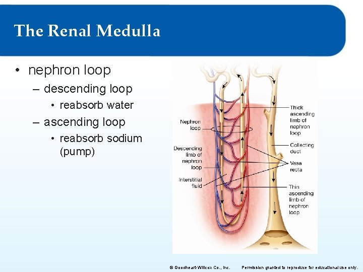
The Renal Medulla • nephron loop – descending loop • reabsorb water – ascending loop • reabsorb sodium (pump) © Goodheart-Willcox Co. , Inc. Permission granted to reproduce for educational use only.
The Countercurrent Mechanism • Blood flows “down and back”, so does filtrate • vasa recta – movement of blood • nephron loop – movement of filtrate © Goodheart-Willcox Co. , Inc. Permission granted to reproduce for educational use only.
Hormonal Regulation of Urine Volume and Composition • aldosterone – Response to drop in Na or rise in K= more aldosterone – Drop in BP= Renin (hormone) reacts to produce angiotensin to increase pressure – Acts on distal tubule to inc sodium in waste= increased osmosis • atrial natriuretic peptide – Response to increased BP ( atrial stretch) – Blocks sodium reabsorption in collecting ducts – causes urine volume to increase (drop pressure, release water) • antidiuretic hormone – causes urine volume to decrease ( hold water, increase pressure) – Dehydration=high ADH=less urine, Hydration= Low ADH=more urine © Goodheart-Willcox Co. , Inc. Permission granted to reproduce for educational use only.
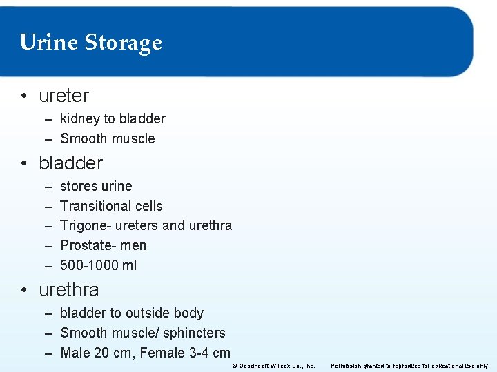
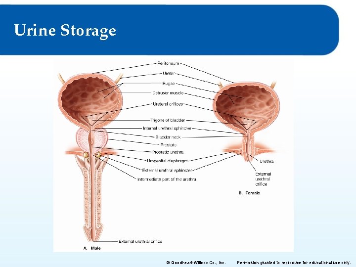
Urine Storage • ureter – kidney to bladder – Smooth muscle • bladder – – – stores urine Transitional cells Trigone- ureters and urethra Prostate- men 500 -1000 ml • urethra – bladder to outside body – Smooth muscle/ sphincters – Male 20 cm, Female 3 -4 cm © Goodheart-Willcox Co. , Inc. Permission granted to reproduce for educational use only.
Urine Storage © Goodheart-Willcox Co. , Inc. Permission granted to reproduce for educational use only.
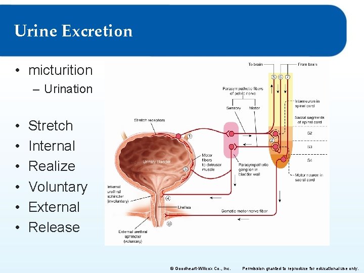
Urine Excretion • micturition – Urination • • • Stretch Internal Realize Voluntary External Release © Goodheart-Willcox Co. , Inc. Permission granted to reproduce for educational use only.
Chapter 14: The Urinary System Lesson 14. 3 Diseases and Disorders of the Urinary System
Assessing Renal Function • physical characteristics of urine – p. H range 4. 5– 8. 0 – sp. G 1. 003 to 1. 035 – 0. 5 -2 L/day • chemical composition of urine – 95% water, 5% waste – Urea, Nitrogen, ions • glomerular filtration rate – estimate with creatinine concentration in blood – Muscle metabolism byproduct, quickly filtered © Goodheart-Willcox Co. , Inc. Permission granted to reproduce for educational use only.
Diabetes • diabetes mellitus – glucose in urine- osmosis is the reason! – type I, type II – Proteinuria, ketoacidosis and renal failure. • diabetes insipidus – large amount of dilute urine (>12 L sp. G <1. 002) – ADH not made or not used – Common in low Hcg (dwarfism)… Remember? © Goodheart-Willcox Co. , Inc. Permission granted to reproduce for educational use only.
Normal Blood Glucose and Blood Insulin Levels • carbohydrate digestion leads to an increase in blood glucose level • increase in blood glucose level causes the pancreas to secrete insulin • increase in blood insulin level causes – – liver glucose uptake liver conversion of glucose to glycogen skeletal muscle glucose uptake skeletal muscle conversion of glucose to glycogen © Goodheart-Willcox Co. , Inc. Permission granted to reproduce for educational use only.
Chronic Kidney Disease • kidney damage • glomerular filtration rate less than 60 ml/min for at least 3 months • develops slowly • Usually diabetes related • When GFR drops to <15 ml/min= renal failure – Waste product accumulation, p. H uncontrolled, only dialysis or transplant will keep alive – Dialysis becomes more frequent, more health effects. Why? © Goodheart-Willcox Co. , Inc. Permission granted to reproduce for educational use only.
Renal Dialysis- remove H 20, urea and Na • hemodialysis – Dialyzer- artificial kidney (external) • peritoneal dialysis – dialysis solution introduced via port to AP cavity – peritoneum acts as dialysis membrane © Goodheart-Willcox Co. , Inc. Permission granted to reproduce for educational use only.
Diseases and Disorders of the Urinary System • kidney stones (pelvis) – – calcium, magnesium, or uric acid <5 mm can pass Lithotripsy- US waves to break stones apart Fluids good…. . • urinary tract infection – – Bacteria up urethra to bladder cystitis more common in women than men Can go to kidneys if untreated= pyelonephritis+ back pain © Goodheart-Willcox Co. , Inc. Permission granted to reproduce for educational use only.