CHAPTER 14 The Foot BONES 26 bones total

CHAPTER 14 The Foot

BONES 26 bones total 7 Tarsal Bones Talus Calcaneus Navicular Cuboid Medial Cuneiform Intermediate Cuneiform Lateral Cuneiform 5 Metatarsal Bones 14 Phalanx Bones

ARCHES OF THE FOOT The foot is supported by ligamentous & bony arrangements that form several arches. Metatarsal Arch Shaped by distal heads of the metatarsal Transverse Arch Extends across the transverse tarsal bones Medial Longitudinal Arch Originates at medial border of the calcaneus & extends forward to distal head of the first metatarsal Lateral Longitudinal Arch Originates at the lateral border of the calcaneus & extends to the distal head of the fifth metatarsal

PES CAVUS & PES PLANUS Pes Cavus : High Arch Pes Planus: Flat Arch The foot is responsible for distributing body weight through the lower extremity. The height of a person’s arch determines how that body weight is distributed. Arches assist the foot in supporting body weight & absorbing shock

PLANTAR APONEUROSIS Plantar Fascia: originates at the medial aspect of the calcaneus at inserts at the distal heads of the metatarsals The plantar fascia supports the foot against downward forces

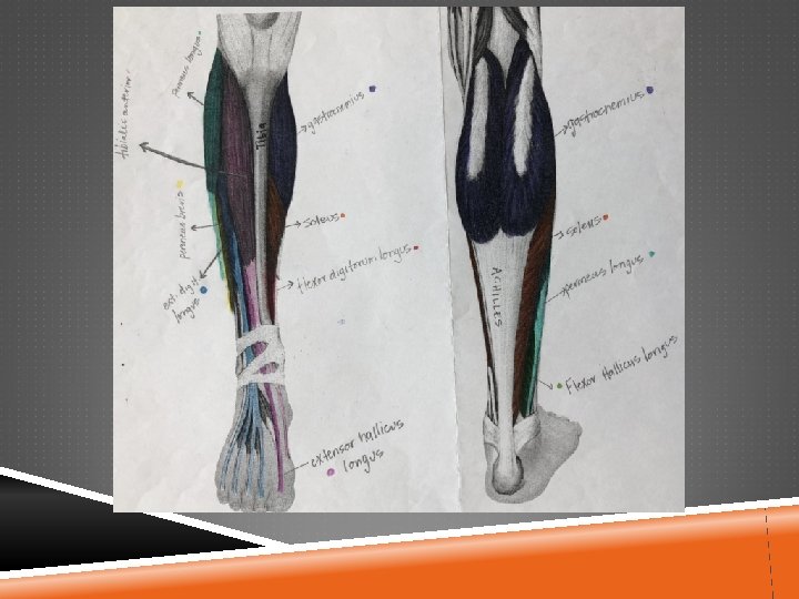
MUSCLES & MOVEMENT OF THE FOOT ADDuction & Supination: Tibialis posterior Flexor digitorum longus, Flexor hallucis longus, Tibialis anterior, Extensor hallucis longus ABDuction & Pronation: Peronus longus & brevis, Peronus tertius, Extensor digitorum longus
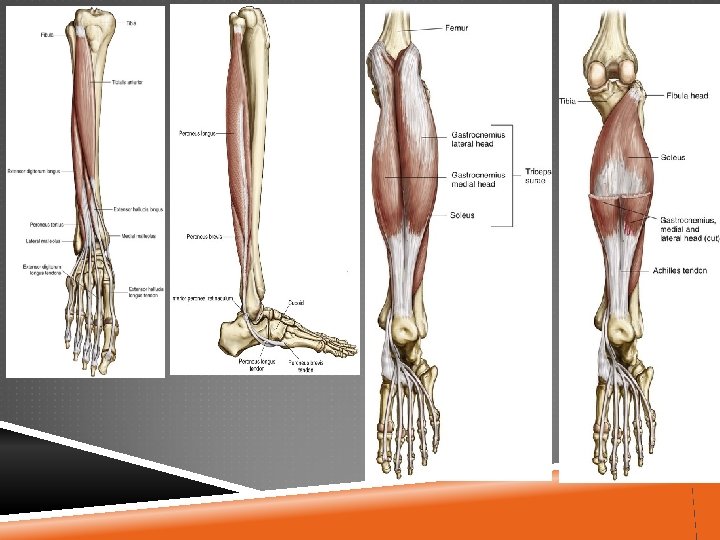

MUSCLES & MOVEMENT Toe Flexion: Flexor digitorum brevis& longus Flexor Hallucis brevis & longus, Flexor digiti minimi brevis Quadratus plantae, lumbricales Toe Extension: extensor digitorum brevis & longus, extensor hallucis longus Toe ADDuction: adductor hallucis, plantar interossi Toe ABDuction: abductor hallucis, dorsal interossi, abductor digiti minimi

1 st Layer 2 nd Layer 3 rd Layer 4 th Layer

PREVENTION OF FOOT INJURIES Proper Footwear: sport & activity appropriate, change footwear when it is worn out Contact Surface Orthotics Taping & Bracing Limit miles Strengthening & Stretching when appropriate Rest when appropriate Proper Foot Hygiene

RETRO-CALCANEAL BURSITIS Cause: Chronic irritation caused by rubbing or pressure from the shoe, if the irritation continues, the patient can develop an extosis (extra bone) Signs & Symptoms: Swelling, warmth, redness & pain in the area Care: pad the area with a doughnut, use a heel lift to shorten the Achilles tendon, ice, take anti-inflammatory medication

SEVER’S DISEASE Cause: Chronic Inflammation at the growth plate which is the attachment of the Achilles Tendon on the Calcaneus. Also known as Calcaneal apophysitis Signs & Symptoms: pain, limping, difficulty walking, stiffness, swelling at heel Care: X-ray to diagnose, REST may include crutches or a boot, physical therapy, heel cup or heel lifts when they return to activity, Ice to control pain & swelling.

HEEL BRUISE Cause: Trauma to the fat pad in the foot Signs & Symptoms: Pain, Difficulty ambulating or weight bearing, ecchymosis (discoloration), swelling Care: pad the area or provide cushion, rest, ice, you may have to use crutches for a period of time

PLANTAR FASCIITIS Cause: Inflexibility of medial arch or gastrocnemius-soleus unit, wearing shoes without supports, running on soft surfaces or lengthened stride during running Signs & Symptoms: Pain in anterior medial heel this pain may eventually move to more centrally to the middle of the plantar fascia, Pain increases with dorsiflexion, Pain usually much worse in the morning Care: Heel cord stretching, Plantar Fascia stretching, Massage, Ice, Heel cup, Arch taping, Orthotics. Night Splints

METATARSAL FRACTURE Cause: Direct Trauma (being stepped on, kicked, or dropping something on the foot), Twisting or Torsion Signs & Symptoms: Swelling, Pain, Point Tenderness, You may see a deformity Care: X-ray to diagnose, PRICE, cast, crutches, boot or shoe

JONES FRACTURE Fracture @ the base of the 5 th metatarsal, particularly at the neck Usually occurs from overuse, acute inversion, or high velocity rotational forces Athlete may complain of sharp pain, a ‘pop’ Typically these injuries result in a non-union due to poor blood supply. If extended immobilization (6 -8 weeks) fails, surgery may be necessary

JONES FRACTURE Dancer’s Fracture

MORTON’S TOE Abnormally short first metatarsal; the second to appears longer Can cause injuries in the second metatarsal b/c body weight is distributed differently

2 ND METATARSAL STRESS FRACTURE Cause: Occur most often in running & jumping sports. Can be caused by structural deformities (Morton’s Toe), training errors, changes in training surfaces, & inappropriate shoes Signs & Symptoms: Athlete usually has pain & point tenderness along the 2 nd Metatarsal & may also feel pain & aching during non-weight bearing activities Care: Remove the stress, REST, Check footwear, slow progress back to activity

2 ND METATARSAL FRACTURE

MORTON’S NEUROMA Cause: Enlargement of the nerve. A mass develops on the common plantar nerve. Typically occurs between the 3 rd & 4 th metatarsal heads Signs & Symptoms: severe intermittent pain from the metatarsal heads to the tips of the toes, pain may be relieved when the patient is non-weight bearing, burning or numbness in the forefoot, Care: metatarsal pad used to open up the joint, shoes with a wide toe box, can be surgically repaired in severe cases

METATARSAL ARCH STRAIN Cause: Typically caused by hypermobile metatarsals, which is secondary to laxity in the ligaments. Hypermobility allows the metatarsals to spread apart (splayed arch) Signs & Symptoms: Athlete may complain of pain or cramping over the metatarsals, point tenderness, & weakness. Pain is most commonly located under the 2 nd & 3 rd metatarsal head. Athlete may also develop a large callus under the area of pain. Care: Apply a pad to elevate the metatarsal head, focus on strengthening foot muscles & heel cord stretching

LONGITUDINAL ARCH STRAIN Cause: Stress caused by hard playing surfaces which causes the longitudinal arch to collapse during weight bearing Signs & Symptoms: Pain & swelling along the medial aspect of the foot. The spring ligament (calcaneonavicular) may be affected with prolonged stress. There may also be pain in the flexor hallucis longus. Care: PRICE, non-weight bearing, Physical Therapy, arch taping or support when return to activity

LISFRANC INJURY Injury tostthe joint at the base nd of the 1 & 2 Metatarsal Rare & often misdiagnosed Complex injury that presents with pain, inability to bear weight & swelling in the midfoot Typically caused by trauma Most of the time surgery is required to repair this injury. Recovery time is approximately one year. Patients must remain nonweight bearing for the first 3 months following surgery

LISFRANC INJURY

LISFRANC INJURY

PHALANX FRACTURES & DISLOCATIONS Cause: Trauma, kicking an object, stubbing a toe, dropping a heavy object on the toe Signs & Symptoms: Swelling, Ecchymosis (discoloration), Pain, Bleeding, Deformity Care: Refer to a physician for X-ray & diagnosis, Buddy Taping, Cast for multiple toes, 3 -4 weeks of inactivity, Wooden shoe may be used

PHALANX DISLOCATION & DISPLACED PHALANX FRACTURE
 at the head of the first metatarsal AKA: Hallux")
BUNION Bony Exostoses (extra bone) at the head of the first metatarsal AKA: Hallux Valgus Deformity Cause: Can be caused by footwear that is too narrow or too small Signs & Symptoms: Tenderness, Swelling, Obvious Deformity Care: Change footwear, use a donut to pad the area, surgery may be required in the later stages of the injury

TURF TOE Cause: Hyperextension of the great toe, from repetitive overuse or trauma. Strain of the muscles & tendon can also be combines with a sprain or the ligaments & jt. Capsule Signs & Symptoms: Pain, Swelling, Loss of motion, Inability to bear weight or push off Care: Rigid shoes that don’t allow for much dorsiflexion, steel shank, taping, ice, rest, PT

TURF TOE

CALLUSES Cause: Caused by pressure points & friction, shoes that are too narrow or too small. Blisters can develop under calluses Signs & Symptoms: Pain, Skin build up Care: File with a callus shaver, but be careful in season. Moisturize the skin to increase elasticity. Use cushioning devices. Check footwear, orthotics may be necessary. Use extra socks to reduce friction.

BLISTERS Cause: A result of shearing forces on the skin, friction. Signs & Symptoms: Fluid accumulates below the outer layer of skin & can be clear or bloody. Athlete will have Pain. Care: Eliminate wrinkles or folds. Skin lubricant. Open blister, clean & pad if fluid is clear. If fluid is bloody, pad/cushion the area. Leave extra skin intact until under layer dries out.

BLISTERS

INGROWN TOENAIL Cause: Side edge of the toenail grows into the soft tissue causing severe inflammation Signs & Symptoms: Redness, Tenderness, Bleeding, warmth & Infection Care: Check nails for length, make sure toe is cleaned properly & covered if needed, cut a ‘V’ in the nail toward the infected side, pack with cotton @ edge, soak in medicated warm water if necessary, once ingrown toenail is resolved be sure to keep nails cut to appropriate length & straight across

SUBUNGUAL HEMATOMA Cause: being stepped on, kicking something, dropping an object on toe or nails hitting the end of shoes/cleats Signs & Symptoms: Bluish-purple nail, pain, may see external bleeding around nail depending on mechanisms Care: Depends on mechanism, Ice, if you think a fracture may be involved the athlete should get an x-ray, if fracture is ruled out then you can drill the nail to release the blood within 24 hours, drilling must be done under sterile conditions
- Slides: 39