
Chapter 14 Seeing and Hearing Functions of the
- Slides: 22
Chapter 14 Seeing and Hearing
Functions of the Eye • The ocular system is responsible for vision. • The combining forms for the eye or sight are opt/i, opt/o, optic/o, ocul/o, and ophthalm/o.
Structures of the Eye • The accessory structures of the eye: – orbit – eye muscles – eyelids or palpebrae • Blephar/o is the combining form.
Structures of the Eye – eyelashes – conjunctiva • Conjunctiv/o is the combining form. – lacrimal apparatus • Lacrim/o and dacry/o are the combining forms.
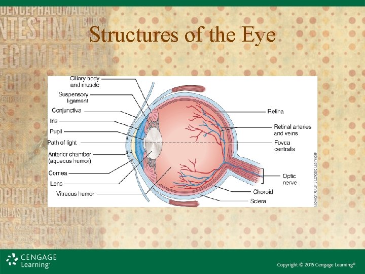
Structures of the Eye • The structures of the eyeball: – sclera = outer layer • Combining form is scler/o. • The transparent anterior portion is called the cornea. – Combining form for cornea is corne/o.
Structures of the Eye • choroid = middle layer – Consists of blood vessels, iris, pupil, lens, and ciliary body – Combining form is choroid/o. • retina = inner layer – Nervous tissue layer that receives images – Combining form is retin/o.
Structures of the Eye
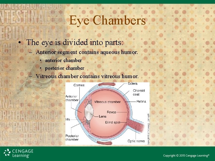
Eye Chambers • The eye is divided into parts: – Anterior segment contains aqueous humor. • anterior chamber • posterior chamber – Vitreous chamber contains vitreous humor.
Vision • Vision occurs when light rays enter the eye through the cornea, pass through the lens, and travel to the retina • The image is focused on the retina and is then transmitted to the optic nerve • Stimulations are transmitted from the optic nerve, to the optic chiasm, to the midbrain, and to the visual cortex of the occipital lobe of the cerebrum
Vision • Accommodation – Adjustment for distance • Refraction – Focusing • Convergence – Maintenance of binocular vision • Acuity – Sharpness
Medical Terms for the Ocular System • Additional terms for ocular system tests, pathology, and procedures can be found in the text. • See this week’s Module for additional images and videos.
Functions of the Ear • The ear is the sensory organ that enables hearing and helps to maintain balance. • The combining forms for ear are audit/o, aud/i, and ot/o. • The combining forms for sound or hearing are acoust/o and acous/o.
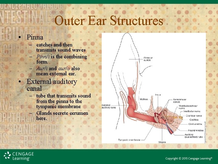
Outer Ear Structures • Pinna – catches and then transmits sound waves – Pinn/i is the combining form. – Aur/i and aur/o also mean external ear. • External auditory canal – tube that transmits sound from the pinna to the tympanic membrane – Glands secrete cerumen here.
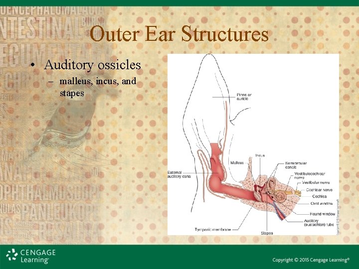
Outer Ear Structures • Auditory ossicles – malleus, incus, and stapes
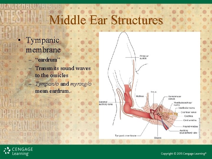
Middle Ear Structures • Tympanic membrane – “eardrum” – Transmits sound waves to the ossicles – Tympan/o and myring/o mean eardrum.
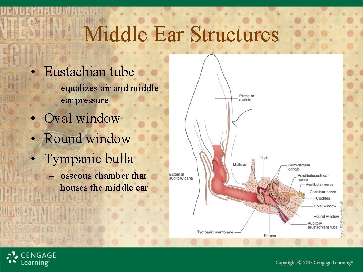
Middle Ear Structures • Eustachian tube – equalizes air and middle ear pressure • Oval window • Round window • Tympanic bulla – osseous chamber that houses the middle ear
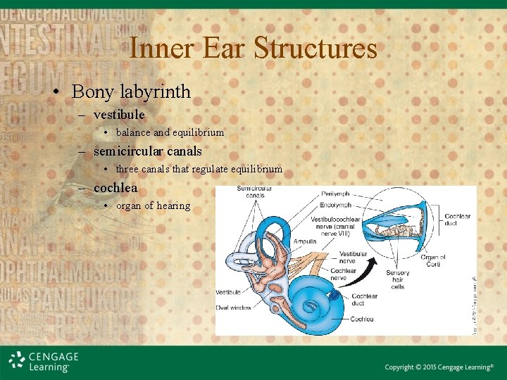
Inner Ear Structures • Bony labyrinth – vestibule • balance and equilibrium – semicircular canals • three canals that regulate equilibrium – cochlea • organ of hearing
Mechanism of Hearing • Sound waves enter the ear through the pinna, travel through the external auditory canal, and strike the tympanic membrane. • As the tympanic membrane vibrates, it moves the ossicles. • The ossicles conduct the sound waves through the middle ear. • Sound vibrations reach the inner ear via the round window.
Mechanism of Hearing • The structures of the inner ear respond to sound waves that displace fluid in the inner ear. • Stimulation of hair cells in the organ of Corti initiates a nerve impulse that is transmitted to the vestibulocochlear nerve. • This nerve impulse is then relayed to the brain.
Mechanism of Equilibrium • the state of balance • maintaining the position of the head relative to gravity – static equilibrium – controlled by organs in the vestibule of the inner ear
Mechanism of Equilibrium • maintaining balance in response to rotational or angular movement – dynamic equilibrium – controlled by the semicircular canals
Medical Terms for the Auditory System • Additional terms for auditory system tests, pathology, and procedures can be found in the text. • See this week’s Module for additional images and videos.