Chapter 14 Peripheral Nervous System SPINAL NERVES Overview


 Lumbar, sacral, and coccygeal nerve roots descend from point of")
 Structure of spinal nerves Each spinal nerve attaches to spinal")
 Ramus ▪ One of several large branches formed after each")
 ▪ Ventral ramus ▪ Structure is more complex than that")
 Nerve plexuses Plexus: complex network formed by the ventral")
 Four major pairs of plexuses ▪ Cervical plexus (Figure")
 ▪ Lumbar plexus (Figure 14 -5) ▪ Located in")
 Dermatomes and myotomes (Figure 14 - 6) Dermatome: region of")
 12 pairs of cranial nerves")

 Olfactory nerve (I) Composed of axons of neurons whose dendrites")
 Oculomotor nerve (III) Fibers originate from cells in the oculomotor")
 Trochlear nerve (IV) Motor fibers originate in cells of the")
 Trigeminal nerve (V) (Figure 14 -10) Has three branches: ophthalmic")
 Abducens nerve (VI) Motor nerve with fibers originating from a")
 Facial nerve (VII) Motor fibers originate from a nucleus in")
 Vestibulocochlear nerve (VIII) Two distinct divisions that are both sensory:")
 Glossopharyngeal nerve (IX) Composed of sensory, motor, and autonomic nerve")
 Accessory nerve (XI) Motor nerve that is an “accessory” to")

 Basic principles of somatic motor pathways")
 Somatic reflexes Nature of a reflex")
 Somatic reflexes of clinical importance: reflexes")


 and efferent (motor) components (the efferent components")
 Structure of the autonomic nervous system Basic plan of")
 Structure of the sympathetic pathways ▪ Sympathetic chain")
 ▪ Preganglionic fiber may take one of three")
 Structure of the parasympathetic pathways ▪ Parasympathetic preganglionic")


 Autonomic neurotransmitters (Figures 14 -18 and 14 -20)")
 Autonomic neurotransmitters (cont. ) ▪ Epinephrine also stimulates")
 Functions of the autonomic nervous system Overview of autonomic")
 Functions of the sympathetic division ▪ Under resting")
- Slides: 59
Chapter 14: Peripheral Nervous System
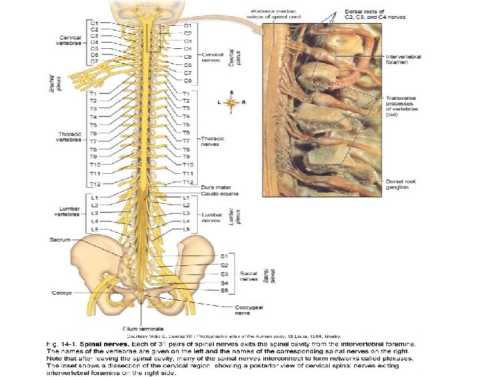
SPINAL NERVES Overview Thirty-one pairs of spinal nerves are connected to the spinal cord (Figure 14 -1) No special names; numbered by level of vertebral column at which they emerge from the spinal cavity ▪ ▪ ▪ Eight cervical nerve pairs (C 1 through C 8) 12 thoracic nerve pairs (T 1 through T 12) Five lumbar nerve pairs (L 1 through L 5) Five sacral nerve pairs (S 1 through S 5) One coccygeal nerve pair
SPINAL NERVES (cont. ) Lumbar, sacral, and coccygeal nerve roots descend from point of origin to the lower end of the spinal cord (level of first lumbar vertebra) before reaching the intervertebral foramina of the respective vertebrae, through which the nerves emerge Cauda equina describes the appearance of the lower end of the spinal cord and its spinal nerves as a horse’s tail
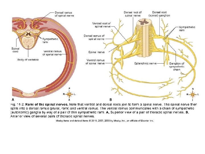
SPINAL NERVES (cont. ) Structure of spinal nerves Each spinal nerve attaches to spinal cord by a ventral (anterior) root and a dorsal (posterior) root Dorsal root ganglion: swelling in the dorsal root of each spinal nerve All spinal nerves are mixed nerves
SPINAL NERVES (cont. ) Ramus ▪ One of several large branches formed after each spinal nerve emerges from the spinal cavity (Figure 14 -2) ▪ Dorsal ramus supplies somatic motor and sensory fibers to smaller nerves that innervate the muscles and skin of the posterior surface of the head, neck, and trunk
SPINAL NERVES (cont. ) ▪ Ventral ramus ▪ Structure is more complex than that of dorsal ramus ▪ Autonomic motor fibers split from the ventral ramus and head toward a ganglion of the sympathetic chain ▪ Some autonomic fibers synapse with neurons that continue on to autonomic effectors through splanchnic nerves; others synapse with neurons whose fibers rejoin the ventral ramus ▪ Sympathetic rami: splitting and rejoining of autonomic fibers ▪ Motor and sensory fibers innervate muscles and glands in the extremities and lateral and ventral portions of neck and trunk
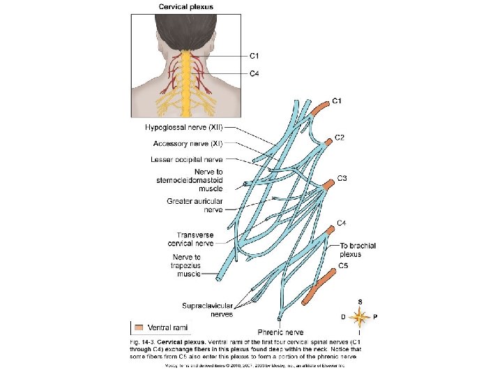
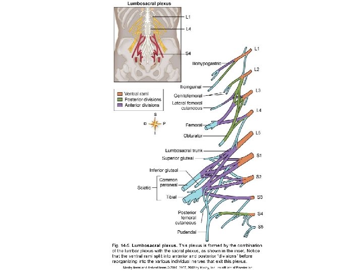
SPINAL NERVES: PLEXUSES (cont. ) Nerve plexuses Plexus: complex network formed by the ventral rami of most spinal nerves (not T 2 through T 12), subdividing and then joining together to form individual nerves Each individual nerve that emerges contains all the fibers that innervate a particular region of the body In plexuses, spinal nerve fibers are rearranged according to their ultimate destination, reducing the number of nerves needed to supply each body part
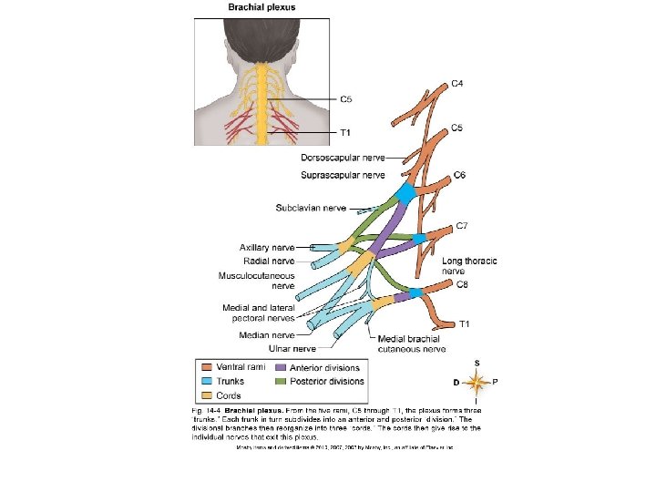
SPINAL NERVES: PLEXUSES (cont. ) Four major pairs of plexuses ▪ Cervical plexus (Figure 14 -3) ▪ Located deep within the neck ▪ Composed of ventral rami of C 1 through C 4 and a branch of the ventral ramus of C 5 ▪ Individual nerves emerging from cervical plexus innervate the muscles and skin of the neck, upper shoulders, and part of the head ▪ Phrenic nerve exits the cervical plexus and innervates the diaphragm ▪ Brachial plexus (Figure 14 -4) ▪ Located deep within the shoulder ▪ Composed of ventral rami of C 5 through T 1 ▪ Individual nerves emerging from brachial plexus innervate the lower part of the shoulder and the entire arm
SPINAL NERVES: PLEXUSES (cont. ) ▪ Lumbar plexus (Figure 14 -5) ▪ Located in the lumbar region of the back in the psoas muscle ▪ Formed by intermingling fibers of L 1 through L 4 ▪ Femoral nerve exits the lumbar plexus, divides into many branches, and supplies the thigh and leg ▪ Sacral plexus and coccygeal plexus (Figure 14 -5) Located in the pelvic cavity in the anterior surface of the piriformis muscle Formed by intermingling of fibers from L 4 through S 4 Tibial, common peroneal, and sciatic nerves exit the sacral plexus and supply nearly all the skin of the leg, posterior thigh muscles, and leg and foot muscles
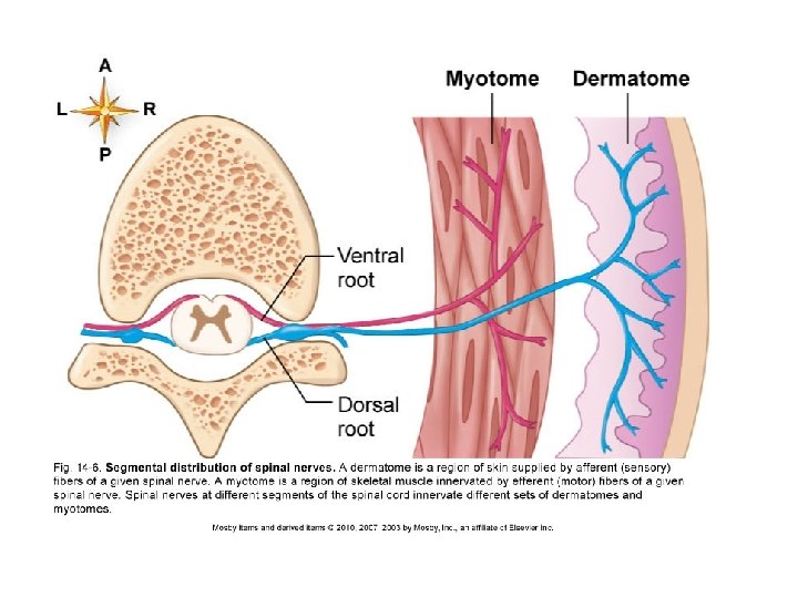
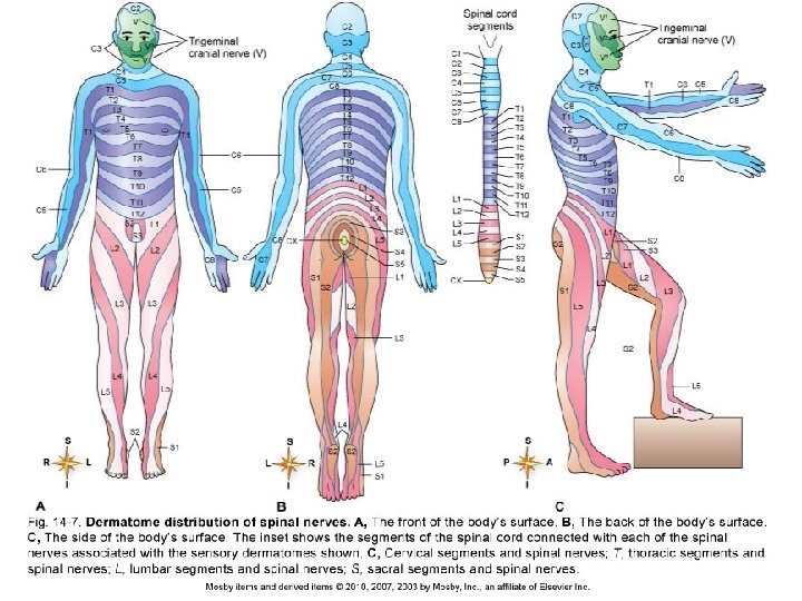
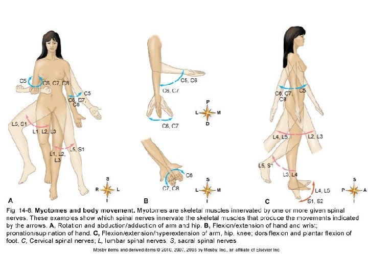
SPINAL NERVES (cont. ) Dermatomes and myotomes (Figure 14 - 6) Dermatome: region of skin surface area supplied by afferent (sensory) fibers of a given spinal nerve (Figure 14 -7) Myotome: skeletal muscle or muscles supplied by efferent (motor) fibers of a given spinal nerve (Figure 14 -8)
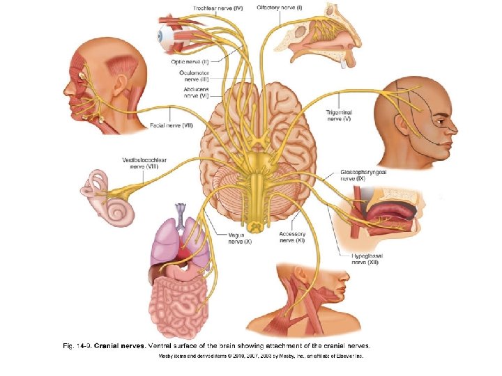
CRANIAL NERVES Overview (Tables 14 -2 and 14 -3) 12 pairs of cranial nerves connect to the brain, mostly the brainstem (Figure 14 -9) Identified by name (determined by either distribution or function) or number (order in which they emerge, anterior to posterior) or both Composed of bundles of axons ▪ Mixed cranial nerve: axons of sensory and motor neurons ▪ Sensory cranial nerve: axons of sensory neurons only ▪ Motor cranial nerve: mainly axons of motor neurons and a small number of sensory fibers (proprioceptors)
Proprioreception the sense of the relative position of one's own parts of the body and strength of effort being employed in movement.
CRANIAL NERVES (cont. ) Olfactory nerve (I) Composed of axons of neurons whose dendrites and cell bodies lie in nasal mucosa and terminate in olfactory bulbs Carries information about sense of smell Optic nerve (II) Composed of axons from the innermost layer of sensory neurons of the retina Carries visual information from the eyes to the brain
CRANIAL NERVES (cont. ) Oculomotor nerve (III) Fibers originate from cells in the oculomotor nucleus and extend to some of the external eye muscles Efferent autonomic fibers are also present, extending to the intrinsic muscles of the eye to regulate amount of light entering eye and help focus on near objects Sensory fibers from proprioceptors in the eye muscles are also present
CRANIAL NERVES (cont. ) Trochlear nerve (IV) Motor fibers originate in cells of the midbrain and extend to the superior oblique muscles of the eye Also contains afferent fibers from proprioceptors in the superior oblique muscles of the eye
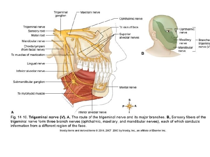
CRANIAL NERVES (cont. ) Trigeminal nerve (V) (Figure 14 -10) Has three branches: ophthalmic nerve, maxillary nerve, and mandibular nerve Sensory neurons carry afferent impulses from skin and mucosa of head and teeth to cell bodies in the trigeminal ganglion Motor fibers originate in trifacial motor nucleus and extend to the muscles of mastication through the mandibular nerve
CRANIAL NERVES (cont. ) Abducens nerve (VI) Motor nerve with fibers originating from a nucleus in the pons on the floor of the fourth ventricle and extending to the lateral rectus muscles of the eye Contains afferent fibers from proprioceptors in the lateral rectus muscles
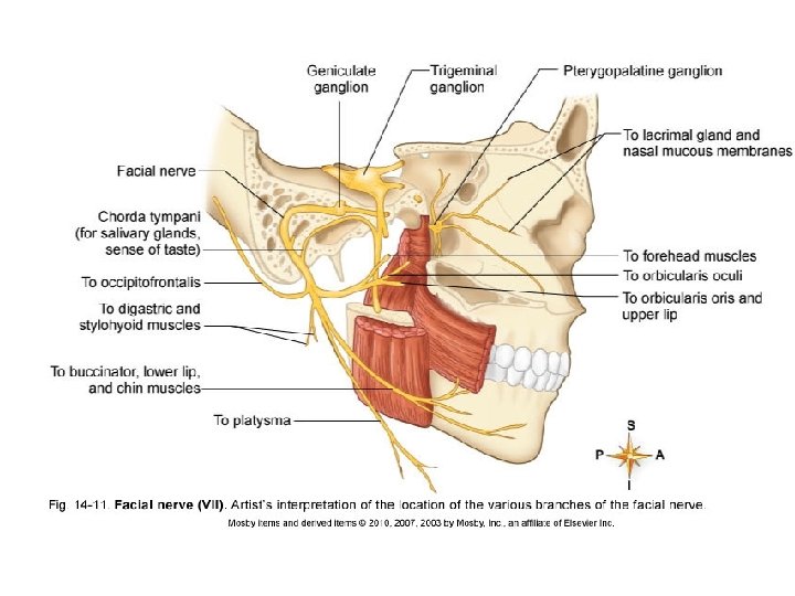
CRANIAL NERVES (cont. ) Facial nerve (VII) Motor fibers originate from a nucleus in lower part of pons and extend to superficial muscles of the face and scalp (Figure 14 -11) Autonomic fibers extend to submaxillary and sublingual salivary glands Also contains sensory fibers from taste buds of anterior two thirds of the tongue
CRANIAL NERVES (cont. ) Vestibulocochlear nerve (VIII) Two distinct divisions that are both sensory: vestibular nerve and cochlear nerve Vestibular nerve fibers originate in the semicircular canals in inner ear and transmit impulses that result in sensations of equilibrium Cochlear nerve fibers originate in organ of Corti in the cochlea of the inner ear and transmit impulses resulting in sensations of hearing
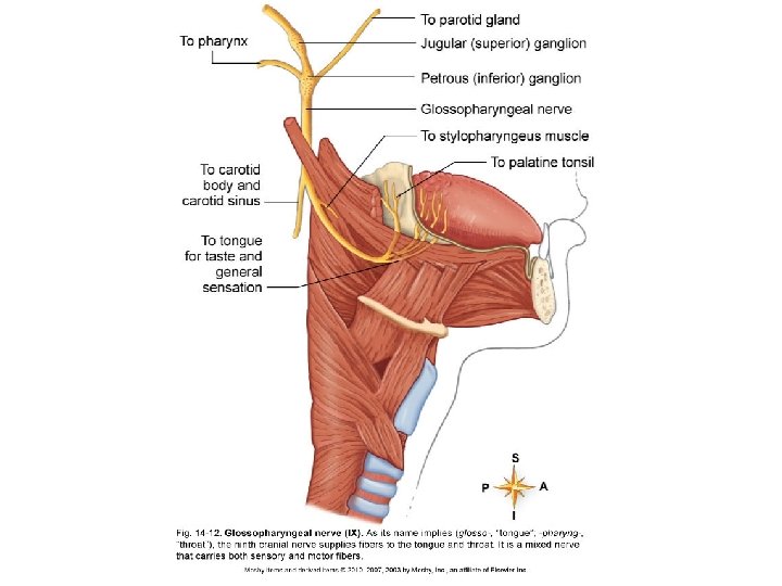
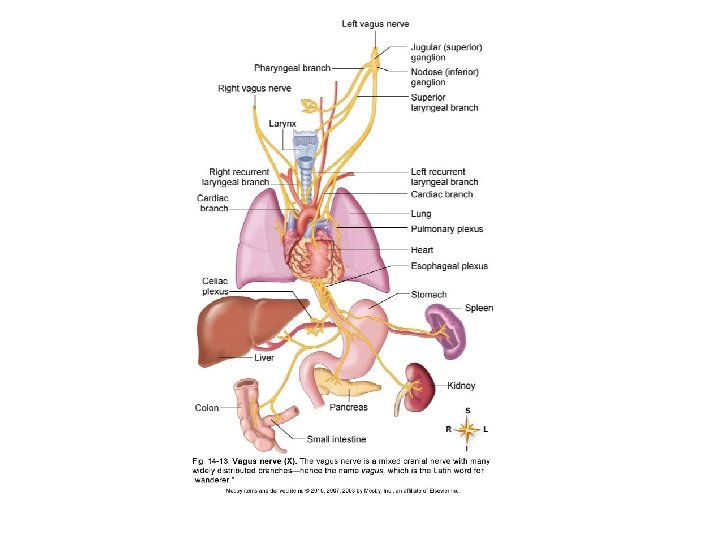
CRANIAL NERVES (cont. ) Glossopharyngeal nerve (IX) Composed of sensory, motor, and autonomic nerve fibers Supplies fibers to tongue, pharynx, and carotid sinus (Figure 14 -12) Vagus nerve (X) Composed of sensory and motor fibers with many widely distributed branches Sensory fibers supply pharynx, larynx, trachea, heart, carotid body, lungs, bronchi, esophagus, stomach, small intestine, and gallbladder (Figure 14 -13) Somatic motor fibers innervate the pharynx and larynx and are mostly autonomic fibers
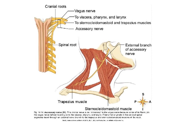
CRANIAL NERVES (cont. ) Accessory nerve (XI) Motor nerve that is an “accessory” to the vagus nerve Innervates thoracic and abdominal viscera, pharynx, larynx, trapezius, and sternocleidomastoid (Figure 14 -14) Hypoglossal nerve (XII) Composed of motor and sensory fibers Motor fibers innervate the muscles of the tongue Contains sensory fibers from proprioceptors in muscles of the tongue
DIVISIONS OF THE PERIPHERAL NERVOUS SYSTEM Two functional divisions of the peripheral nervous system Afferent (sensory) division Efferent (motor) division Efferent division is divided further into the somatic motor nervous system and the efferent portions of the autonomic nervous system
DIVISIONS OF THE PERIPHERAL NERVOUS SYSTEM (cont. ) Basic principles of somatic motor pathways Somatic nervous system includes all voluntary motor pathways outside the central nervous system Somatic effectors: skeletal muscles
DIVISIONS OF THE PERIPHERAL NERVOUS SYSTEM (cont. ) Somatic reflexes Nature of a reflex ▪ Reflex: action that results from a nerve impulse passing over a reflex arc; predictable response to a stimulus ▪ Cranial reflex: center of reflex arc is in the brain ▪ Spinal reflex: center of reflex arc is in the spinal cord ▪ Reflex consists of either muscle contraction or glandular secretion ▪ Somatic reflex: contraction of skeletal muscles ▪ Autonomic (visceral) reflex: either contraction of smooth or cardiac muscle or secretion by glands Q?
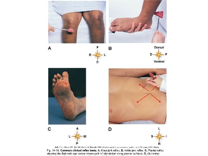
DIVISIONS OF THE PERIPHERAL NERVOUS SYSTEM (cont. ) Somatic reflexes of clinical importance: reflexes deviate from normal in certain diseases, and reflex testing is a valuable diagnostic aid (Figure 14 -16) ▪ Abdominal reflex: drawing in of the abdominal wall in response to stroking the side of the abdomen; superficial reflex; mediated by arcs with sensory and motor fibers in T 9 through T 12 and centers in these segments of the cord; decreased or absent reflex may involve lesions of pyramidal tract upper motor neurons
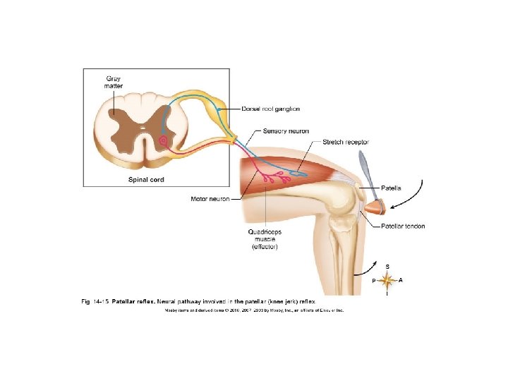
Other examples of reflexes Other reflexes that provide us with biofeedback (information about what’s wrong with the nerves based off of their reflex action) Knee jerk Plantar reflex Corneal reflex Spinal cord Many others
Ganglia In vertebrates the ganglion is a cluster of neural bodies outside the central nervous system. A spinal ganglion, for instance, is a cluster of nerve bodies positioned along the spinal cord at the dorsal and ventral roots of a spinal nerve.
Ganglia
AUTONOMIC NERVOUS SYSTEM Overview Contains afferent (sensory) and efferent (motor) components (the efferent components are emphasized here) Carries fibers to and from the autonomic effectors Major function: to regulate heartbeat, smooth muscle contraction, and glandular secretions to maintain homeostasis Two efferent divisions: sympathetic division and parasympathetic division Sympathetic division consists of neural pathways separate from parasympathetic pathways Many autonomic effectors are dually innervated, which allows remarkably precise control of effector
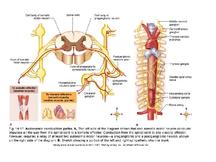
AUTONOMIC NERVOUS SYSTEM (cont. ) Structure of the autonomic nervous system Basic plan of efferent autonomic pathways (Figure 1417) ▪ Each pathway is composed of autonomic nerves, ganglia, and plexuses, which are made of efferent autonomic neurons ▪ All autonomic neurons function in reflex arcs ▪ Efferent autonomic regulation ultimately depends on feedback from sensory receptors ▪ Relay of two efferent autonomic neurons conducts information from central nervous system to autonomic effectors ▪ Preganglionic neuron: conducts impulses from the central nervous system to an autonomic ganglion ▪ Postganglionic neuron: efferent neuron with which a preganglionic neuron synapses within autonomic ganglion
AUTONOMIC NERVOUS SYSTEM: STRUCTURE (cont. ) Structure of the sympathetic pathways ▪ Sympathetic chain ganglia ▪ Most ganglia of the sympathetic division lie along either side of the anterior surface of the vertebral column and are joined with the other ganglia located on the same side ▪ Each chain runs from the second cervical vertebra to the level of the coccyx ▪ 22 sympathetic chain ganglia are usually on each side of vertebral column: three cervical, 11 thoracic, four lumbar, and four sacral ▪ Thoracolumbar division ▪ Sympathetic preganglionic neurons with dendrite and cell bodies in lateral gray horns of the thoracic and lumbar segments of the spinal cord ▪ Axons leave the cord by way of the ventral roots of the thoracic and first four lumbar spinal nerves and split away from other spinal nerve fibers by the white ramus to a sympathetic chain ganglion
AUTONOMIC NERVOUS SYSTEM: STRUCTURE (cont. ) ▪ Preganglionic fiber may take one of three paths once inside the sympathetic chain ganglion ▪ Synapse with sympathetic postganglionic neuron ▪ Send ascending or descending branches through the sympathetic trunk to synapse with postganglionic neurons in other chain ganglia ▪ Pass through one or more chain ganglia without synapsing ▪ Sympathetic postganglionic neurons ▪ Dendrites and cell bodies are mostly in sympathetic chain ganglia or collateral ganglia ▪ Gray ramus: short branch by which some postganglionic axons return to a spinal nerve ▪ In the sympathetic division, preganglionic neurons are relatively short, and postganglionic neurons are relatively long ▪ Axon of one sympathetic preganglionic neuron synapses with many postganglionic neurons, terminating in widely spread organs (Figure 14 -18)
AUTONOMIC NERVOUS SYSTEM: STRUCTURE (cont. ) Structure of the parasympathetic pathways ▪ Parasympathetic preganglionic neurons: cell bodies are located in nuclei in the brainstem or lateral gray columns of the sacral cord; extend a considerable distance before synapsing with postganglionic neurons ▪ Parasympathetic postganglionic neurons: dendrites and cell bodies are located in parasympathetic ganglia, which are embedded in or near autonomic effectors ▪ Parasympathetic postganglionic neurons synapse with postganglionic neurons that each lead to a single effector (Figure 14 -18)
Q?
Para v sympathetic Sympathetic is responsible for the response commonly referred to as "fight or flight, " while parasympathetic is referred to as "rest and digest. BOTH parts of autonomic nervous system
THE BIG PICTURE: THE PERIPHERAL NERVOUS SYSTEM AND THE WHOLE BODY The peripheral nervous system is composed of all the afferent nervous pathways coming into the central nervous system and all the efferent pathways going out of the central nervous system Peripheral pathways lead from the integrator central nervous system to the effectors Peripheral motor pathways serve as an informationcarrying network that allows the central nervous system to communicate regulatory information to all the nervous effectors in the body Every major organ is influenced, directly or indirectly, by peripheral nervous system output
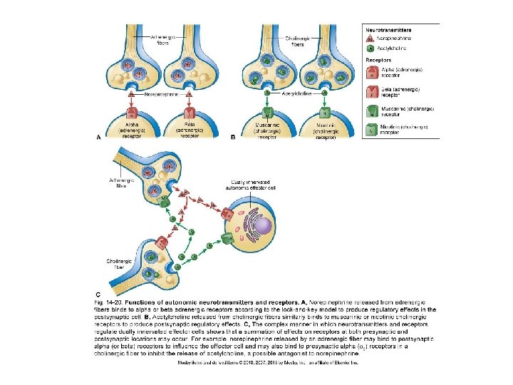
AUTONOMIC NERVOUS SYSTEM: STRUCTURE (cont. ) Autonomic neurotransmitters (Figures 14 -18 and 14 -20) ▪ Axon terminal of autonomic neurons releases either of two neurotransmitters: norepinephrine or acetylcholine ▪ Adrenergic fibers: release norepinephrine; axons of postganglionic sympathetic neurons ▪ Cholinergic fibers: release acetylcholine; axons of preganglionic sympathetic neurons and of preganglionic and postganglionic parasympathetic neurons ▪ Norepinephrine affects visceral effectors by first binding to one of two types of adrenergic receptors in plasma membranes: alpha receptors or beta receptors ▪ Binding of norepinephrine to alpha receptors in smooth muscle of blood vessels is stimulating, causing the vessels to constrict ▪ Binding of norepinephrine to beta receptors in smooth muscle of blood vessels is inhibitory, causing blood vessels to dilate; in cardiac muscle, has stimulating effect
AUTONOMIC NERVOUS SYSTEM: STRUCTURE (cont. ) Autonomic neurotransmitters (cont. ) ▪ Epinephrine also stimulates adrenergic receptors, enhancing and prolonging effects of sympathetic stimulation ▪ Effect of a neurotransmitter on any postsynaptic cell is determined by characteristics of the receptors, not by the neurotransmitter ▪ Termination of actions of norepinephrine and epinephrine ▪ Monoamine oxidase (MAO): enzyme that breaks up neurotransmitter molecules taken back up by the synaptic knobs ▪ Catechol-O-methyl transferase (COMT): enzyme that breaks down the remaining neurotransmitter ▪ Acetylcholine binds to two types of cholinergic receptors: nicotinic receptors and muscarinic receptors ▪ Termination of acetylcholine is by the enzyme acetylcholinesterase ▪ Autonomic neurotransmitters and receptors may influence different types of presynaptic and postsynaptic receptors at synapses with dually innervated effectors; this summation of effects increases precision of control
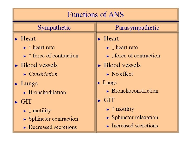
AUTONOMIC NERVOUS SYSTEM (cont. ) Functions of the autonomic nervous system Overview of autonomic function ▪ The autonomic nervous system functions to regulate effectors in ways that tend to maintain or quickly restore homeostasis ▪ Sympathetic and parasympathetic divisions are tonically active, often exerting antagonistic influences on visceral effectors ▪ Doubly innervated effectors continually receive both sympathetic and parasympathetic impulses; summation of the two determines the controlling effect
AUTONOMIC NERVOUS SYSTEM: FUNCTIONS (cont. ) Functions of the sympathetic division ▪ Under resting conditions, the sympathetic division can act to maintain the normal functioning of doubly innervated autonomic effectors ▪ Sympathetic impulses function to maintain normal tone of the smooth muscle in blood vessel walls ▪ Major function of sympathetic division is as an “emergency” system—the “fight or flight” reaction (Table 14 -7) Functions of the parasympathetic division (Table 14 -6) ▪ Dominant controller of most autonomic effectors most of the time ▪ Acetylcholine: slows heartbeat and promotes digestion and elimination