Chapter 14 Gene Expression From Gene to Protein


 RNA")


 codes for the genetic")


 5` Schematic model with m. RNA and t. RNA. A t. RNA")


 You.")


- Slides: 67
Chapter 14: Gene Expression: From Gene to Protein Central Dogma Song Sing along How did a single faulty gene result in Albie?
What is the function of DNA? • The information content of DNA – is in the form of specific sequences of nucleotides along the DNA strands – It codes for the production of proteins • Aka: Protein Synthesis
• The DNA inherited by an organism – leads to specific traits by dictating the synthesis of proteins • The process by which DNA directs Protein Synthesis, Gene expression – includes two stages: • Transcription • Translation
Where does this process take place? • The ribosome – Is part of the cellular machinery for translation, translation polypeptide synthesis in all living organisms Figure 17. 1: A model of a ribosome
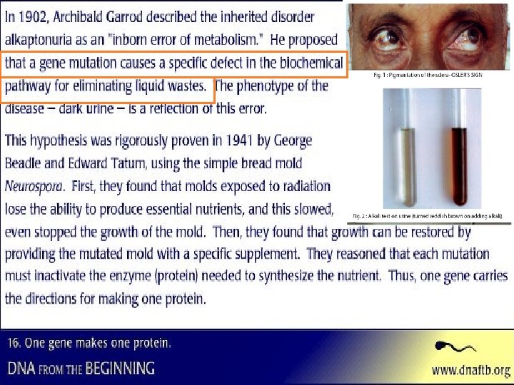
Evidence from the Study of Metabolic Defects • In 1909, British physician Sir Archibald Garrod – Was the first to suggest that genes dictate phenotypes through enzymes that catalyze specific chemical reactions in the cell – An inheritable disease result from the inability to produce a certain enzyme – He referred to certain diseases as caused by “inborn errors of metabolism”
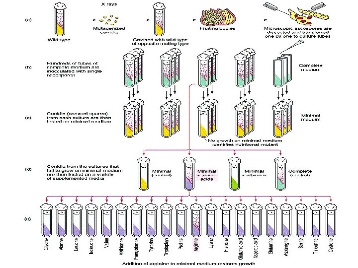
Nutritional Mutants in Neurospora: Scientific Inquiry • Beadle and Tatum causes bread mold to mutate with X-rays – Creating mutants that could not survive on minimal medium – Disabled genes one by one & looked for changes in each mutant’s phenotype thus seeing the normal function of the gene – Preview
• Using genetic crosses – they determined that their mutants fell into three classes, each mutated in a different gene EXPERIMENT RESULTS Working with the mold Neurospora crassa, George Beadle and Edward Tatum had isolated mutants requiring arginine in their growth medium and had shown genetically that these mutants fell into three classes, each defective in a different gene. From other considerations, they suspected that the metabolic pathway of arginine biosynthesis included the precursors ornithine and citrulline. Their most famous experiment, shown here, tested both their one gene–one enzyme hypothesis and their postulated arginine pathway. In this experiment, they grew their three classes of mutants under the four different conditions shown in the Results section below. The wild-type strain required only the minimal medium for growth. The three classes of mutants had different growth requirements Wild type Minimal medium (MM) (control) MM + Ornithine MM + Citrulline Figure 17. 2 MM + Arginine (control) Class I Mutants Class III Mutants
CONCLUSION Gene A From the growth patterns of the mutants, Beadle and Tatum deduced that each mutant was unable to carry out one step in the pathway for synthesizing arginine, arginine presumably because it lacked the necessary enzyme. Because each of their mutants was mutated in a single gene, they concluded that each mutated gene must normally dictate the production of one enzyme Their results supported the one gene–one enzyme hypothesis and also confirmed the arginine pathway. (Notice that a mutant can grow only if supplied with a compound made after the defective step. ) Wild type Class I Mutants (mutation in gene A) Precursor A A A Ornithine B B B Citrulline C C C Arginine Enzyme A Ornithine Gene B Enzyme B Citrulline Gene C Enzyme C Arginine George Beadle speaks to you Class II Mutants (mutation in gene B) Class III Mutants (mutation in gene C)
• Beadle and Tatum developed the: – “one gene–one enzyme hypothesis” • Which states that the function of a gene is to dictate the production of a specific enzyme • As researchers learned more about proteins – they made minor revision to the one gene–one enzyme hypothesis • Genes code for polypeptide chains or for RNA molecules which code for the production of enzymes
Basic Principles of Transcription and Translation • Transcription – Is the synthesis of messenger RNA (m. RNA) under the direction of DNA • Translation – Is the actual synthesis of a polypeptide, which occurs under the direction of m. RNA – Occurs on ribosomes of all cells
• In prokaryotes – Transcription and translation occur together (no nuclear envelope) RNA polymerase TRANSCRIPTION DNA m. RNA Ribosome TRANSLATION m. RNA Polyribosome RNA polymerase Direction of transcription 0. 25 m DNA Polypeptide Polyribosome Polypeptide (amino end) (a) Prokaryotic cell. In a cell lacking a nucleus, m. RNA produced by transcription is immediately translated without additional processing. Figure 17. 3 a Ribosome m. RNA (5 end) Prokaryotic Transcription & Translation occurs simultaneously
• In eukaryotes – RNA transcripts are modified before becoming true m. RNA Nuclear envelope DNA – Pre-m. RNA – m. RNA - Polypeptide DNA TRANSCRIPTION Pre-m. RNA PROCESSING m. RNA Ribosome TRANSLATION Polypeptide Figure 17. 3 b (b) Eukaryotic cell. The nucleus provides a separate compartment for transcription. The original RNA transcript, called pre-m. RNA, is processed in various ways before leaving the nucleus as m. RNA.
• Cells are governed by a cellular chain of command DNA RNA Protein Francis Crick's “Central Dogma” of molecular biology: “DNA makes RNA makes protein. ” This general rule emphasized the order of events from transcription through translation history. nih. gov/exhibits/nirenberg/glossa ry. htm
The Genetic Code The order of the nucleotides (Nitrogen bases) codes for the genetic code How many bases correspond to an amino acid? If only 2 bases in a codon, there would only be 16 codons (42) – Is encoded as a sequence of nonoverlapping base triplets, or codons – 43 = 64
• During transcription – The gene determines the sequence of bases along the length of an m. RNA molecule Gene 2 DNA molecule Gene 1 Gene 3 DNA strand (template) 3 A C C A A A C C G A G T U G G U U U G G C U A 5 TRANSCRIPTION m. RNA 5 C Codon TRANSLATION Protein Figure 17. 4 Trp Amino acid Phe Gly Ser 3
Cracking the Code • A codon in messenger RNA – Is either translated into an amino acid or serves as a translational stop signal UUU Phe UUC U UUA Leu UUG Second m. RNA base C A UAU UCU Tyr UAC UCA Ser UAA Stop UAG Stop UCG UGU Cys UGC UGA Stop UGG Trp CUU CUC C CUA CUG CCU CCC CCA CCG CGU CGC CGA CGG G U C Arg A G Thr AAU Asn AAC AAA Lys AAG AGU AGC AGA AGG U Ser C A Arg G Ala GAU GAC Asp GAA Glu GAG U GGU C GGC Gly GGA A GGG G Figure 17. 5 AUU AUC A AUG GUU GUC G GUA GUG Leu lle Met or start Val ACU ACC ACA ACG GCU GCC GCA GCG Pro CAU CAC CAA CAG His Gln G U C A Third m. RNA base (3 end) First m. RNA base (5 end) U
The Genetic Code In the Genetic Code, there are 43 = 64 (# of N-bases# of bases in a codon) possible codons
Evolution of the Genetic Code • The genetic code is nearly universal – Shared by organisms from the simplest bacteria to the most complex animals – UUU in bacteria & a blue whale codes for Phenylalanine
• In laboratory experiments – Genes can be transcribed and translated after being transplanted from one species to another – All three organisms below are expressing a firefly gene inserted into their genome Figure 17. 6
Transcription is the DNA-directed synthesis of m. RNA DNA
Molecular Components of Transcription • RNA synthesis – Is catalyzed by RNA polymerase, which pries the DNA strands apart and hooks together the RNA nucleotides Follows the same basepairing rules as DNA, except that in RNA, Uracil substitutes for Thymine
Synthesis of an RNA Transcript • The stages of transcription are – Initiation – Elongation – Termination Promoter Transcription unit 5 3 Start point 3 5 DNA Initiation After RNA polymerase binds to 1 Initiation. the promoter, the DNA strands unwind, and the polymerase initiates RNA synthesis at the start point on the template strand. RNA polymerase 5 3 Unwound DNA Template strand of DNA Elongation transcript RNA 2 Rewound 3 5 The polymerase moves downstream, unwinding the DNA (3` to 5`) and elongating the RNA transcript 5 3 . In the wake of transcription, the DNA strands re-form a double helix. RNA 5 3 3 5 RNA transcript Termination Eventually, the RNA 3 Termination. transcript is released, and the polymerase detaches from the DNA. 5 3 3 5 5 Figure 17. 7 Completed RNA transcript 3
General Initiation of Transcription 3` to 5` on DNA 5` to 3` for m. RNA
Eukaryotic Initiation of Transcription
RNA Polymerase Binding and Initiation of Transcription • Promoters signal the initiation of RNA synthesis – Series of T’s & A’s (TATA box) going in a 3` to 5` direction on DNA Eukaryotic promoters 1 TRANSCRIPTION DNA RNA PROCESSING Pre-m. RNA TRANSLATION Ribosome Polypeptide 5 3 Promoter T A A AA AT A T T TATA box 3 5 UTR Start point Several transcription factors 2 • Transcription factors – Help eukaryotic RNA polymerase recognize promoter sequences – Complex of transcription factors and RNA polymerase II bound to the promoter is called a transcription initiation complex. Figure 17. 8 5 3 UTR Template DNA strand 3 5 UTR 3 Additional transcription factors Transcription factors 3 5 5 RNA transcript Transcription initiation complex
Elongation Non-template strand of DNA RNA nucleotides RNA polymerase A 3 T C C C A A T U 3 end G T A U C A T E A G G C A G T A A T Direction of transcription (“downstream”) 5 Newly made RNA G 5 T G C A Template strand of DNA
Elongation of the RNA Strand • As RNA polymerase moves along the DNA – it continues to untwist the double helix, exposing about 10 to 20 DNA bases at a time for pairing with RNA nucleotides – A single gene can be transcribed simultaneously by several molecules of RNA polymerase following each other.
• The mechanisms of termination – are different in prokaryotes and eukaryotes • Eukaryotic cells modify RNA after transcription – Enzymes in the eukaryotic nucleus modify pre-m. RNA or the primary transcript in specific ways before the genetic messages are dispatched to the cytoplasm
Alteration of m. RNA Ends • Each end of a pre-m. RNA molecule is modified in a particular way – The 5 end receives a modified nucleotide cap (GTP cap – Guanine triphosphate) – The 3 end gets a Poly-A tail – Facilitates the export of mature m. RNA from the nucleus – Protects m. RNA from breakdown by hydrolytic enzymes – Helps ribosomes attache to the 5` end of m. RNA in the cytoplasm – UTR are untranslated regions A modified guanine nucleotide added to the 5 end TRANSCRIPTION 50 to 250 adenine nucleotides added to the 3 end DNA Pre-m. RNA PROCESSING m. RNA 5 Protein-coding segment Polyadenylation signal 3 G P P P AAUAAA AAA…AAA Ribosome TRANSLATION Polypeptide Figure 17. 9 5 Cap 5 UTR Start codon Stop codon 3 UTR (Untranslated region) region Poly-A tail
Split Genes and RNA Splicing • Average gene ~ 1, 200 nucleotides (400 Amino Acids) • ~ length of transcription unit is 8, 000 nucleotides therefore it needs to be cut down. • Most eukaryotic genes have long noncoding stretches of nucleotides, introns, between coding sections, exons • RNA splicing – Removes introns and joins exons (coding regions) – Animation TRANSCRIPTION RNA PROCESSING DNA Pre-m. RNA 5 Exon Intron Pre-m. RNA 5 Cap 1 30 Exon 31 Coding segment m. RNA Ribosome Exon Intron 3 Poly-A tail 104 105 146 Introns cut out and exons spliced together TRANSLATION Polypeptide m. RNA 5 Cap 1 3 UTR Figure 17. 10 Poly-A tail 146 3 UTR
Role of Splicesomes - animation • Is carried out by spliceosomes in some cases – Made up of Small nuclear ribonucleic acid (sn. RNA) w/ specific proteins Figure 17. 11
Ribozymes • Ribozymes – Are catalytic RNA molecules that function as enzymes and can splice r. RNA – Pre-r. RNA actually can remove its own introns.
• Proteins often have a modular architecture – consisting of discrete structural and functional regions called domains • In many cases – different exons code for the different domains in a protein – By the removal of introns between the domains, the polypeptide can then be produced Gene DNA Exon 1 Intron Exon 2 Intron Exon 3 Transcription RNA processing Translation Domain 3 Domain 2 Domain 1 Polypeptide
Many genes can give rise to two or more polypeptides depending on which segments are treated as exons during RNA processing – Alternate RNA splicing
Why Introns and Split Genes? 1. Though proteins synthesized in a linear fashion, they are actually modular w/ various functional regions called domains 2. Allows for additional crossing over of chromosomes 3. Genes of our evolutionary past from prokaryotic organisms (they don’t have introns) 4. Promoters & terminal signals for turning on and off genes
Molecular Components of Translation • A cell translates a m. RNA message into proteinpolypeptide – With the help of transfer RNA (t. RNA)
Translation: the basic concept TRANSCRIPTION DNA m. RNA Ribosome TRANSLATION Polypeptide Amino acids Polypeptide Ribosome Trp Phe t. RNA with amino acid attached Gly t. RNA C A A A U G G U U U G G Codons 5 Figure 17. 13 G m. RNA C G Anticodon C 3
t. RNA & anticodon Amino Acid t. RNA Anticodon Actually a single RNA strand ~ 80 nucleotides long with nitrogenous bases H bonding to each other causing the strand to fold on itself into a cloverleaf
The Structure and Function of Transfer RNA • Molecules of t. RNA are not all identical (45 total) – Each carries a specific amino acid on one end A – Each has an anticodon on the other end C • A t. RNA molecule – Consists of a single RNA strand that is only about 80 nucleotides long 3 A Amino acid C attachment site C – Is roughly L-shaped (or t) A 5 C G G C C G U A A U U C A G * C A G UA * U C C G * * GU GU * G Figure 17. 14 a C C GA G C A G G * * U * GA * G C Two-dimensional structure. The four base-paired regions and Hydrogen (a) G C three loops are characteristic of all t. RNAs, as is the base U A bonds sequence of the amino acid attachment site at the 3 end. The * G A anticodon triplet is unique to each t. RNA type. (The asterisks A* C mark bases that have been chemically modified, a characteristic U * A G of t. RNA. ) A Anticodon
Wobble w/ 3 rd anticodon. U can bond with A or G and still not change the coded amino acid (AGA & AGG codes for arginine
t. RNA’s three dimensional structure 5 Amino acid attachment site 3 Hydrogen bonds A AG 3 5 Anticodon (b) Three-dimensional structure Figure 17. 14 b (c) Symbol used in this book
• A specific enzyme called an aminoacyl-t. RNA synthetase – joins each amino acid to the correct t. RNA – There is one for each amino acid Figure 17. 15
Ribosomes – Facilitate the specific coupling of t. RNA anticodons with m. RNA codons during protein synthesis – m. RNA bonds first to the small subunit of the ribosomes then the large subunit attaches
• The ribosomal subunits – Are constructed of proteins and RNA molecules named ribosomal RNA or r. RNA TRANSCRIPTION DNA TRANSLATION m. RNA Ribosome Polypeptide Exit tunnel Growing polypeptide t. RNA molecules Large subunit E P A Small subunit 5 m. RNA 3 (a) Computer model of functioning ribosome. This is a model of a bacterial ribosome, showing its overall shape. The eukaryotic ribosome is roughly similar. A ribosomal subunit is an aggregate of ribosomal RNA molecules and proteins. Figure 17. 16 a
• The ribosome has three binding sites for t. RNA – The P site – primary bonding site – The A site – adjacent bonding site – The E site – exit site P site (Peptidyl-t. RNA binding site) A site (Aminoacylt. RNA binding site) E site (Exit site) E P A m. RNA binding site Figure 17. 16 b Large subunit Small subunit (b) Schematic model showing binding sites. A ribosome has an m. RNA binding site and three t. RNA binding sites, known as the A, P, and E sites. This schematic ribosome will appear in later diagrams.
3` (c) 5` Schematic model with m. RNA and t. RNA. A t. RNA fits into a binding site when its anticodon basepairs with an m. RNA codon. The P site holds the t. RNA attached to the growing polypeptide. The A site holds the t. RNA carrying the next amino acid to be added to the polypeptide chain. Discharged t. RNA leaves via the E site through an exit tunnel. **Translation of t. RNA is always in the 5` to 3` direction of m. RNA Figure 17. 16 c **Protein is built in the 3` to 5` direction (like DNA!)
• Certain antibiotics can inactivate bacterial ribosomes without interferring with eukaryotic ribosomes & normal protein synthesis. • Tetracycline and Streptomycin. • Bacterial ribosomes are smaller than eukaryotic ribosomes.
Building a Polypeptide • We can divide translation into three stages – Initiation – Elongation – Termination
Ribosome Association and Initiation of Translation • The initiation stage of translation – brings together m. RNA, t. RNA bearing the first amino acid of the polypeptide to the P site, and two subunits of a ribosome – GTP needed to bind the t. RNA to m. RNA & form Peptide bond Large t Me 3 U A C 5 5 A U G 3 Initiator t. RNA t Me GTP GDP E m. RNA 5 Start codon m. RNA binding site Figure 17. 17 ribosomal subunit P site 3 Small ribosomal subunit 5 A 3 Translation initiation complex 2 The arrival of a large ribosomal subunit completes 1 A small ribosomal subunit binds to a molecule of the initiation complex. Proteins called initiation m. RNA. In a prokaryotic cell, the m. RNA binding site factors (not shown) are required to bring all the on this subunit recognizes a specific nucleotide translation components together. GTP provides sequence on the m. RNA just upstream of the start the energy for the assembly. The initiator t. RNA is codon. An initiator t. RNA, with the anticodon UAC, in the P site; the A site is available to the t. RNA base-pairs with the start codon, AUG. This t. RNA bearing the next amino acid. carries the amino acid methionine (Met).
Elongation of the Polypeptide Chain • In the elongation stage of translation – Amino acids are added one by one to the preceding amino acid. Energy in the form of GTP is needed to form peptide bonds TRANSCRIPTION Amino end of polypeptide DNA m. RNA Ribosome TRANSLATION Polypeptide m. RNA Ribosome ready for next aminoacyl t. RNA E 3 P A site 5 1 Codon recognition. The anticodon of an incoming aminoacyl t. RNA base-pairs with the complementary m. RNA codon in the A site. Hydrolysis of GTP increases the accuracy and efficiency of this step. 2 GTP 2 GDP E E P Figure 17. 18 3 Translocation. The ribosome translocates the t. RNA in the A site to the P site. The empty t. RNA in the P site is moved to the E site, where it is released. The m. RNA moves along with its bound t. RNAs, bringing the next codon to be translated into the A site. P A GDP GTP E P A A 2 Peptide bond formation. An r. RNA molecule of the large subunit catalyzes the formation of a peptide bond between the new amino acid in the A site and the carboxyl end of the growing polypeptide in the P site. This step attaches the polypeptide to the t. RNA in the A site.
Termination of Translation • The final stage of translation is termination – When the ribosome reaches a stop codon (UAG, UAA, or UGA) in the m. RNA Release factor Free polypeptide 5 3 3 5 5 GTP 3 Stop codon (UAG, UAA, or UGA) 1 When a ribosome reaches a stop 2 The release factor hydrolyzes 3 The two ribosomal subunits codon on m. RNA, the A site of the bond between the t. RNA in and the other components of ribosome accepts a protein called the P site and the last amino the assembly dissociate. a release factor instead of t. RNA. acid of the polypeptide chain. The polypeptide is thus freed from the ribosome. Figure 17. 19
Polyribosomes • A number of ribosomes can translate a single m. RNA molecule simultaneously in prokaryotes – forming a polyribosome Completed polypeptide Growing polypeptides Incoming ribosomal subunits Start of Polyriboso me End of m. RNA (5 end) (3 end) (a) An m. RNA molecule is generally translated simultaneously by several ribosomes in clusters called polyribosomes. Ribosomes m. RNA 0. 1 µm Figure 17. 20 a, b (b) This micrograph shows a large polyribosome in a prokaryotic cell (TEM).
Completing and Targeting the Functional Protein • Polypeptide chains – Undergo modifications after the translation process – Proteins may be modified in ways that affect their three-dimensional shape – Chaparonins and shaping proteins
Targeting Polypeptides to Specific Locations • Two populations of ribosomes are evident in cells – Free and bound • Free ribosomes in the cytosol for proteins that stay in the cytosol (like for glycolysis)
• RER ribosomes make proteins destined for the endomembrane system or for secretion – must be transported into the ER – have signal peptides to which a signal-recognition particle (SRP) binds, enabling the translation ribosome to bind to the ER
The signal mechanism for targeting proteins to the ER 1 Polypeptide synthesis begins on a free ribosome in the cytosol. 2 An SRP binds 3 The SRP binds to a 4 The SRP leaves, and receptor protein in the ER the polypeptide resumes to the signal growing, meanwhile peptide, halting membrane. This receptor is part of a protein complex translocating across the synthesis (a translocation complex) membrane. (The signal momentarily. that has a membrane pore peptide stays attached and a signal-cleaving enzyme. to the membrane. ) 5 The signalcleaving enzyme cuts off the signal peptide. 6 The rest of the completed polypeptide leaves the ribosome and folds into its final conformation. Ribosome m. RNA Signal peptide Signalrecognition particle (SRP) SRP receptor CYTOSOL protein ERLUMEN Figure 17. 21 Translocation complex Signal peptide removed ER membrane Protein
A summary of transcription and translation in a eukaryotic cell DNA TRANSCRIPTION 1 RNA is transcribed from a DNA template. 3 Po 5 RNA transcript RNA PROCESSING 2 In eukaryotes, the RNA transcript (prem. RNA) is spliced and modified to produce m. RNA, which moves from the nucleus to the cytoplasm. A ly- RNA polymerase Exon RNA transcript (pre-m. RNA) Intron Aminoacyl-t. RNA synthetase p Ca NUCLEUS Amino acid t. RNA FORMATION OF INITIATION COMPLEX CYTOPLASM 3 After leaving the nucleus, m. RNA attaches to the ribosome. Growing polypeptide m. RNA -A Activated amino acid ly Po AMINO ACID ACTIVATION 4 Each amino acid attaches to its proper t. RNA with the help of a specific enzyme and ATP. Ribosomal subunits Ca 5 p C A C E A UA A UGGUU UA U G Figure 17. 26 A y- l Po Codon Ribosome C TRANSLATION 5 A succession of t. RNAs add their amino acids to the polypeptide chain Anticodon as the m. RNA is moved through the ribosome one codon at a time. (When completed, the polypeptide is released from the ribosome. )
Songs & animations • • You. Tube - Protein Synthesis (Song: Poker Face) You. Tube - Protein Synthesis Song - Kathleen June Standford Protein Synthesis video – Psychodelic! Mc Graw Hill Transcription Mc. Graw Hill. Animation: How Translation Works Mc. Graw Hill Protein Synthesis Cool 3 D animation
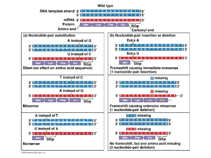
Mutations • Point mutations can affect protein structure and function • Mutations – Are changes in the genetic material of a cell – Gene Mutations vs Chromosomal mutations – Mutations or alterations of the DNA, RNA or protein produced is a Gene mutation & are inheritable • Point mutations – Are changes in just one base pair of a gene due to a substitution of nucleotides – Could Lead to the production of an abnormal protein – Aka: Base - substitution mutation
Point Mutation Wild-type hemoglobin DNA 3 Mutant hemoglobin DNA 5 C T 3 5 T C m. RNA A T In the DNA, the mutant template strand has an A where the wild-type template has a T. m. RNA G A A 5 G 3 U A 5 3 Normal hemoglobin Sickle-cell hemoglobin Glu Val Figure 17. 23 The mutant m. RNA has a U instead of an A in one codon. The mutant (sickle-cell) hemoglobin has a valine (Val) instead of a glutamic acid (Glu).
Substitutions – THE FAT CAT ATE THE RAT THE FAT BAT ATE THE RAT – Is the replacement of one nucleotide and its partner with another pair of nucleotides – Can cause missense or nonsense Wild type m. RNA Protein A U 5 G A U Lys Met U U G Phe G C U A A 3 Gly Stop Amino end Carboxyl end Base-pair substitution Silent mutation: No effect on amino acid sequence U instead of C A U G A A G U Lys Met U Nonsense Figure 17. 24 G G U U Gly A A Stop A instead of G G A A G U Lys Met A U Phe Missense A U U U A Phe G U U Ser A A Stop U instead of A U Met G U A Stop G U U U G G C U A A
Insertions and Deletions – THE FAT CAT ATE THE RAT THE ATC ATA TET HER AT– Are additions or losses of nucleotide pairs in a gene – May produce Frameshift mutations Wild type m. RNA 5 Protein A U G A Met A G U U U G G C U A A Lys Gly Phe Amino end Base-pair insertion or deletion Stop Carboxyl end Frameshift causing immediate nonsense Extra U A U G U Met Frameshift causing extensive missense A A G U U U G G C U A Stop U Missing A U G A A G U U G G C U A A Met Lys Leu Ala Insertion or deletion of 3 nucleotides: no frameshift but extra or missing amino acid A A G Missing A U G U U U G G C U A A Figure 17. 25 Met Phe Gly Stop 3
Point vs Frameshift mutations
New definition of a GENE A gene is a region of DNA that can be expressed to produce a final functional product that is either a polypeptide or an RNA molecule