Chapter 14 Blood Blood connective tissue transports vital

Chapter 14: Blood

Blood • connective tissue • transports vital substances • maintains stability of interstitial fluid • distributes heat
 • form mostly in red bone marrow • 3")
Blood Cells (Formed Elements ) • form mostly in red bone marrow • 3 types 1. red blood cells 2. white blood cells 3. platelets (cell fragments)

Blood Volume • varies with • body size • changes in fluid concentration • changes in electrolyte concentration • amount of adipose tissue • about 8% of body weight • about 5 liters 4

Blood Composition Hematocrit: Cell volume compared to total volume 45% Formed Elements 55% Plasma 5

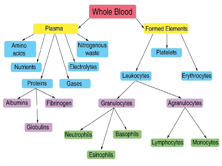
Origin of Blood Cells 6

Erythrocytes - Red Blood Cells • biconcave discs • one-third hemoglobin • oxyhemoglobin • deoxyhemoglobin • can readily squeeze through capillaries • lack nuclei and mitochondria 7

Red Blood Cell Counts • number of RBCs in a cubic millimeter of blood • reflects blood’s oxygen carrying capacity Ø 4, 600, 000 – 6, 200, 000 in males Ø 4, 200, 000 – 5, 400, 000 in adult females Ø 4, 500, 000 – 5, 100, 000 in children 8

Red Blood Cell Production • low blood oxygen causes kidneys and liver to release erythropoietin which stimulates RBC production • vitamin B 12, folic acid and iron necessary for RBC production 9

Dietary Factors Affecting Red Blood Cell Production 10

Life Cycle of Red Blood Cell • circulate for about 120 days • macrophages in spleen and liver destroy worn out RBCs • hemoglobin is broken down into heme and globin • iron from heme returns to red bone marrow • bilirubin and biliverdin excreted in bile 11

Types of Anemia 12

Anemia Normal RBCs of person with hypochromic anemia 13

Destruction of Red Blood Cells 14

Antigens and Antibodies Agglutination – clumping of red blood cells in response to a reaction between an antibody and an antigen Antigens – a chemical that stimulates cells to produce antibodies Antibodies – a protein that reacts against a specific antigen 15

Antigens and Antibodies 16

Agglutination 17

ABO Blood Group Based on the presence or absence of two major antigens on red blood cell membranes • antigen A • antigen B 18

ABO Blood Group 19

Blood Types for Transfusion 20

Rh Blood Group Rh positive – presence of antigen D or and other Rh antigens on the red blood cell membranes Rh negative – lack of these antigens 21

Rh Blood Group 22

White Blood Cells • leukocytes • protect against disease • hormones stimulate development • interleukins • colony-stimulating factors 23

White Blood Cells 1. granulocytes • neutrophils • eosinophils • basophils 2. Agranulocytes • lymphocytes • monocytes 24

Neutrophils • light purple granules in acid-base stain • lobed nucleus • other names • segs • polymorphonuclear leukocyte • bands (young neutrophils) • first to arrive at infections • phagocytic • 54% - 62% of leukocytes • elevated in bacterial infections 25

Eosinophils • deep red granules in acid stain • bilobed nucleus • moderate allergic reactions • defend against parasitic worm infestations • 1% - 3% of leukocytes • elevated in parasitic worm infestations and allergic reactions 26

Basophils • deep blue granules in basic stain • release histamine • release heparin • less than 1% of leukocytes • similar to eosinophils in size and shape of nuclei 27

Monocytes • largest blood cell • spherical, kidney-shaped, oval or lobed nuclei • leave bloodstream to become macrophages • 3% - 9% of leukocytes • phagocytize bacteria, dead cells, and other debris 28

Lymphocytes • slightly larger than RBC • large spherical nucleus surrounded by thin rim of cytoplasm • T cells and B cells • important in immunity • B cells produce antibodies • 25% - 33% of leukocytes 29

Diapadesis • leukocytes squeeze between the cells of a capillary wall and enter the tissue space outside the blood vessel 30

Positive Chemotaxis • movement of leukocytes toward the damaged tissue region because of the chemicals that were released by damaged cells 31

White Blood Cell Counts • Normal = 5, 000 – 10, 000 per cubic millimeter of blood • leukopenia • low WBC count (below 5, 000) • typhoid fever, flu, measles, mumps, chicken pox, AIDS • leukocytosis • high WBC count (above 10, 000) • acute infections, vigorous exercise, great loss of body fluids • differential WBC count • lists percentages of types of leukocytes • may change in particular diseases 32

White Blood Cell Counts 33

Blood Platelets • thrombocytes • cell fragments of megakaryocytes • 130, 000 – 360, 000 per cubic millimeter of blood • helps control blood loss from broken vessels 34

Blood Platelets 35

Blood Plasma straw colored • liquid portion of blood • 55% of blood • 92% water • 36

Blood Plasma 37

Plasma Proteins 38

Gases and Nutrients Gases • oxygen • carbon dioxide Nutrients • amino acids • simple sugars • nucleotides • lipids 39

Nonprotein Nitrogenous Substances • molecules containing nitrogen but are not proteins • urea – product of protein catabolism; about 50% of NPN substances • uric acid – product of nucleic acid catabolism • amino acids – product of protein catabolism • creatine – stores phosphates • creatinine – product of creatine metabolism • BUN – blood urea nitrogen; indicate health of kidney 40

Plasma Electrolytes • absorbed from the intestine or released as by-products of cellular metabolism • sodium • potassium • calcium • magnesium • chloride • bicarbonate • phosphate • sulfate • sodium and chloride are most abundant 41

Hemostasis • stoppage of bleeding Blood Vessel Spasm • triggered by pain receptors, platelet release, or serotonin • smooth muscle in vessel contracts Platelet Plug Formation • triggered by exposure of platelets to collagen • platelets adhere to rough surface to form a plug Blood Coagulation • triggered by cellular damage and blood contact with foreign surfaces • blood clot forms 43

Platelet Plug Formation 44

Blood Coagulation • hemostatic mechanism • causes the formation of a blot clot via a series of reactions which activates the next in a cascade • occurs extrinsically or intrinsically 45

Blood Coagulation Extrinsic Clotting Mechanism • chemical outside of blood triggers blood coagulation • triggered by thromboplastin (not found in blood) • triggered when blood contacts damaged tissue Intrinsic Clotting Mechanism • chemical inside blood triggers blood coagulation • triggered by Hageman factor (found inside blood) • triggered when blood contacts a foreign surface 46

Blood Coagulation 47

Blood Coagulation 48

Fate of Blood Clots • After forming, a blood clot retracts and pulls the edges of a broken vessel together while squeezing the fluid serum from the clot • Platelet-derived growth factor stimulates smooth muscle cells and fibroblasts to repair damaged blood vessel walls • Plasmin digests blood clots • thrombus – abnormal blood clot • embolus – blood clot moving through blood 49

Prevention of Coagulation • The smooth lining of blood vessels discourages the accumulation of platelets and clotting factors • As a clot forms, fibrin adsorbs thrombin and prevents the clotting reaction from spreading • Antithrombin inactivates additional thrombin by binding to it and blocking its action on fibrinogen • Some cells, such as basophils and mast cells secrete heparin (an anticoagulant) 50

Prevention of Coagulation 51

Clinical Application Leukemia Myeloid Leukemia • bone marrow produces too many immature granulocytes • leukemia cells crowd out other blood cells • anemia • bleeding • susceptible to infections Lymphoid Leukemia • lymphocytes are cancerous • symptoms similar to myeloid leukemia Treatments • drugs • marrow and umbilical cord transplants • chemotherapy regimens 52
- Slides: 52