Chapter 13 The Respiratory System Organs Nose Pharynx


 • Muscular passage from nasal cavity to larynx • Three regions from")


 • Function: – Routes air and food into proper channels –")


 • 4 inch long tube that connects larynx with bronchi • Walls")
 Figure 13. 3 a")
 The yellow structures are cilia. The orange structures are goblet cells that")
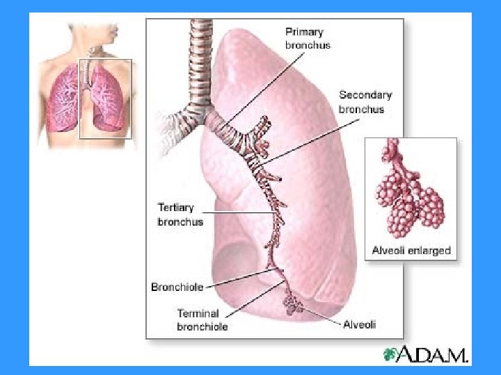
 Bronchi • Formed by division of the trachea • Right bronchus is")


 Tree Divisions • All but the smallest of these passageways have reinforcing")

 • Walls of alveoli are thin squamous epithelial layer (much")
 Figure 13. 6 (1 of 2)")
 Figure 13. 6 (2 of 2)")
- Slides: 28
Chapter 13: The Respiratory System
Organs • • • Nose Pharynx Larynx Trachea Bronchi Lungs
Functions • Gas exchanges between the blood and environment – Occurs in the alveoli of the lungs • Passageways to the lungs purify, humidify, and warm the incoming air
The Nose • Only externally visible part of the respiratory system • Air enters the nose through the external nostrils (nares) • Interior of the nose consists of a nasal cavity divided by a nasal septum
Inside the nasal cavity: • Olfactory receptors in mucosa on superior surface for sense of smell • Respiratory mucosa everywhere else produces sticky mucus – Moisten air – Trap incoming foreign particles • Cilia = tiny hair-like structures that move contaminated mucus towards the pharynx where it is swallowed and digested by stomach juices • Projections called conchae on lateral walls – Increase surface area & air turbulence
Figure 13. 2
• The nasal cavity is separated from the oral cavity by the palate – Anterior part = hard palate (bone) – Posterior part = soft palate (muscle)
Paranasal Sinuses • Cavities within bones surrounding the nasal cavity • Located in the following bones: – Frontal – Sphenoid – Ethmoid – Maxillary • Functions: – Lighten the skull – Act as resonance chambers for speech – Produce mucus
Pharynx (Throat) • Muscular passage from nasal cavity to larynx • Three regions from superior to inferior: – Nasopharynx – Oropharynx – Laryngopharynx • Passageways for air and food
• Pharyngotympanic tubes that drain middle ear open into the nasopharynx – Ear infections may follow sore throat • Tonsils: – Pharyngeal tonsil (adenoids) in the nasopharynx – Palatine tonsils in the oropharynx – Lingual tonsils at the base of the tongue
Figure 13. 2
Larynx (Voice Box) • Function: – Routes air and food into proper channels – Plays a role in speech • Structure: – Eight rigid hyaline cartilages • Thyroid cartilage is largest = Adam’s apple – Epiglottis =spoon-shaped flap of elastic cartilage • Rises to covers larynx when you swallow so that liquid and food go to the esophagus instead of airway – Vocal folds (aka true vocal cords) • Vibrate with expelled air to create sound – Glottis = opening between vocal cords
Anterior View of Larynx Posterior View of Larynx http: //www. youtube. com/watch? v=Qv. GYv. K 6 q. Sc. E
Upper Respiratory Tract: Larynx Figure 13. 2 http: //www. youtube. com/watch? v=9 MDn 5 Ggyxy. U&feature=related
Trachea (Windpipe) • 4 inch long tube that connects larynx with bronchi • Walls are reinforced with C-shaped hyaline cartilage • Lined with ciliated mucosa – Beat continuously in the opposite direction of incoming air – Expel mucus loaded with dust and other debris away from lungs
Trachea (Windpipe) Figure 13. 3 a
Trachea (Windpipe) The yellow structures are cilia. The orange structures are goblet cells that secrete mucus and have microvilli. Figure 13. 3 b
Main (Primary) Bronchi • Formed by division of the trachea • Right bronchus is wider, shorter, and straighter than left – More common site for inhaled objects to get stuck • Subdivide into smaller and smaller branches
Main Bronchi Figure 13. 1
Lungs • Occupy most of the thoracic cavity – Heart occupies central portion called mediastinum • Apex (superior part) is near the clavicle • Base (inferior part) rests on the diaphragm • Each lung is divided into lobes by fissures – Left lung has two lobes – Right lung has three lobes
Lungs Figure 13. 4 a
• Covering of lungs: – Serosa covers the outer surface of the lungs • Pulmonary (visceral) pleura covers the lung surface • Parietal pleura lines the walls of the thoracic cavity – Pleural fluid fills the area between layers of pleura to allow gliding = less friction
Bronchial (Respiratory) Tree Divisions • All but the smallest of these passageways have reinforcing cartilage in their walls • Largest to smallest: – Primary bronchi – Secondary bronchi – Tertiary bronchi – Bronchioles – Terminal bronchioles
Respiratory Zone • Structures from largest to smallest: – Respiratory bronchioles – Alveolar ducts – Alveolar sacs – Alveoli (air sacs) – 40 x more surface area than skin • Site of gas exchange = alveoli only
Respiratory Membrane (Air-Blood Barrier) • Walls of alveoli are thin squamous epithelial layer (much thinner than tissue paper!) • Alveolar pores connect neighboring air sacs incase mucus blocks other paths • Pulmonary capillaries cover external surfaces of alveoli • One side of membrane is air and the other side is blood flowing past • Also has: – Alveolar macrophages (“dust cells”) that remove bacteria and debri – Cuboidal cells that make surfactant (prevent alveoli from collapsing)
Respiratory Membrane (Air-Blood Barrier) Figure 13. 6 (1 of 2)
Respiratory Membrane (Air-Blood Barrier) Figure 13. 6 (2 of 2)