Chapter 13 Meiosis and Sexual Life Cycles Power


- Slides: 80

Chapter 13 Meiosis and Sexual Life Cycles Power. Point Lectures for Biology, Seventh Edition Neil Campbell and Jane Reece Lectures by Chris Romero Copyright © 2005 Pearson Education, Inc. publishing as Benjamin Cummings

• Overview: Hereditary Similarity and Variation • Living organisms – Are distinguished by their ability to reproduce their own kind Copyright © 2005 Pearson Education, Inc. publishing as Benjamin Cummings

• Heredity – Is the transmission of traits from one generation to the next • Variation – Shows that offspring differ somewhat in appearance from parents and siblings Figure 13. 1 Copyright © 2005 Pearson Education, Inc. publishing as Benjamin Cummings

• Genetics – Is the scientific study of heredity and hereditary variation Copyright © 2005 Pearson Education, Inc. publishing as Benjamin Cummings

• Concept 13. 1: Offspring acquire genes from parents by inheriting chromosomes Copyright © 2005 Pearson Education, Inc. publishing as Benjamin Cummings

Inheritance of Genes • Genes – Are the units of heredity – Are segments of DNA Copyright © 2005 Pearson Education, Inc. publishing as Benjamin Cummings

• Each gene in an organism’s DNA – Has a specific locus on a certain chromosome • We inherit – One set of chromosomes from our mother and one set from our father Copyright © 2005 Pearson Education, Inc. publishing as Benjamin Cummings

Comparison of Asexual and Sexual Reproduction • In asexual reproduction – One parent produces genetically identical offspring by mitosis Parent Bud Figure 13. 2 Copyright © 2005 Pearson Education, Inc. publishing as Benjamin Cummings 0. 5 mm

• In sexual reproduction – Two parents give rise to offspring that have unique combinations of genes inherited from the two parents Copyright © 2005 Pearson Education, Inc. publishing as Benjamin Cummings

• Concept 13. 2: Fertilization and meiosis alternate in sexual life cycles • A life cycle – Is the generation-to-generation sequence of stages in the reproductive history of an organism Copyright © 2005 Pearson Education, Inc. publishing as Benjamin Cummings

Sets of Chromosomes in Human Cells • In humans – Each somatic cell has 46 chromosomes, made up of two sets – One set of chromosomes comes from each parent Copyright © 2005 Pearson Education, Inc. publishing as Benjamin Cummings

• A karyotype – Is an ordered, visual representation of the chromosomes in a cell Pair of homologous chromosomes Centromere Sister chromatids Figure 13. 3 Copyright © 2005 Pearson Education, Inc. publishing as Benjamin Cummings 5 µm

Figure 13. 3 Preparing a Karyotype APPLICATION A karyotype is a display of condensed chromosomes arranged in pairs. Karyotyping can be used to screen for abnormal numbers of chromosomes or defective chromosomes associated with certain congenital disorders, such as Down syndrome. TECHNIQUE Karyotypes are prepared from isolated somatic cells, which are treated with a drug to stimulate mitosis and then grown in culture for several days. A slide of cells arrested in metaphase is stained and then viewed with a microscope equipped with a digital camera. A digital photograph of the chromosomes is entered into a computer, and the chromosomes are electronically rearranged into pairs according to size and shape. RESULTS This karyotype shows the chromosomes from a normal human male. The patterns of stained bands help identify specific chromosomes and parts of chromosomes. Although difficult to discern in the karyotype, each metaphase chromosome consists of two, closely attached sister chromatids (see diagram). Copyright © 2005 Pearson Education, Inc. publishing as Benjamin Cummings Pair of homologous chromosomes Centromere Sister chromatids 5 µm

Figure 13. 3 Preparation of a human karyotype (Layer 2)

Figure 13. 3 Preparation of a human karyotype (Layer 3)

Figure 13. 3 Preparation of a human karyotype (Layer 4)

Figure 13. x 2 Human female chromosomes shown by bright field G-banding

Figure 13. x 3 Human female karyotype shown by bright field G-banding of chromosomes

Figure 13. x 4 Human male chromosomes shown by bright field G-banding

Figure 13. x 5 Human male karyotype shown by bright field G-banding of chromosomes

• Homologous chromosomes – Are the two chromosomes composing a pair – Have the same characteristics – May also be called autosomes Copyright © 2005 Pearson Education, Inc. publishing as Benjamin Cummings

• Sex chromosomes – Are distinct from each other in their characteristics – Are represented as X and Y – Determine the sex of the individual, XX being female, XY being male Copyright © 2005 Pearson Education, Inc. publishing as Benjamin Cummings

• A diploid cell – Has two sets of each of its chromosomes – In a human has 46 chromosomes (2 n = 46) Copyright © 2005 Pearson Education, Inc. publishing as Benjamin Cummings

• In a cell in which DNA synthesis has occurred – All the chromosomes are duplicated and thus each consists of two identical sister chromatids Key Maternal set of chromosomes (n = 3) 2 n = 6 Paternal set of chromosomes (n = 3) Two sister chromatids of one replicated chromosome Centromere Figure 13. 4 Two nonsister chromatids in a homologous pair Copyright © 2005 Pearson Education, Inc. publishing as Benjamin Cummings Pair of homologous chromosomes (one from each set)

• Unlike somatic cells – Gametes, sperm and egg cells are haploid cells, containing only one set of chromosomes Copyright © 2005 Pearson Education, Inc. publishing as Benjamin Cummings

Behavior of Chromosome Sets in the Human Life Cycle • At sexual maturity – The ovaries and testes produce haploid gametes by meiosis Copyright © 2005 Pearson Education, Inc. publishing as Benjamin Cummings

• During fertilization – These gametes, sperm and ovum, fuse, forming a diploid zygote • The zygote – Develops into an adult organism Copyright © 2005 Pearson Education, Inc. publishing as Benjamin Cummings

• The human life cycle Key Haploid gametes (n = 23) Haploid (n) Diploid (2 n) Ovum (n) Sperm Cell (n) FERTILIZATION MEIOSIS Ovary Testis Mitosis and development Figure 13. 5 Multicellular diploid adults (2 n = 46) Copyright © 2005 Pearson Education, Inc. publishing as Benjamin Cummings Diploid zygote (2 n = 46)

The Variety of Sexual Life Cycles • The three main types of sexual life cycles – Differ in the timing of meiosis and fertilization Copyright © 2005 Pearson Education, Inc. publishing as Benjamin Cummings

• In animals – Meiosis occurs during gamete formation – Gametes are the only haploid cells Key Haploid Diploid n n Gametes n MEIOSIS Zygote 2 n Figure 13. 6 A Diploid multicellular organism FERTILIZATION 2 n Mitosis (a) Animals Copyright © 2005 Pearson Education, Inc. publishing as Benjamin Cummings

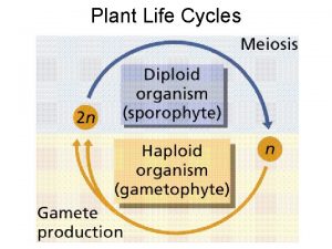
• Plants and some algae – Exhibit an alternation of generations – The life cycle includes both diploid and haploid multicellular stages Haploid multicellular organism (gametophyte) n Mitosis n n n Spores Gametes MEIOSIS Diploid multicellular organism (sporophyte) Figure 13. 6 B 2 n (b) Plants and some algae Copyright © 2005 Pearson Education, Inc. publishing as Benjamin Cummings FERTILIZATION 2 n Mitosis Zygote

• In most fungi and some protists – Meiosis produces haploid cells that give rise to a haploid multicellular adult organism – The haploid adult carries out mitosis, producing cells that will become gametes Haploid multicellular organism Mitosis n n n Gametes MEIOSIS FERTILIZATION 2 n Figure 13. 6 C Zygote (c) Most fungi and some protists Copyright © 2005 Pearson Education, Inc. publishing as Benjamin Cummings n

• Concept 13. 3: Meiosis reduces the number of chromosome sets from diploid to haploid • Meiosis – Takes place in two sets of divisions, meiosis I and meiosis II Copyright © 2005 Pearson Education, Inc. publishing as Benjamin Cummings

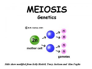
The Stages of Meiosis • An overview of meiosis Interphase Homologous pair of chromosomes in diploid parent cell Chromosomes replicate Homologous pair of replicated chromosomes Sister chromatids Diploid cell with replicated chromosomes Meiosis I 1 Homologous chromosomes separate Haploid cells with replicated chromosomes Meiosis II 2 Sister chromatids separate Figure 13. 7 Copyright © 2005 Pearson Education, Inc. publishing as Benjamin Cummings Haploid cells with unreplicated chromosomes

• Meiosis I – Reduces the number of chromosomes from diploid to haploid • Meiosis II – Produces four haploid daughter cells Copyright © 2005 Pearson Education, Inc. publishing as Benjamin Cummings

• Interphase and meiosis I MEIOSIS I: Separates homologous chromosomes INTERPHASE PROPHASE I Centrosomes (with centriole pairs) Sister chromatids METAPHASE I Chiasmata ANAPHASE I Sister chromatids remain attached Centromere (with kinetochore) Spindle Nuclear envelope Metaphase plate Homologous Microtubule chromosomes Tetrad attached to Chromatin separate kinetochore Pairs of homologous Chromosomes duplicate Tertads line up chromosomes split up Homologous chromosomes (red and blue) pair and exchange Figure 13. 8 segments; 2 n = 6 in this example Copyright © 2005 Pearson Education, Inc. publishing as Benjamin Cummings

• Telophase I, cytokinesis, and meiosis II MEIOSIS II: Separates sister chromatids TELOPHASE I AND CYTOKINESIS PROPHASE II METAPHASE II Cleavage furrow Figure 13. 8 Two haploid cells form; chromosomes are still double ANAPHASE II Sister chromatids separate TELOPHASE II AND CYTOKINESIS Haploid daughter cells forming During another round of cell division, the sister chromatids finally separate; four haploid daughter cells result, containing single chromosomes Copyright © 2005 Pearson Education, Inc. publishing as Benjamin Cummings

A Comparison of Mitosis and Meiosis • Meiosis and mitosis can be distinguished from mitosis – By three events in Meiosis l Copyright © 2005 Pearson Education, Inc. publishing as Benjamin Cummings

• Synapsis and crossing over – Homologous chromosomes physically connect and exchange genetic information Copyright © 2005 Pearson Education, Inc. publishing as Benjamin Cummings

• Tetrads on the metaphase plate – At metaphase I of meiosis, paired homologous chromosomes (tetrads) are positioned on the metaphase plates Copyright © 2005 Pearson Education, Inc. publishing as Benjamin Cummings

• Separation of homologues – At anaphase I of meiosis, homologous pairs move toward opposite poles of the cell – In anaphase II of meiosis, the sister chromatids separate Copyright © 2005 Pearson Education, Inc. publishing as Benjamin Cummings

• A comparison of mitosis and meiosis MITOSIS MEIOSIS Chiasma (site of crossing over) Parent cell (before chromosome replication) MEIOSIS I Prophase Chromosome replication Duplicated chromosome (two sister chromatids) Chromosome replication Tetrad formed by synapsis of homologous chromosomes 2 n = 6 Metaphase Chromosomes positioned at the metaphase plate Anaphase Telophase Sister chromatids separate during anaphase 2 n Tetrads positioned at the metaphase plate Homologues separate during anaphase I; sister chromatids remain together Metaphase I Anaphase I Telophase I Haploid n=3 Daughter cells of meiosis I 2 n MEIOSIS II Daughter cells of mitosis n n n Daughter cells of meiosis II Figure 13. 9 Copyright © 2005 Pearson Education, Inc. publishing as Benjamin Cummings Sister chromatids separate during anaphase II n

• Concept 13. 4: Genetic variation produced in sexual life cycles contributes to evolution • Reshuffling of genetic material in meiosis – Produces genetic variation Copyright © 2005 Pearson Education, Inc. publishing as Benjamin Cummings

Origins of Genetic Variation Among Offspring • In species that produce sexually – The behavior of chromosomes during meiosis and fertilization is responsible for most of the variation that arises each generation Copyright © 2005 Pearson Education, Inc. publishing as Benjamin Cummings

Independent Assortment of Chromosomes • Homologous pairs of chromosomes – Orient randomly at metaphase I of meiosis Copyright © 2005 Pearson Education, Inc. publishing as Benjamin Cummings

• In independent assortment – Each pair of chromosomes sorts its maternal and paternal homologues into daughter cells independently of the other pairs Key Maternal set of chromosomes Possibility 1 Possibility 2 Two equally probable arrangements of chromosomes at metaphase I Metaphase II Daughter cells Figure 13. 10 Combination 1 Combination 2 Copyright © 2005 Pearson Education, Inc. publishing as Benjamin Cummings Combination 3 Combination 4

Crossing Over • Crossing over – Produces recombinant chromosomes that carry genes derived from two different parents Prophase I of meiosis Nonsister chromatids Tetrad Metaphase I Chiasma, site of crossing over Metaphase II Daughter cells Figure 13. 11 Copyright © 2005 Pearson Education, Inc. publishing as Benjamin Cummings Recombinant chromosomes

Random Fertilization • The fusion of gametes – Will produce a zygote with any of about 64 trillion diploid combinations Copyright © 2005 Pearson Education, Inc. publishing as Benjamin Cummings

Evolutionary Significance of Genetic Variation Within Populations • Genetic variation – Is the raw material for evolution by natural selection Copyright © 2005 Pearson Education, Inc. publishing as Benjamin Cummings

• Mutations – Are the original source of genetic variation • Sexual reproduction – Produces new combinations of variant genes, adding more genetic diversity Copyright © 2005 Pearson Education, Inc. publishing as Benjamin Cummings

1. How do cells at the completion of meiosis compare with cells that have replicated their DNA and are just about to begin meiosis? 1) They have twice the amount of cytoplasm and half the amount of DNA. 2) They have half the number of chromosomes and half the amount of DNA. 3) They have the same number of chromosomes and half the amount of DNA. 4) They have half the number of chromosomes and onefourth the amount of DNA. 5) They have half the amount of cytoplasm and twice the amount of DNA. Copyright © 2005 Pearson Education, Inc. publishing as Benjamin Cummings

1. Which number represents G 2? * 1) I 2) II 3) III 4) IV 5) V Copyright © 2005 Pearson Education, Inc. publishing as Benjamin Cummings

1. Which number represents the DNA content of a sperm cell? 1) I 2) II 3) III 4) IV 5) V Copyright © 2005 Pearson Education, Inc. publishing as Benjamin Cummings

1. The DNA content of a diploid cell in the G 1 phase of the cell cycle is measured. If the DNA content is x, then the DNA content of the same cell at metaphase of meiosis I would be 1) 0. 25 x 2) 0. 5 x 3) x. 4) 2 x. 5) 4 x. Copyright © 2005 Pearson Education, Inc. publishing as Benjamin Cummings

1. The DNA content of a diploid cell in the G 1 phase of the cell cycle is measured. If the DNA content is x, then the DNA content at metaphase of meiosis II would be 1) 0. 25 x. 2) 0. 5 x. 3) x. 4) 2 x. 5) 4 x. Copyright © 2005 Pearson Education, Inc. publishing as Benjamin Cummings

1. The DNA content of a cell is measured in the G 2 phase. After meiosis I, the DNA content of one of the two cells produced would be 1) equal to that of the G 2 cell. 2) twice that of the G 2 cell. 3) one-half that of the G 2 cell. 4) one-fourth that of the G 2 cell. 5) impossible to estimate due to independent assortment of homologous chromosomes. Copyright © 2005 Pearson Education, Inc. publishing as Benjamin Cummings

1. Which of the following would not be considered a haploid cell? 1) daughter cell after meiosis II 2) gamete 3) daughter cell after mitosis in gametophyte generation of a plant 4) cell in prophase I 5) cell in prophase II Copyright © 2005 Pearson Education, Inc. publishing as Benjamin Cummings

1. A cell in G 2 before meiosis compared with one of the four cells produced by that meiotic division has 1) twice as much DNA and twice as many chromosomes. 2) four times as much DNA and twice as many chromosomes. 3) four times as much DNA and four times as many chromosomes. 4) half as much DNA but the same number of chromosomes. 5) half as much DNA and half as many chromosomes. Copyright © 2005 Pearson Education, Inc. publishing as Benjamin Cummings

Children with Down Syndrome

Down Syndrome—As a function of mother’s age l l l 1 in 1000 births in U. S. 1 in 12 births at age 50 Most frequent genetic cause of mental retardation l I. Q. = 20 -50

Down Syndrome—As a function of mother’s age

Human Somatic Cells have 46 Chromosomes l l How many chromosomes in human gametes? How do you know?

Down Syndrome l l How many chromosomes are in the egg? Sperm? How do you know? D. S. is due to an error in Meiosis

Down Syndrome Karyotype l l Down syndrome due to an error in meiosis l What’s meiosis? What is wrong with the Karyotype? Why are most trisomies fatal? Trisomies involving the sex chromo’s sex chromosomes are not fatal.

Human Life Cycle 1. 2. Role of mitosis? Meiosis l l l 3. Sperm (23 C) Fertilization produces gametes A reductive division (46 23 chromosomes) Don’t confuse meiosis with mitosis What if gametes were made by mitosis? Egg (23 C) Fertilization Zygote (46 C) Meiosis Mitosis Adult Human (Somatic cells: 46 C) Meiosis

Normal Meiosis followed by Fertilization Meiosis II Fertilization Normal sperm Chromosomes Normal diploid zygote Both daughter cells have one copy of each chromosome Product of Meiosis: Haploid Egg Cell

Genetic Basis of Down Syndrome Nondisjunction of chromosome pair #21 during meiosis leads to Down Syndrome No copy of chromosome 21 Diploid minus one copy of chromosome 21: Zygote dies Normal sperm Chromosome pair #21 Are misaligned Two copies of chromosome 21 Diploid plus one extra copy of chromosome #21: Down syndrome

Down Syndrome—a function of mom’s age • Why is the incidence of Down Syndrome a function of mom’s age and not that of the dad?

Egg formation in humans 1. All pre-egg cells present before girls are born Girls are born with about a 1000 pre-egg cells 2. – – 3. At birth all Pre-egg cells are stuck in metaphase I Homologous chromosomes are held in the middle of pre-egg cell by spindle fibers After Reaching Puberty, each month one pre-egg cell finishes meiosis I & II – – Meiosis produces one egg The other 3 cells are called polar bodies and die

Mom’s Pre-egg Cells form Prenatally 1. At about 12 years, women start ovulating one egg each month for the next 40 years. 2. A 50 yr old woman has had her eggs sitting with chromosomes aligned in metaphase I for over 50 years! 3. Egg spindle fibers degenerate with age—Causes. . – – Chromosomes move to one side Results in nondisjunction 4. Nondisjunction is rare with the larger chromosomes, #’s 1 – 20—Why? ? – Usually lethal because too much genetic imbalance.

Why doesn’t the age of the father influence the incidence of Down syndrome? 1. Sex cell formation in Males – – Sperm formation starts at puberty and continues daily for life Each pre-sperm cell divides twice to produce 4 sperm • 200 -300 million sperm produced per day! 2. Sperm forming cells do not stop in meiosis I of metaphase – – Sperm cells don't get old Therefore, no nondisjunction

Screening for Down Syndrome: Amniocentesis 1. Fetus located using ultrasound, needle inserted to remove amniotic fluid • 2. 3. 4. Not performed until 16 th week of pregnancy Fluid contains fetal biochemicals and fetal cells from skin, respiratory tract, urinary tract Culture cells for 1 -2 weeks Make Karyotype to detect abnormal chromosome numbers Amniocentesis

Screening for Down Syndrome: Chorionic Villi Sampling (CVS) 1. Catheter inserted vaginally and chorionic tissue removed Perform 9 -11 weeks after conception Make Karyotype CVS— 2. 3. 4. • Done earlier in pregnancy - • • 5. Less chance of complications if end pregnancy Slightly riskier for fetus Greater chance of infection Amniocentesis—have results. . . • • Done later in pregnancy Slightly safer than CVS

Mitosis 1. 2. 3. 4. 5. 6. No cross-over Produces 2 genetically identical somatic cells Involves only 1 division Chromosomes align single file in the middle of the cell during metaphase Sister chromatids separate during anaphase Daughter cells have the same number of chromosomes as the parent cell vs. Meiosis Cross-over during prophase I Produces 4 genetically different gametes Involves 2 divisions Homologous pairs align in pairs during metaphase I Homologous pairs separate during anaphase 1 1. 2. 3. 4. 5. • 6. Sister chromatids separate during anaphase 2 Daughter cells have half the number of chromosomes as the parent cell

Why Meiosis Causes Genetic Variation Independent Assortment 1. Homologous pairs of chromosomes align independently of one another during metaphase I • • 2. Maternal and paternal chromosomes are shuffled during meiosis 223 or 8, 388, 608 different combinations for each parent Fertilization gives 70 trillion possible genetic combinations

One Cause of Genetic Variation: Independent Assortment Possibility 1 Two possible arrangements of chromosomes during metaphase I Possibility 2

Cross-over—the 2 nd Reason for Genetic Variation • • Homologous chromosomes exchange parts during Prophase I Cross over results in thousands of genetically different gametes

Cross-over results in genetically different gametes 1. Cross-over 2. Regions exchanged Meiosis I Paternal copy Crossing over 3. Products of meiosis Meiosis II Parental Recombinant Homologous chromosomes Maternal copy Recombinant Parental

Homologous chromosomes Parent cell Sister chromatids Review of Mitosis Product of mitosis: Two genetically identical diploid daughter cells DNA Replication Metaphase: Chromosomes align single file Anaphase Separates sister chromatids 1. How do chromo’s align at metaphase? 2. What separates at anaphase? 3. How do the daughter cells compare genetically?

Review of Meiosis Parent cell 1. 2. 3. 4. Crossing over • Product of • meiosis: • Four genetically • different • haploid • daughter cells DNA Replication Metaphase I (homologous Chromosomes are paired) Anaphase I separates homologous chromosomes Alignment without replication How do the chromo’s align at metaphase I? What separates at anaphase I How do the chromo’s align at metaphase II? What separates at anaphase II? Anaphase II separates sister chromatids
 Metaphase ii
 Chapter 13 meiosis and sexual life cycles
 Crossing over occurs during
 Section quick check chapter 10 section 1 meiosis answer key
 Chapter 10 section 1: meiosis
 Sexual reproduction and genetics section 1 meiosis
 Chapter 10 section 3 gene linkage and polyploidy
 Difference between meiosis 1 and meiosis 2
 Style fashion and fad life cycles
 Plant life cycles and alternation of generations
 Vapour power cycles
 Meiosis i vs meiosis ii
 Meisis 1 and 2
 Image of prophase 2
 Alliteration for cycle
 Rvguev
 Life cycle of a bird
 Section 26.3 life cycles of stars
 What is charted on an hr diagram?
 Sac fungi life cycle
 Cycling of matter in an ecosystem
 The real lesson 21
 Compare and contrast carbon and nitrogen cycles
 Chapter 10 sexual reproduction and genetics
 Chapter 10 sexual reproduction and genetics
 Mesa and trading market cycles pdf
 Interphase of meiosis
 Sexual reproduction
 Mandarin cycles
 Biogeochemical cycles of water
 Model for improvement
 Precession milankovitch cycles
 Transaction cycle flow chart
 Cpi cycles per instruction
 Biogeochemical cycles water cycle
 Apes cycles
 Abiotic cycles
 Biogeochemical cycles quiz
 Enam securities portfolio
 Joke cycles
 Joke cycles
 Biogeochemical cycles class 9 ppt
 Sleep wake cycle
 Biogeochemical cycles foldable
 Design science research cycles
 Biogeochemical cycles poster
 Pdsa cycles
 Alphonse mucha advertising
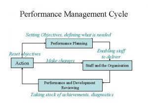
 Performance management cycle definition
 Water cycles of matter
 Isagenix presidents pack
 Multiplication of cycles
 Cpi in cpu
 Tidal patterns
 4 major biogeochemical cycles
 Water cycle pearson education
 Mandarin cycles
 Tsw cycles
 Business cycles economics
 Calculate real gdp per person
 Milankovitch cycles
 End-to-end procurement life cycle
 Biogeochemical cycles
 Generalization in database
 Milankovitch cycles
 Milankovitch cycle
 3-3 cycles of matter
 Minerals vs elements
 Air refrigeration cycle
 Cpi cycles per instruction
 Gersmehl diagram
 ……………types of bus cycles / operations in 80386dx
 아두이노 로고
 Lesson 2 cycles of matter answer key
 Rocks cycle
 Solid rock cycles
 Nutrient cycles in marine ecosystems
 Business cycles occur in free enterprise systems because
 Kondratieff cycles geography
 Practice geochemical cycles answer key
 Abiotic cycles
