
CHAPTER 13 2 2 Electrochemical Impulse 2 Movement



- Slides: 17
CHAPTER 13. 2 -2 Electrochemical Impulse #2
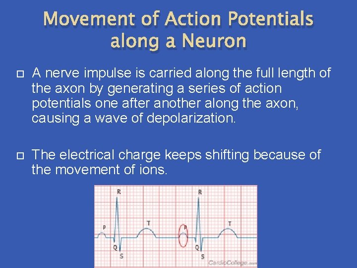
Movement of Action Potentials along a Neuron A nerve impulse is carried along the full length of the axon by generating a series of action potentials one after another along the axon, causing a wave of depolarization. The electrical charge keeps shifting because of the movement of ions.
Saltatory Conduction When axons are myelinated, nerve impulses travel by a process called saltatory conduction. In myelinated axons, the gated ion channels of K+ and Na+ are concentrated at the nodes of Ranvier between the Schwann cells. The flow of ions across the cell membrane can only happen at the nodes and so therefore action potentials can only happen at the nodes as well so the action potentials have to “jump” from node to node. This causes a nerve signal to be transmitted down an axon much faster.
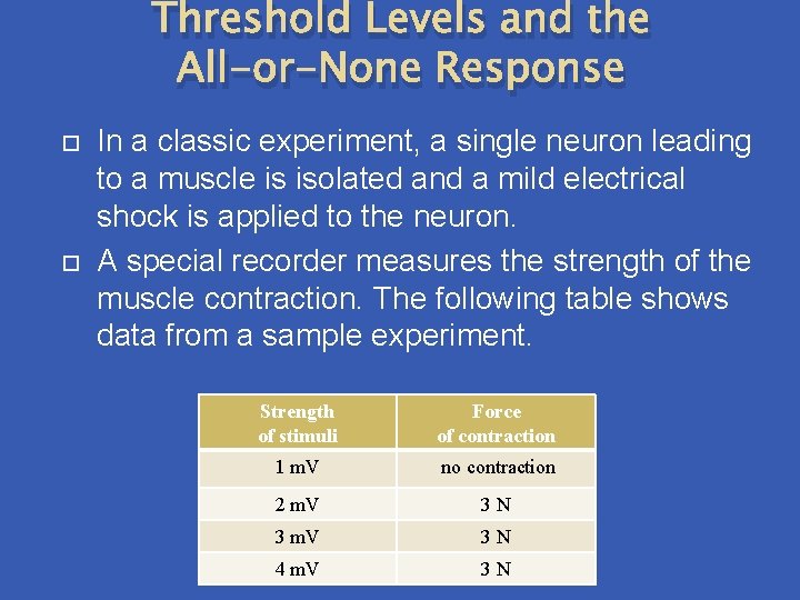
Threshold Levels and the All-or-None Response In a classic experiment, a single neuron leading to a muscle is isolated and a mild electrical shock is applied to the neuron. A special recorder measures the strength of the muscle contraction. The following table shows data from a sample experiment. Strength of stimuli Force of contraction 1 m. V no contraction 2 m. V 3 N 3 m. V 3 N 4 m. V 3 N
In this example, stimuli of less than 2 m. V does not produce any muscle contraction, whereas anything 2 mv and over produces the same force of muscle contraction. This experiment shows us two important things: 1. All neurons have a threshold level or a minimum level that must be reached in order for an action potential to be generated. In our example 2 m. V is the minimum voltage required to produce a response from the muscle. Stimuli below threshold levels do not initiate a response. Threshold levels are different for each neuron.
2. Increasing the intensity of the stimuli above threshold level value will not produce an increased response. This is known as the all-or none response It doesn’t matter if the stimuli is 2 m. V or 10 m. V the muscle produced exactly the same response. So just to clarify: Threshold level – the minimum level of a stimulus required to produce a result. All-or-None Response – a nerve or muscle fibre responds to a stimulus completely or not at all.
Difference in Stimuli If the all or none response is true and our neurons either fire action potentials or don’t, how can we tell the difference between the intensity of stimuli? The result of the stimulus remains the same, however the frequency of the nerve impulses changes.
The more intense a stimulus (hand on stove), the greater the frequency of impulses. There are more impulses travelling at a faster rate. The less intense a stimulus (hand under warm water) the lower the frequency of impulses. The nerve impulses will be fewer and travelling slower. Our brain interprets the number of impulses and rate at which they enter the brain in order to relay to us how much pain or sensation to feel.
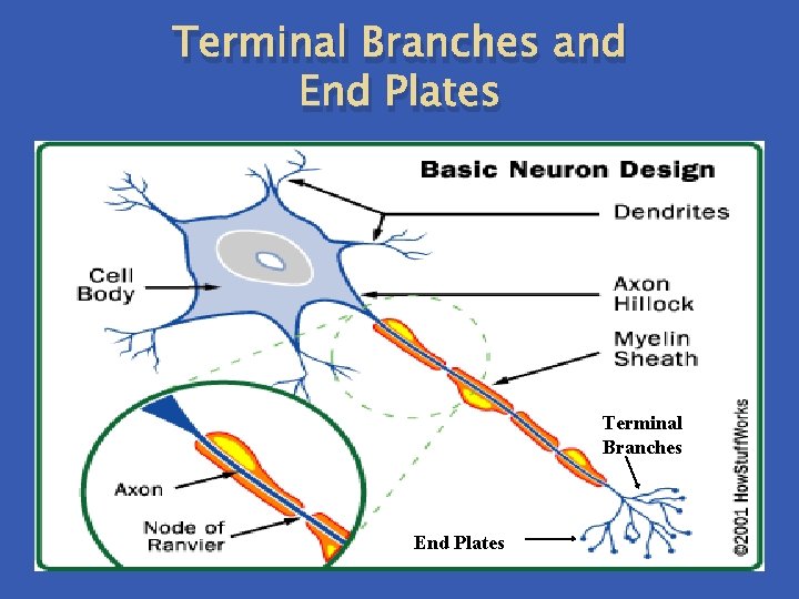
Transmission Across Synapses Remember that our nervous system is made up of billions of nerve cells (glial cells and neurons). Action potentials are used to carry the message along one neuron, however action potentials end when the message reaches the end of the neuron. The end of the neuron is made up of terminal branches with end plates. In order to carry the message across the gap to the next neuron, a chemical called a neurotransmitter is used.
Terminal Branches and End Plates Terminal Branches End Plates
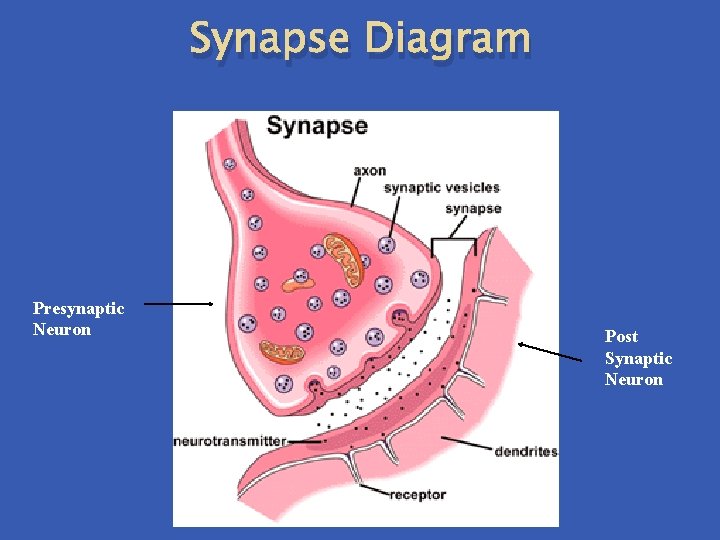
The gap between two neurons or a neuron and an effector is known as a synapse. The neuron carrying the impulse into the synapse is called the presynaptic neuron. The neuron leaving the synapse is called the postsynaptic neuron. The neurotransmitters that carry the impulse across the synapse are contained in small vesicles located in the end plates of axons.
The transmitter diffuses across the synaptic gap and binds to a receptor on the postsynaptic neuron. Here another action potential is produced and the signal continues on through the next neuron. Once the signal has been delivered the transmitter must be removed so that new signals may be received. In some cases the transmitter is broken down by an enzyme in the synapse. In other cases the transmitter is recycled- it is transported back into the presynaptic neuron to be used again.
Synapse Diagram Presynaptic Neuron Post Synaptic Neuron
Neurotransmitters 2 main classes of neurotransmitters: 1. Excitatory – speed nerve transmission on post synaptic neuron. Ex. Acetlycholine – opens Na+ channels on post synaptic neuron so the neuron becomes depolarized an action potential occurs immediately. Cholinesterase – enzyme that destroys acetylcholine, so the Na+ channels close.
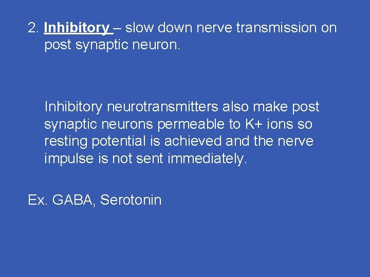
2. Inhibitory – slow down nerve transmission on post synaptic neuron. Inhibitory neurotransmitters also make post synaptic neurons permeable to K+ ions so resting potential is achieved and the nerve impulse is not sent immediately. Ex. GABA, Serotonin
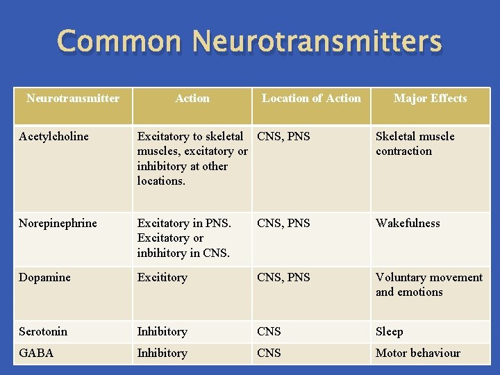
Common Neurotransmitters Neurotransmitter Action Location of Action Major Effects Acetylcholine Excitatory to skeletal CNS, PNS muscles, excitatory or inhibitory at other locations. Skeletal muscle contraction Norepinephrine Excitatory in PNS. Excitatory or inbihitory in CNS, PNS Wakefulness Dopamine Excititory CNS, PNS Voluntary movement and emotions Serotonin Inhibitory CNS Sleep GABA Inhibitory CNS Motor behaviour
Exit slip 1. How are the lessons? q Too fast? long? slow? Boring? 2. How are you finding the pace of the course? Difficult? Easy? Challenging? 3. What do you wish we could do more of in class? Less of in class? 4. Did you go on the website last night?