Chapter 12 The Nervous System Biology 3201 Unit

Chapter 12 The Nervous System Biology 3201 Unit I - Maintaining Homeostasis II

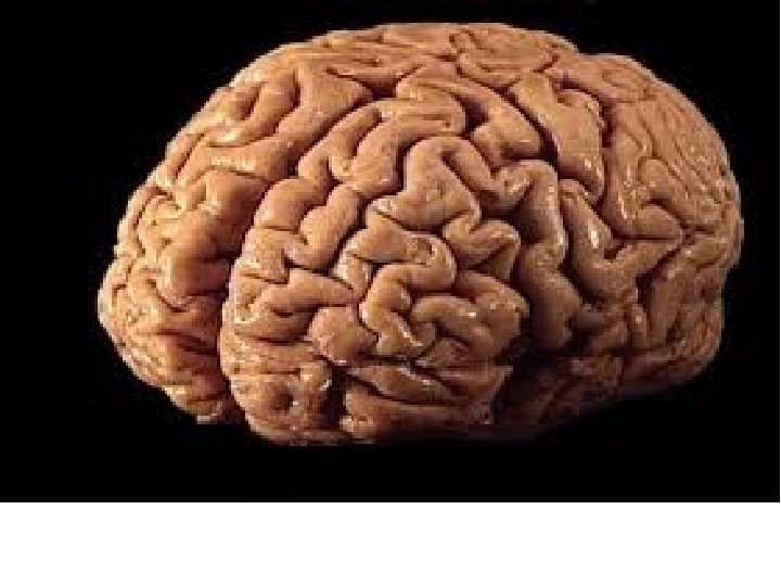
Interesting Brain Facts • The average adult human brain weighs three pounds, has a texture like firm jelly and is made up of 75 percent water. • Every time your heart beats, your arteries carry 20 to 25 percent of your blood to the brain. • There are 100 billion neurons (nerve cells) in the brain • There are 100, 000 miles of blood vessels in the brain. The distance around the world at the equator is 24, 900 miles
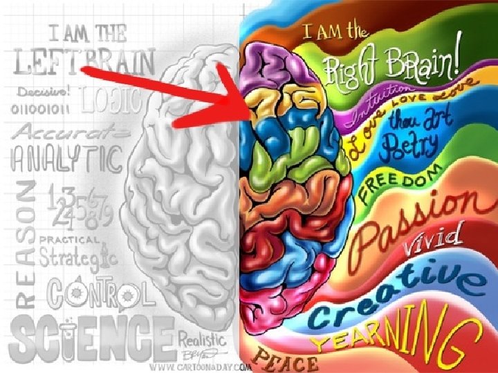

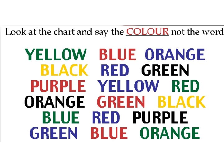
Left-Right Conflict • Right Brain tries to say the color • Left Brain insists you say the word This example demonstrates how both sides of your brain are connected, although one side tends to take the lead.

Turn to Page 3 of Booklet Worksheet: Phineas Gage Video: Phineas Gage https: //www. youtube. com/watch? v=Mvp. IRN 9 D 4 D 4

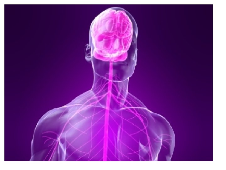
What is the nervous system? • The NS is made up of your brain, spinal cord, and all the nerves in your body. • It is the control center for your entire body.
 It maintains homeostasis in living organisms Homeostasis")
Why is the nervous system important? 1) It maintains homeostasis in living organisms Homeostasis – The process that maintains a relatively constant internal environment despite changing external conditions Ex: when you get too hot, you sweat
 It transmits electrical impulses throughout the body 3) It coordinates movement")
2) It transmits electrical impulses throughout the body 3) It coordinates movement
 – Made")
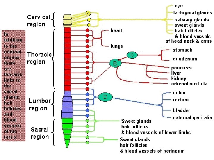
Two Major Parts of the Nervous System 1. Central Nervous System (CNS) – Made up of the brain and spinal cord 2. Peripheral Nervous System (PNS) – Made up of all the nerves that lead into and out of the CNS. See Figure 12. 2 - Page 392

CNS

http: //www. brainline. org/multimedia/interactive_brain/the_human_brain. html? g clid=CLn 4 gvu. Cq 7 ICFTN 0 Mgod 4 WAAIA
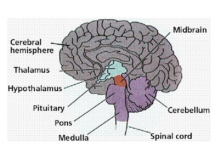

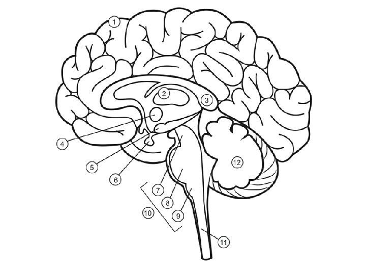
Turn to Page 4 of Booklet Parts of the Brain (Figure 12. 11 p. 399) 1. Cerebrum • Sorts and interprets all the information from our senses • Center for human consciousness Note: The cerebrum an be divided into 2 hemispheres (left and right) or into 4 lobes (frontal, parietal, temporal, occipital)

2. Thalamus • It sorts sensory information • It receives sensations of touch, pain, heat and cold • It regulates consciousness and sleep If the thalamus becomes impaired due to a head injury, illness or other trauma, it can result in the person entering a comatose state. Without a thalamus we wouldn't be able to stay awake and in turn we wouldn't be able walk, talk, sit, stand or even think.

3. Corpus Callosum • Controls eye movement • Connects the left and right sides of the brain • Important for maintaining attention
 • Endocrine control • Maintains homeostasis")
4. Hypothalamus • Autonomic control (fight or flight) • Endocrine control • Maintains homeostasis (hunger, body temperature, sleep-wake cycle, metabolism) • Aggression http: //www. everwell. com/fun/stu mp_the_doc/teeth_chattering. php

5. YOU DO NOT NEED TO KNOW! Hippocampus - involved in memory forming, organizing, and storing of information

6. Pituitary Gland • Called the “Master Gland” • Releases hormones (We’ll learn about it in Chapter 13)

http: //www. midbrain. com/ 7. Midbrain • Involved in sight and hearing • Has reflex centers to move your head

8. Pons • Controls your face, blinking, chewing/swallowing • Regulates breathing rate • Head movement

9. Medulla Oblongata • Controls autonomic functions, such as breathing, digestion and heart rate • Coughing, swallowing, sneezing, vomiting, hiccuping Damage to this part of the brain is usually fatal

You DO NOT need to memorize the brainstem 10. Brainstem – connects the cerebrum to the spinal cord. It is made up of the midbrain, pons and medulla

11. Spinal Cord – relays information from the brain to the rest of the body

12. Cerebellum – controls muscle coordination and balance. Video: Damage to the cerebellum http: //www. youtube. com/watch? v=5 e. Bwn 22 Bnio https: //www. youtube. co m/watch? v=BTH 2 Kv. FLc. Ho

Practice Question: • Rose is 6 -years old little girl who suffers from the following symptoms: double vision, inability to close her eyelids completely, facial weakness, headaches, and problems chewing and swallowing. Doctors preformed a CT scan, which showed a tumor in her brain. Where is this tumor located? Explain your reasoning.

Practice – see page 5 in Booklet 1. As a person drinks alcohol, they, at first, experience slurred speech, then they begin to stagger, finally they pass out and their breathing rate slows. Identify which parts of the brain are progressively being affected and explain your choice.

http: //www. youtube. com/watch? v=6 Zd 6 IECe. Ks&feature=related

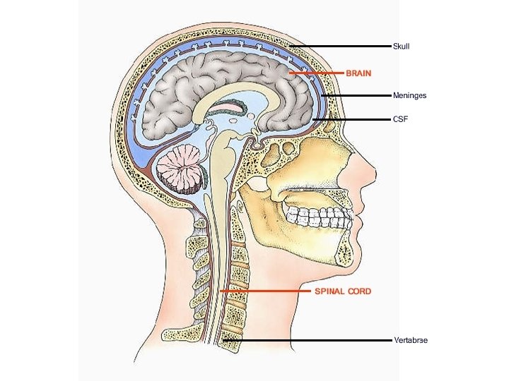
How is the CNS protected? 1. Meninges - Protective membranes that surround the brain and spinal cord 2. Cerebrospinal fluid – fluid that fills the spaces between the brain and meninges, which cushions the brain and spinal cord 3. Bone – hard physical barrier. The skull protects the brain and vertebrae protects the spinal cord

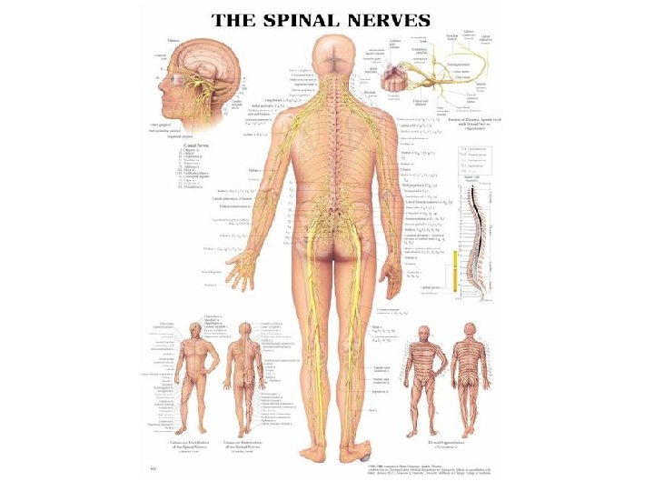
Structure of the Spinal Cord • The function of the spinal cord is to provide communication between the brain and the peripheral nervous system (PNS). • It is protected by vertebrae • Spinal nerves pass through the vertebrae and out to the PNS
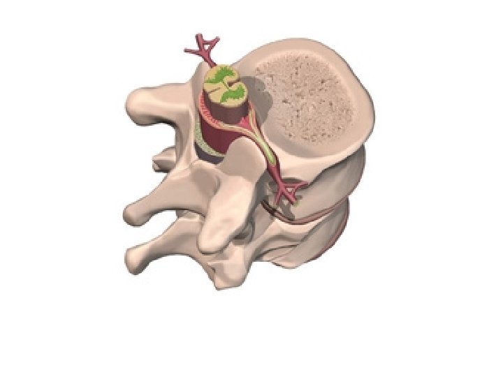

• Spinal Cord Anatomy http: //www. spineuniverse. com/anatomy/spinalanatomy-animation
 https: //www. youtube. co m/watch?")
Peripheral Nervous System • Made up of nerves (neurons) https: //www. youtube. co m/watch? v=Od. OOIxc. Uj. As • The PNS is made up of two subsystems: 1. Autonomic Nervous System (think of an automatic car) 2. Somatic Nervous System (think of a standard car) The Nervous System Simplified Video: http: //www. youtube. com/watch? v=sq 9 qf. Sus. Nm. A

Explain the difference between the Somatic vs. Autonomic NS Somatic Autonomic

Explain how your body responds during fight or flight. Sympathetic Response Parasympathetic Response
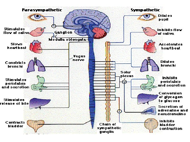

Sympathetic Nervous System • The sympathetic nervous system sets off what is known as a “fight - or - flight” reaction. This prepares the body to deal with an immediate threat. • Stimulation of the sympathetic nervous system causes a number of things to occur in the body: 1. Heart rate increases 2. Breathing rate increases 3. Blood sugar is released from the liver to provide energy which will be needed to deal with the threat.

Parasympathetic Nervous System • The parasympathetic nervous system has an opposite effect to that of the sympathetic nervous system. • It returns the body to its normal state of rest • The parasympathetic system does this by: 1. Decreasing heart rate 2. Decreasing breathing rate 3. A message is sent to the liver to stop releasing blood sugar since less energy is needed by the body

Fight or Flight Response http: //www. youtube. com/watch? v=m 2 Gywo. S 77 qc https: //www. youtube. com/watch? v=mt. Rrx. NTnyh 8

Somatic Nervous System • The somatic nervous system is under conscious control. • It is made up of – sensory nerves - carry information from the sense organs – motor neurons – controls/moves the muscles – Interneurons – found in CNS (brain & spinal cord) • The somatic nervous system will involves reflex actions that are not under conscious control.

What is required for the Somatic Nervous System to work? 1. A way to detect stimuli in the skin, eye and ears (called the receptor) 2. A way to send the information to the spinal cord 3. A way to interpret and analysis the information 4. A way to respond or move your muscle (called neurons) (called the integrator or brain or spinal cord) (called the effector)

Explain what happens in a reflex. Use the diagram to explain your answer.

What is the difference between a motor neuron, interneuron and sensory neuron?

Three types of reflexes: Where are these reflexes? How do they work? Knee Jerk Achilles Tendon Babinski

https: //www. youtube. com/watch? v=vy. Nk. Au. X 29 OU

STRUCTURE AND FUNCTION OF THE NEURON • Cell Body: decides if a stimulus has reached threshold. It has a nucleus (brains of neuron)
 from other neurons.")
• Dendrites: receives information (signals) from other neurons.

• Axon: Transmits a signal, when the impulse that is strong enough.

• Axon Terminal: contains neurotransmitters which get released into the synapse when the neuron is stimulated
: a fatty layer that wraps around the axon.")
• Myelin Sheath (Schwann cells): a fatty layer that wraps around the axon. This speeds up the transmission of an impulse.

• Node of Ranvier - the gap between each Schwann cell. It allows the impulse to jump from one node to the next.

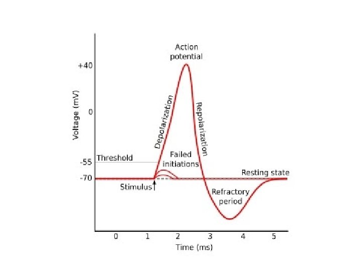
Action Potential: How does a Neuron Work? • Neurons are like little batteries because they generate electrical signals. When a neuron is stimulated, an electrical signal passes from dendrites, to the cell body, to the axon and the axon terminal. This is called a wave of depolarization. Video: How a neuron fires http: //www. youtube. com/watch? v=C 4 Gt 322 -Xx. I
")
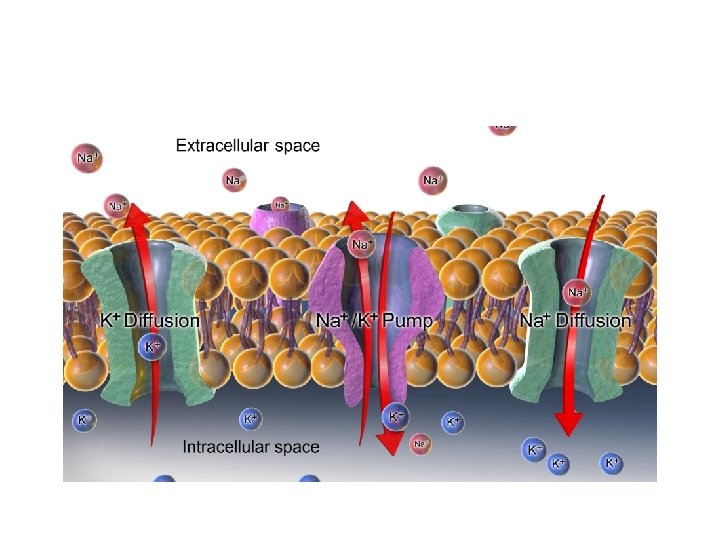
• This is possible because of the movement of two ions, sodium (NA+) and potassium (K+), across the membrane of the axon. These are specialized channels or gates, which allow for the movement of ions. Cl- remains trapped inside the cell.

What stimulates sensory neurons? 1. Chemicals 2. Light 3. Heat 4. Electrical current

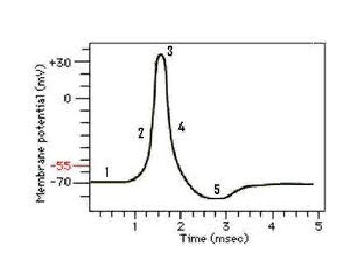
STEP 1: Resting Potential • The neuron is at rest • There is unequal distribution of charges on the axon – Outside the axon is positive (Na+ concentration is high) – Inside the axon is negative (Cl- concentration is high, and Na+ is low)

• The resting potential is generated and maintained with the help of a Na+/K+ pump, which is ALWAYS pumping 3 Na+ out and 2 K+ inside the axon • If the inside of a resting neuron was measure it would be -70 m. V (milli volts)

Draw a Neuron at Rest

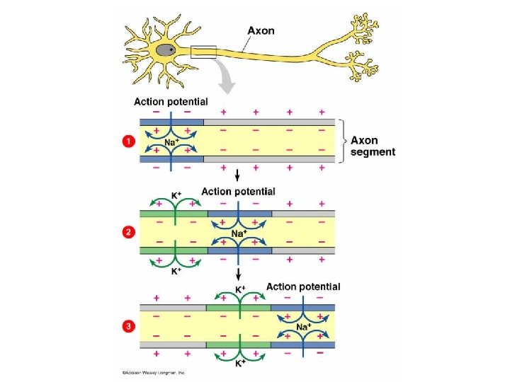
STEP 2: Depolarization • the neuron is stimulated and a nerve impulse is produced • There is a change in the charge of a neuron: – Outside becomes negative – Inside become positive • This change in charge is called action potential http: //www. youtube. com/watch? v=5 VWa. OKw. WE-o&safe=active

Why does this happen? • The stimulus causes the Na+ gates to open. The gates open one after another, down the length of the axon (like dominoes). This causes a reverse in the polarity of the axon, which causes a ‘spark’ of electricity.

Draw a diagram for Depolarization

STEP 3: Repolarization • the neuron starts to go back to resting potential • Immediately after depolarization has occurred, the K+ gates open and the Na+ gates close. This allows K+ ions to move out of the axon.

• This repolarizes the neuron so it returns to its original resting state: – Outside the axon is positive again – Inside the axon is negative again • The Na+/K+ pump is also pumping 3 Na+ ions out of the axon and 2 K+ inside, which help to return the neuron to resting potential faster.

Draw a diagram for Repolarization https: //www. youtube. com/watch? v=OZG 8 M_ld. A 1 M

• Refractory Period – the period of time between the triggering of an impulse and when it is available for the next impulse. This takes about 0. 001 seconds. • All-or-None Principle – In order for the neuron to send a wave of depolarization down its axon, the stimulus must reach a certain level. If the stimulus does not reach this level, no message is sent. • The level of stimulus needed is about -50 m. V, this is known as threshold potential.

Public Exam Question • Due to a mutation, the sodium binding site on a sodium potassium pump has changed such that it now binds with chloride ions (Cl-). Sodium ions (Na+) remain inside the membrane. What happens to nerve transmission? Give two reasons to justify your answer.

Public Exam Question • The graph below shows the potential difference between the inside and the outside of a motor neuron. Did an action potential pass down the neuron? Justify your answer?

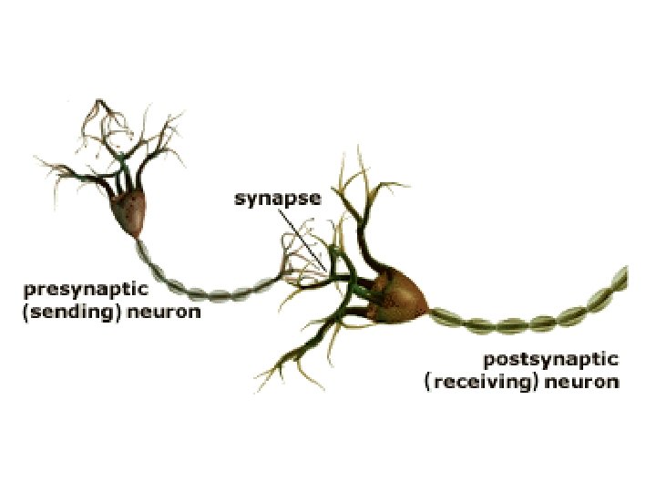
Synapse

What is the synapse? • The space between neurons • An action potential must cross a synapse and can cross in one direction only

Presynaptic fibre • A fibre which carries an impulse toward a synapse

Postsynaptic fibre • A fibre which receives the impulse after it crosses the synapse.

What happens in the Synapse? 1. A stimulus in the presynaptic fiber causes neurotransmitters to be released into the synapse. 2. The neurotransmitters cross the synaspe and attach to the receptor site on the postsynaptic fiber (dendrite)

3. When the neurotransmitter binds the receptor, it causes sodium gates to open and depolarization occurs in the neuron

Draw the Synapse
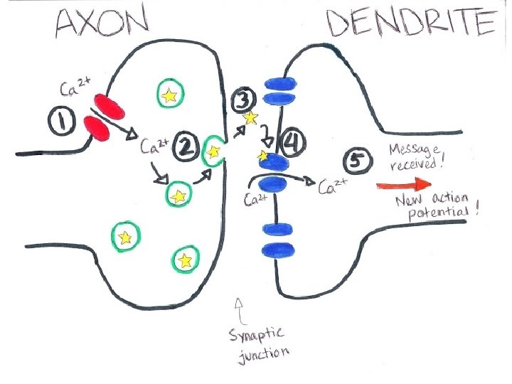

• The neurotransmitter diffuses into the synapse

Synapse Structure and Function - video • http: //www. youtube. com/watch? v=r. Wrnz-Ci. M 7 A

• The neurotransmitter will then either excite or inhibit the neuron • The excitory response causes the next neuron to be stimulated • The inhibitory response will prevent the next neuron from being stimulated. Instead, chloride channels open up which make the neuron more negative inside and raise threshold of stimulus.

What happens to the neurotransmitters in the synapse? • Once a neurotransmitter has attached to the receptor site of the postsynaptic neuron 1. The enzyme cholinesterase may be released from the presynaptic neuron to break down the neurotransmitter OR 2. The neurotransmitter may be reabsorbed into the vesicles of the axon terminal.


Chemical neurotransmission - video http: //www. mind. ilstu. edu/flash/synapse_1. swf Chemical Synapse - video http: //highered. mcgrawhill. com/sites/0072495855/student_view 0/chapter 14/animat ion__chemical_synapse__quiz_1_. html

How the Synapse works - video • http: //www. youtube. com/watch? v=Zuclw. AOJFh 8
 – Relaxes heart muscles")
Types of Neurotransmitters: 1. Acetylcholine – It stimulates muscles (excitatory) – Relaxes heart muscles (inhibitory) – Main neurotransmitter of the somatic and parasympathetic NS
 – Increases heart rate and glucose – Reduced levels are associated")
2. Noradrenaline (norepinephrine) – Increases heart rate and glucose – Reduced levels are associated with depression – Main neurotransmitter of the sympathetic NS

3. Glutamate • Accounts for 75% of excitatory transmissions in the brain • Oversupply can cause migraines or seizures

4. GABA • brain’s most common inhibitory neurotransmitter • Under supply causes seizures or insomnia

5. Dopamine • elevates mood; controls muscles • Affects sleep, cognition and attention and learning 6. Seratonin • alertness, sleepiness, mood

Nervous Disorders See page 14 in Booklet

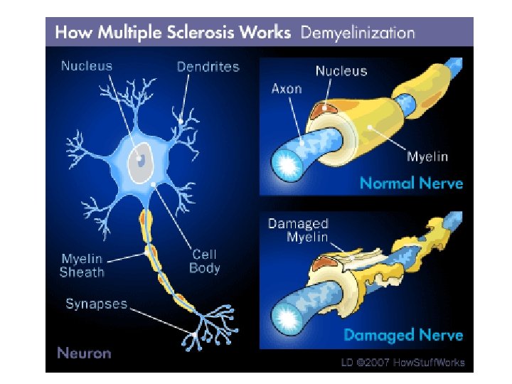
Multiple Sclerosis What is it? • An autoimmune disease

Symptoms:

What causes it? • Inflammation and deterioration of the myelin sheath. • This causes the transmission of messages to be slow or stopped

Treatment: • Autoimmune suppressants http: //biologyanimations. blogspot. ca/search/label/mul tiple%20 sclerosis

Alzheimer's Disease • first identified by German physician, Alois Alzheimer, in 1906 • What is it? – a progressive, neurodegenerative disease

• Symptoms: • impaired memory, thinking, and behavior • confusion • personality and behavior changes
 deposits in the brain which disrupts pathways. It also")
What causes it? Protein (amyloid) deposits in the brain which disrupts pathways. It also results in low levels of acelylcholine.
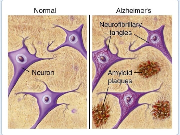
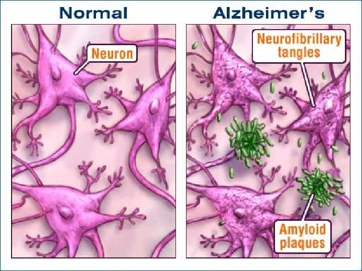
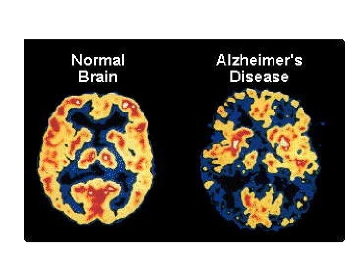

• Treatment: – No cure – Treatment: Medication to slow the disease http: //biologyanimations. blogspot. ca/search/label/Alzheimer% 27 s%20 disease http: //www. onenewspage. com/video/20150326/2 690311/Ultrasound-Shows-Promise-as-Treatment -for-Alzheimer. htm

Parkinson’s Disease • What is it? – It is a chronic movement disorder

Parkinson’s Disease

Symptoms include: • lack of coordination • Tremors • stiff muscles and joints • difficulty moving
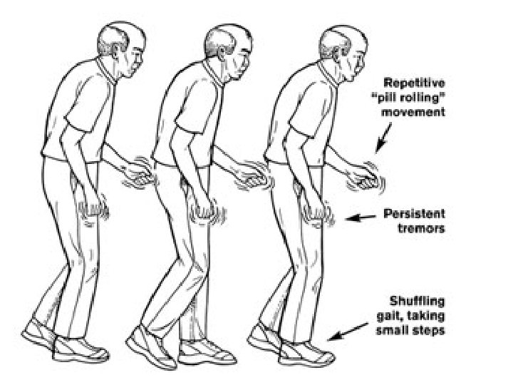

• What causes it? – Gradual death of neurons that produce dopamine.

• Treatment: Medications to increase dopamine levels http: //biologyanimations. blogspot. ca/search/label/parkinso ns%20 disease Michael J Fox https: //www. youtube. com/watch? v=ko. L 0 PWCJ 4 lo

Meningitis • What is it? – An infection of the meninges membranes which surround the brain and spinal cord.

Symptoms • • • Headache Fever Stiff neck Light sensitivity Vomiting Drowsiness

What causes it? • It is caused by a viral or bacterial infection. • Viral meningitis is common in children and usually clears up in 7 to 10 days. Bacterial meningitis can be fatal if not treated immediately.
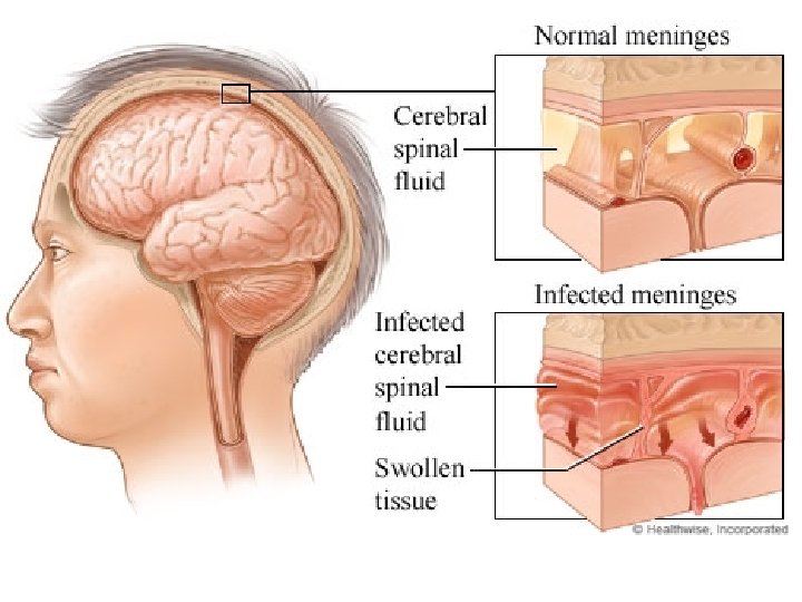
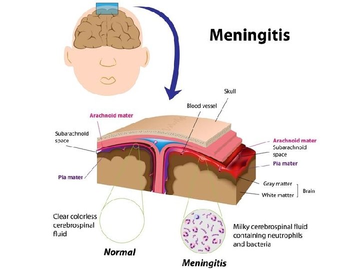

• Treatment: – Antibiotics. Vaccinations are available for bacterial meningitis http: //www. youtube. com/watch? v=e. Vswu. Wrxif 8 Amy Purdy http: //www. bing. com/videos/search? q=amy%20 p urdy%20 amputee&qs=n&form=QBVR&pq=amy%2 0 purdy%20 amputee&sc=1 -17&sp=1&sk=#view=detail&mid=4 A 1 EAC 017 D 9 ABF 6 C 201 14 A 1 EAC 017 D 9 ABF 6 C 2011
 • Huntington’s chorea (greek for “dance”) • about")
Huntington’s Disease • George Huntington (1872) • Huntington’s chorea (greek for “dance”) • about 1 in every 10, 000

• What is it? – This is a fatal disorder in which nerve cells in certain parts of the brain degenerate.

Symptoms: • Memory loss • Dementia • Involuntary twitching • Clumsiness • Personality changes – The symptoms usually progress for around a 15 year period until death occurs.

• What causes it? – The death of nerve cells – Parents with this disease have a 50% chance of passing it on to their child.
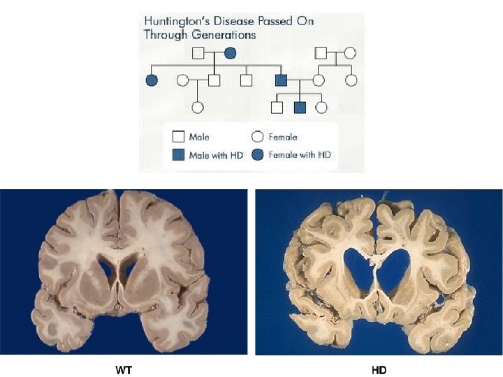

Treatment: • No cure • No treatment http: //www. bing. com/videos/search? q=huntington's&qs =n&form=QBVR&pq=huntington's&sc=8 -12&sp=1&sk=#view=detail&mid=8 AA 98 A 859 B 8770 B 276 E 78 A A 98 A 859 B 8770 B 276 E 7

Page 15 in Booklet

Stroke and Spinal Cord Injuries • Levels of Function in Spinal Cord Injury http: //www. youtube. com/watch? v=Pse. Uxlt. Iw _U&safe=active • Example: Amy van Dyken https: //www. youtube. com/watch? v=L 84 Z 4 Fbwq. WA

• Therapeutic cloning explained https: //www. youtube. com/watch? v=ev. H 0 I 7 Coc 54

Page 15 in Booklet 1. Explain either two reasons for supporting or two reasons for opposing the use of therapeutic cloning to replace damaged cells in the spinal cord.

2. After a snowmobile accident a person cannot move his legs and losses the ability to feel pain in his legs. Give two possible reasons for the loss of these abilities.

Describe how each of the following technologies work? 1. MRI 2. EEG 3. CAT scan 4. PET scan
")
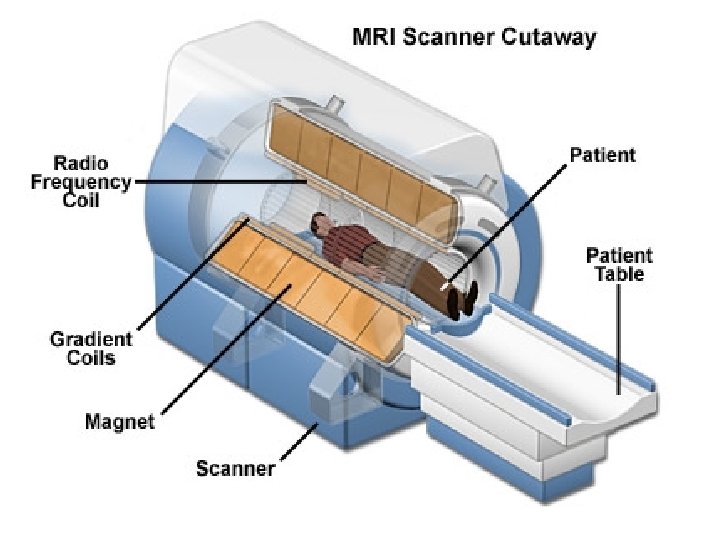
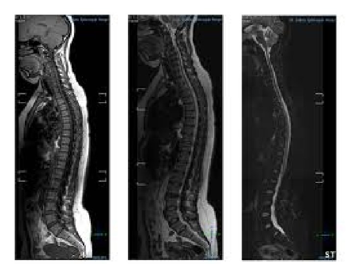
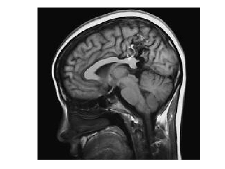
Magnetic Resonance Imaging (MRI)

How does a MRI work? • The patient lies in a large, hollow tube surrounded by magnets • The magnet spins and produces 2 D and 3 D images of various organs, showing any abnormalities, identify tumors, and detect early signs of disease.

MRI • https: //www. youtube. com/watch? v=Aw. XJNX Nc. LNs
")
EEG (electroencephalograph)
 are measured using electrodes placed")
How does an EEG work? • Brain messages (electricity) are measured using electrodes placed on the head. • A printout will show which parts of the brain are functioning. can be used to diagnose disorders (ex: epilepsy, tumours, sleep disorders)
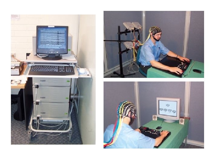

EEG Printout of Brain Waves

• EEG http: //www. youtube. com/watch? v=M 9 XVmks 1 ME
 scans")
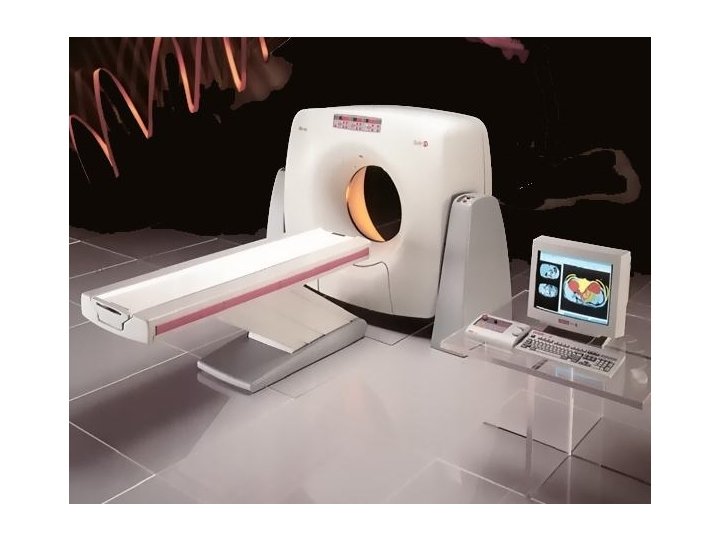
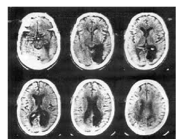
Computerized tomography (CAT) scans

How does a CAT scan work? • A scanner moves around the patient’s body, emitting x-ray beams at many different angles. • It produces a detailed, 3 D view of the body’s internal structures.


• CAT scan https: //www. youtube. com/watch? v=e 0 x. ZFka. RZ SU
 Scan")
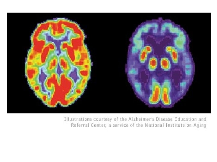
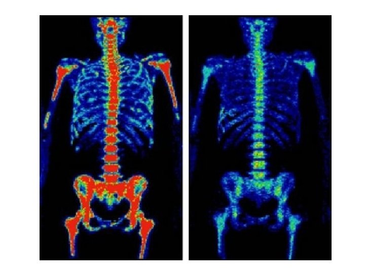
Positron Emission Tomography (PET) Scan

How does a PET scan work? • Identifies which areas of the brain are most active when the subject performs certain tasks • Patients are injected with a radioactive dye, which can be detected by a computer

• Some of the things PET measures: – brain abnormalities (tumours, memory disorders and seizures) – normal human brain and heart function. – helps doctors evaluate how well organs and tissues are functioning (blood flow, oxygen use)

PET/CT scan https: //www. youtube. com/watch? v=oy. Svkmezd o 0

Overview of Technologies Structure • CAT • MRI Function • PET • EEG

Check it out!! http: //www. pbs. org/wnet/brain/

Drugs and Homeostasis • Drugs interfere with the body’s homeostasis • It disrupts the body’s neurotransmitters and can damage neurons

Types of Drugs 1. Stimulants – speed the body up. They increase dopamine and noradrenaline levels. 2. Depressants – slow/relax the body. They increase GABA levels. 3. Pain killers – numb the body’s ability to feel pain, by blocking receptors on the dendrites

Designer Drugs What are they? When pre-existing drugs are altered chemically

• Why are they so dangerous? They are prepared by untrained chemists (called cookers). There is poor quality control (dangerous)
- Slides: 163