Chapter 12 The Cellular Basis of Reproduction Inheritance

Chapter 12 The Cellular Basis of Reproduction Inheritance and Power. Point Lectures for Biology: Concepts & Connections, Sixth Edition Campbell, Reece, Taylor, Simon, and Dickey Lecture by Richard L. Myers Copyright © 2009 Pearson Education, Inc.

CHAPTER 12 NEW GENETICS

Topics Discussed in this chapter Cell Division sexual and asexual reproduction Binary fission Eukaryotic Cell Cycle chromatin and chromosomes Mitosis and Meiosis phases of mitosis phases of meiosis tetrads, synapsis and crossing over somatic cells and sex cells autosomes and sex chromosomes

Cell Division and Reproduction Copyright © 2009 Pearson Education, Inc.

Methods of Reproduction Living organisms reproduce by two methods 1. Asexual reproduction – Offspring are identical to the original cell or organism – Involves inheritance of all genes from one parent – Prokaryotes reproduce asexually by binary fission. 2. sexual reproduction Involves inheritance of unique sets of genes from two parents Offspring are similar to parents, but show variations in traits

Prokaryotes reproduce by binary fission Binary fission means “dividing in half” – Occurs in prokaryotic cells – Two identical cells arise from one cell – Steps in the process: – A single circular chromosome duplicates, and the copies begin to separate from each other – The cell elongates, and the chromosomal copies separate further – The plasma membrane grows inward at the midpoint to divide the cells

Plasma membrane Prokaryotic chromosome Cell wall Binary fission in Prokaryotes 1 Duplication of chromosome and separation of copies Binary fission of a prokaryotic cell 2 Continued elongation of the cell and movement of copies 3 Division into two daughter cells

Eukaryotic Cell Division and Cell Cycle The cell cycle is an ordered sequence of events for cell division. Cells divide when they reach a certain size. INTERPHASE The cell cycle consists of two stages 1. Interphase: Includes G 1, S, and G 2 phases during which cell contents are duplication. G 1: first gap phase, growth and prepares for S-phase (DNA synthesis) s to Cy MIT PHA OTIC SE (M) M 2. Mitotic phase: (the M phase) involves mitosis and cytokinesis. is es n ki ito G 2: second gap phase, growth and preparation for division S G 1 si S: DNA synthesis phase, duplication of chromosomes, each becomes two sister chromatids G 2 Mitosis: division of the chromosomes Cytokinesis: division of cytoplasm Copyright © 2009 Pearson Education, Inc. The eukaryotic cell cycle

Eukaryotic chromosomes • The chromosomes carry the genetic information. • Eukaryotic chromosomes contain DNA and protein • The chromosomes are so named because they may be stained by certain dyes • When cells are not dividing, the genetic material is decondensed and is called chromatin • When cells are dividing, the genetic material is condensed and is called chromosome condensed Chromatin chromosome decondensed

Chromosome Organization

Chromosomes, Mitosis and Meiosis Human chromosomes karyotype Human chromosomes metaphase spread

The large, complex chromosomes of eukaryotes duplicate with each cell division – Early in the division process, chromosomes duplicate in Sphase. – Each chromosome appears as two sister chromatids containing identical DNA molecules. – Sister chromatids are joined at a narrow region called the centromere.

Sister chromatids Chromosome duplication Sister chromatids Centromere Electron micrograph of a duplicated chromosome Chromosome distribution to daughter cells Chromosome duplication and distribution

Mitosis • Identical chromosomes are distributed to each daughter cell • Mitosis preserves chromosome number in eukaryotic cell

Stages of Mitosis: progresses through a series of stages: 1. Prophase: Chromatin condenses into duplicated chromosomes (pair of sister chromatids) and chromosomes become visible. 2. Prometaphase: Chromosomes begin to move toward cell’s midplan. 3. Metaphase: Chromosomes align on cell’s midplane on top of each 4. Anaphase: other. Sister chromatids separate, move to opposite poles. Each former chromatid is now a chromosome. 5. Telophase: Cytokinesis: Chromosomes decondensed. Cytokinesis begins Cytoplasmic division. Often overlaps telophase

PROPHASE INTERPHASE Chromatin Nucleolus Nuclear Envelope Early mitotic spindle PROMETAPHASE Fragments of Nuclear envelope Centromere Plasma Membrane Chromosome, consisting of two sister chromatids Kinetochore Spindle Microtubules

Cell division is a continuum of dynamic changes Interphase – In the cytoplasm – Cytoplasmic contents double – In the nucleus Chromosomes duplicate during the S phase

METAPHASE ANAPHASE Cleavage furrow Metaphase plate Spindle TELOPHASE AND CYTOKINESIS Nucleolus Forming Daughter chromosomes Nuclear envelope Forming

Cytokinesis differs for plant and animal cells Cytokinesis – Cleavage in animal cells – A cleavage furrow forms from a contracting ring of microfilaments, interacting with myosin – The cleavage furrow deepens to separate the contents into two cells – Cytokinesis in plant cells – A cell plate forms in the middle from vesicles containing cell wall material – The cell plate grows outward to reach the edges, dividing the contents into two cells – Each cell has a plasma membrane and cell wall

Cleavage furrow Cytokinesis in animal cells Contracting ring of Microfilaments Daughter cells

Wall of parent cell Cell plate forming Daughter nucleus Cytokinesis in plant cells Cell wall New cell wall Vesicles containing cell wall material Cell plate Daughter cells
")
Growth (in an onion root)

Mitosis prophase Metaphase Telophase Early Anaphase Midi Anaphase Late Anaphase Telophase

MEIOSIS

Chromosomes are matched in homologous pairs Somatic cells(all body cells except sex cells, sperm and ovum) have pairs of homologous chromosomes, receiving one member of each pair from the father and one from the mother Homologous chromosomes are matched in – Length – Centromere position – Gene locations – A locus (plural, loci) is the position of a gene – Different versions of a gene may be found at the same locus on maternal (mother) and paternal (father) chromosomes Copyright © 2009 Pearson Education, Inc.

Chromosomes are matched in homologous pairs Homologous pair ofchromosomes The human sex chromosomes X and Y differ in size and genetic composition Pairs of autosomes(all chromosomes other than sex chromosomes, X &Y) have the same size and genetic composition Centromere Sister chromatids One duplicated chromosome A homologous pair of chromosomes

Gametes have a single set of chromosomes Meiosis is a process that converts diploid nuclei to haploid nuclei – Diploid cells have two homologous sets of chromosomes (2 n) – Haploid cells have one set of chromosomes (1 n) – Meiosis occurs in the sex organs (testes and ovaries) producing gametes (sperm and eggs) Fertilization is the union of sperm and egg – The zygote has a diploid chromosome number, one set from each parent
 n Egg cell n Sperm cell Meiosis Fertilization Diploid")
Haploid gametes (n = 23) n Egg cell n Sperm cell Meiosis Fertilization Diploid zygote (2 n = 46) 2 n Multicultural diploid Adults (2 n = 46) Mitosis and development The human life cycle

Meiosis reduces the chromosome number from diploid to haploid Like mitosis, meiosis is preceded by interphase – Chromosomes duplicate during the S phase Unlike mitosis, meiosis has two divisions – During meiosis I, homologous chromosomes separate – The chromosome number is reduced by half 2 n→ 1 n – During meiosis II, sister chromatids separate – The chromosome number remains the same 1 n

Meiosis reduces the chromosome number from diploid to haploid Events in the nucleus during meiosis I –Prophase I – Chromosomes coil and become compact – Homologous chromosomes come together as pairs by synapsis – Each pair, with four chromatids, is called a tetrad – Nonsister chromatids exchange genetic materials by crossing over –Metaphase I tetrads (duplicated homologous chromosomes) line up on metaphase plate side by side –Anaphase I • homologous chromosomes separate distributed to different nuclei • Each nucleus contains haploid number of chromosomes • Each chromosome has 2 chromatids TELOPHASE I –Telophase I and cytokinesis
 PROPHASE I Sites of")
MEIOSIS I: Homologous chromosomes separate INTERPHASE Centrosomes (with Centriole pairs) PROPHASE I Sites of crossing over ( ANAPHASE I METAPHASE I Microtubules attached to Kinetochore Metaphase Plate Sister chromatids remain attached Spindle Nuclear Envelope Sister Chromatin Chromatids Tetrad Centromere (with kinetochore) The stages of miosis I Homologous chromosomes separate

Meiosis II • Sister chromatids of each chromosome separate • one distributed to each daughter cell • Each former chromatid is now called a chromosome

MEIOSIS II: Sister chromatids separate TELOPHASE I AND CYTOKINESIS PROPHASE II METAPHASE II ANAPHASE II TELOPHASE II AND CYTOKINESIS Cleavage furrow Sister chromatids Separate The stages of miosis II Haploid daughter cells forming

MEIOSIS A B C D E F G H I A. B. C. D. E. F. G. H. I. PROPHASE I METAPHASE I ANAPHASE I TELOPHASE I PROPHASE II METAPHASE II ANAPHASE II TELOPHASE II TETRAD

Chapter 9 Patterns of Inheritance Power. Point Lectures for Biology: Concepts & Connections, Sixth Edition Campbell, Reece, Taylor, Simon, and Dickey Lecture by Richard L. Myers Copyright © 2009 Pearson Education, Inc.

Topics Discussed in this chapter Mendel’s laws Mendel’s monohybrid pea crosses. True breeding phenotype, genotype Gene, locus, allele dominant allele, recessive allele, homozygous, heterozygous A pedigree Exceptions to Mendel’s laws Incomplete dominance, co-dominance Multiple alleles, polygene Pleiotropy Sex determination in different species

The Basic Principles of Heredity MENDEL’S LAWS Copyright © 2009 Pearson Education, Inc.

Experimental genetics began in a garden Gregor Mendel discovered principles of genetics in experiments with the garden pea Mendel showed that parents pass heritable factors to offspring (heritable factors are now called genes) Advantages of using pea plants Controlled matings Self-fertilization or cross-fertilization ﺍ Observable characteristics with two distinct forms True-breeding strains Copyright © 2009 Pearson Education, Inc.
 One of Mendel’s pea crosses. F 1 generation Fertilization among")
P generation (true-breeding parents) One of Mendel’s pea crosses. F 1 generation Fertilization among F 1 plants (F 1 F 1) F 2 generation

Mendel’s law of segregation describes the inheritance of a single character Example of a monohybrid cross Parental generation: Tall plant Short plant F 1 generation: all plants were tall F 2 generation: Tall plants and short plants Mendel needed to explain Why one trait seemed to disappear in the F 1 generation Why that trait reappeared in one quarter of the F 2 offspring

Questions: Why one trait seemed to disappear in the F 1 generation? Why that trait reappeared in one quarter of the F 2 offspring? Answers: The questions were answered by Mendel’s Principle of Segregation which states that: v Each trait is controlled by two factors ( now known as alleles). v During gametes formation (meiosis) the two alleles segregate(separate), so that each gamete (sperm or ovum) have one allele only.
� TT tt t T Gametes T t t")
T P plants Genetic makeup (alleles)� TT tt t T Gametes T t t All T T F 1 plants (hybrids) All. Tt t T 1 – 2 Gametes T T Phenotypic ratio 3 purple : 1 white F 2 plants Genotypic ratio 1 BB : 2 Bb : 1 bb T Eggs t 1 – 2 Sperm T t T TT Tt tt Explanation of the crosses in previous figure t t t

Learning Objective Define the terms phenotype, genotype locus, allele dominant allele, recessive allele homozygous, and heterozygous

Genes information units in chromosomes. There are two copies of each gene. One on the father chromosome and one on the mother chromosome. Each copy is called allele Locus site of a gene on the chromosome Alleles: Copy of a gene (each gene has 2 copies, one on each of the homologous chromosomes), same loci on homologous chromosomes

Gene Pairs Diploid individuals: Individual whose cells contain 2 sets of chromosome (23 from the mother egg+23 from the father sperm). Consequently, genes on these homologous chromosomes are in pairs. One from the father and one from the mother. Each copy is called alleles. Homozygous Two identical alleles e. g. AA or aa. Heterozygous Two different alleles e. g. Aa.

Gene loci Dominant allele Genotype: P a B P a b PP aa Bb Homozygous for the dominant allele Homozygous for the recessive allele Recessive allele Heterozygous Matching gene loci on homologous chromosomes

Gene Expression Dominant allele Alleles that is expressed in the heterozygous and it masks expression of a recessive allele Recessive allele Alleles that is not expressed in the heterozygous Phenotype appearance Genotype genetic constitution

Genetic traits in humans can be tracked through family pedigrees A pedigree Shows the inheritance of a trait in a family through multiple generations Can also be used to deduce genotypes of family members. Important in genetic counseling. Female Affected Normal Copyright © 2009 Pearson Education, Inc. Symbols used in pedigree analysis

Examples of single-gene inherited traits in humans Earlobe Dominant Traits Recessive Traits Genotype FF or Ff ff Phenotype Free earlobe Attached earlobe
 Ff Second generation (parents, aunts, and uncles) FF or Ff ff")
First generation (grandparents) Ff Second generation (parents, aunts, and uncles) FF or Ff ff Ff Ff ff Third generation (two sisters) Female ff Male FF or Ff Affected Unaffected Pedigree showing inheritance of attached versus free earlobe in a hypothetical family
")
Parents Normal Dd Sperm D Offspring Eggs D d DD Normal Dd Normal (carrier) dd Deaf Offspring produced by parents who are both carriers for Deafness which is a recessive diorder

Exceptions to Mendel’s laws

Variations to Mendel’s Laws Traits inheritance is not always dominant or recessive, or controlled by one gene. Some of the exceptions to Mendel’s Laws are: 1. Incomplete dominance: heterozygote has intermediate phenotype 2. Codominance: heterozygote expresses phenotypes of both homozygotes. 3. Multiple alleles: Three or more alleles in a population for the same 4. Pleiotropy: locus. Diploid individual has any two alleles. the phenomenon of one gene mutation being responsible for or affecting more than one phenotypic characteristic. 5. Polygenes. Multiple independent pairs of genes may have similar and additive effects on the phenotype.

Incomplete dominance results in intermediate phenotypes Incomplete dominance Neither allele is dominant over the other Expression of both alleles is observed as an intermediate phenotype in the heterozygous individual

P generation Red RR White rr Gametes r R F 1 generation Pink Rr Incomplete dominance in snapdragon color. Gametes 1 – 2 R 1 – 2 r Sperm R r RR RR r. R 1 – 2 F 2 generation 1 – 2 R 1 – 2 r Eggs Rr 1 – 2 rr

Exceptions to to Mendel Laws When Mendel’s laws/results may not be observed Genetic Occurrence Definition Polygenic inheritance More than one gene can affect a single trait Pleiotropy A single gene can affect more than one trait Multiple alleles for one gene Genes may have more than two alleles Dominance is not always complete Environmental factors • In incomplete dominance the heterozygote is Examples • 4 genes are involved in determining eye color. • Human height • A pleiotropic allele dominant for yellow fur in mice is recessive for lethal developmental defect. • Sickle cell anemia ABO blood types in humans intermediate. • In co-dominance no single allele is dominant, and the heterozygote shows some aspect of both homozygotes. • Human blood groups Genes may be affected by the environment. Siamese cats

Sex determination in different species X-Y system in mammals, fruit flies XX = female XY = male X-O system in grasshoppers and roaches XX = female XO = male Z-W in system in birds, butterflies, and some fishes ZW = female ZZ = male Chromosome number in ants and bees Diploid = female haploid = male

Female 22 + XX 76 + ZW 32 X-O system X-O Z-W system Z-W male 22 + X 76 + ZZ 16 Chromosome number system
 44 + XY 22 + X (female) Parents’ diploid cells 44 + XX")
(male) 44 + XY 22 + X (female) Parents’ diploid cells 44 + XX 22 + Y Sperm Egg X-Y system 44 + XX Offspring (diploid) 44 + XY

Chapter 10 Molecular Biology of the Gene Power. Point Lectures for Biology: Concepts & Connections, Sixth Edition Campbell, Reece, Taylor, Simon, and Dickey Lecture by Richard L. Myers Copyright © 2009 Pearson Education, Inc.

Topics Discussed in this chapter Molecular Structure of DNA and RNA Base pairing rule DNA replication Flow of genetic information from DNA to protein Transcription Codon Translation Characteristics of the genetic code Types of mutations and their effects Cloning of plants and animals

Molecular Structure of The Genetic Material

DNA and RNA are polymers of nucleotides The monomer unit of DNA and RNA is the nucleotide, containing – 5 -carbon sugar – Phosphate group – Nitrogenous base A sugar-phosphate backbone is formed by covalent bonding between the phosphate of one nucleotide and the sugar of the next nucleotide Nitrogenous bases extend from the sugar-phosphate backbone

Sugar-phosphate backbone Phosphate group Nitrogenous base Sugar DNA nucleotide Nitrogenous base (A, G, C, or T) Phosphate group Thymine (T) (T ) Sugar (deoxyribose) DNA nucleotide DNA polynucleotide The structure of DNA polynucleotide

DNA is a double-stranded helix DNA is composed of two polynucleotide chains joined together by hydrogen bonding between bases, twisted into a helical shape – The sugar-phosphate backbone is on the outside – The nitrogenous bases are perpendicular to the backbone in the interior – Base-pairing rule : – – A pairs with T, forming two hydrogen bonds A=T _ G pairs with C, forming three hydrogen bonds G=C

Hydrogen bond Base pair Ribbon model Partial chemical structure Three presentations of DNA Computer model

DNA replication is semiconservative DNA replication follows a semiconservative model – The two DNA strands separate – Each strand is used as a pattern to produce a complementary strand, using specific base pairing rule – Each new DNA helix has one old strand with one new strand

Untwisting and replication of DNA semiconservatively Nucleotides Parental molecule of DNA Both parental strands serve as templates Two identical daughter molecules of DNA A template mode for DNA replication

The flow of genetic information from DNA ▬► RNA ▬► protein

The DNA genotype is expressed as proteins, which provide the molecular basis for phenotypic traits A gene is a sequence of DNA that directs the synthesis of a specific protein – DNA is transcribed into RNA – RNA is translated into protein The presence and action of proteins determine the phenotype of an organism

DNA Genotype Transcription Nucleus RNA Cytoplasm Translation Phenotype Protein Flow of genetic information in a eukaryotic cell

Genetic information written in codons is translated into amino acid sequences The sequence of nucleotides in DNA provides a code for constructing a protein – Protein construction requires a conversion of a nucleotide sequence to an amino acid sequence – Transcription rewrites the DNA code into RNA, using the same nucleotide “language” Each “word” is a codon, consisting of three nucleotides – Translation involves switching from the nucleotide “language” to amino acid “language” – Each amino acid is specified by a codon – 64 codons are possible – Some amino acids have more than one possible codon Copyright © 2009 Pearson Education, Inc.

DNA molecule Gene 1 Transcription & translations of codons Gene 2 Gene 3 DNA strand Transcription RNA Translation ﺍ Codon ﻭﺣﺪﺓ ﺍﻟﺸﻔﺮﺓ Polypeptide Amino acid

Characteristics of the genetic code – Triplet: Three nucleotides specify one amino acid – 61 codons correspond to amino acids – AUG codes for methionine and signals the start of transcription – 3 “stop” codons signal the end of translation – Redundant: More than one codon for same amino acids – Unambiguous: Any codon for one amino acid does not code for any other amino acid – Does not contain spacers or punctuation: Codons are adjacent to each other with no gaps in between – Nearly universal
")
Third base First base Second base Dictionary of the genetic code (RNA codons)

Deciphering the genetic information in DNA Strand to be transcribed DNA Transcription RNA Stop codon Start codon Translation Polypeptide Met Lys Phe

Mutations can change the meaning of genes A mutation is a change in the nucleotide sequence of DNA – Base substitutions: replacement of one nucleotide with another – Effect depends on whethere is an amino acid change that alters the function of the protein – Deletions or insertions: Alter the reading frame of the m. RNA, so that nucleotides are grouped into different codons – Lead to significant changes in amino acid sequence downstream of mutation Cause a nonfunctional polypeptide to be produced

Mutations can change the meaning of genes Mutations can be – Spontaneous: due to errors in DNA replication or recombination – Induced by mutagens – High-energy radiation – Chemicals

Normal hemoglobin DNA m. RNA Mutant hemoglobin DNA * m. RNA Normal hemoglobin Glu Sickle-cell hemoglobin * Val The molecular basis of Sickle-cell disease

Normal gene m. RNA Protein Met Lys Phe Gly Ala Lys Phe Ser Ala Base substitution ﺍ Types of mutations and their effects Met Base deletion Met Missing Lys Leu Ala His

Cloning of plants and animals Copyright © 2009 Pearson Education, Inc.

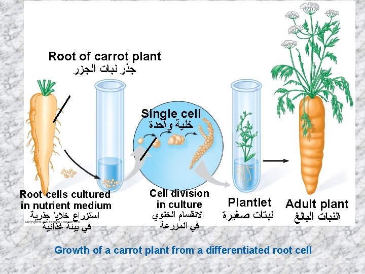
Plant cloning shows that differentiated cells may retain all of their genetic potential Most differentiated cells retain a full set of genes, even though only a subset may be expressed Evidence is available from – – Plant cloning (A root cell can divide to form an adult plant) Animal limb regeneration Remaining cells divide to form replacement structures Involved dedifferentiation followed by redifferentiation into specialized cells Copyright © 2009 Pearson Education, Inc.

Nuclear transplantation can be used to clone animals Nuclear transplantation – Replacing the nucleus of an egg cell or zygote with a nucleus from an adult somatic cell – Early embryo (blastocyst) cells (stem cells) can be used in: – Reproductive cloning – Implant embryo in surrogate mother for development – New animal is genetically identical to nuclear donor – Therapeutic cloning – Remove embryonic stem cells and grow in culture for medical treatments – Induce stem cells to differentiate to different tissues (cardiac, nervous, pancreas ----etc. ) Copyright © 2009 Pearson Education, Inc.

Donor Cell Remove nucleus from egg cell Nucleus from donor cell Add somatic cell from adult donor Grow in culture to produce an early embryo (blastocyst) Reproductive Cloning Implant blastocyst in surrogate mother Clone of donor is born Therapeutic Cloning Remove embryonic stem cells from blastocyst and grow in culture Induce stem cells to form specialized cells

Chapter 12 DNA Technology Power. Point Lectures for Biology: Concepts & Connections, Sixth Edition Campbell, Reece, Taylor, Simon, and Dickey Lecture by Mary C. Colavito Copyright © 2009 Pearson Education, Inc.
 Gene splicing")
Topics Discussed in this chapter Recombinant DNA Techniques Restriction endonuclease plasmids (vector) Gene splicing and production of recombinant DNA molecule Cloning Protein products of recombinant dna technology. Genetically modified (GM) plant and animals Transgenic organisms

Recombinant DNA Techniques Gene splicing, which results in recombinant DNA, is one of the most exciting aspects of genetic engineering. The DNA molecule can be cut at precise points and specific genes removed and transferred from one cell to another, even from such widely divergent species as plants beings and bacteria.

Steps to produce a recombinant DNA in the colon bacteria E. Coli (by genetic engineering technolog) ( )ﺗﻘﻨﻴﺔ ﺍﻟﻬﻨﺪﺳﺔ ﺍﻟﻮﺭﺍﺛﻴﺔ E. Coli ( ﻓﻲ ﺑﻜﺘﻴﺮﻳﺎ ﺍﻟﻘﻮﻟﻮﻥ recombinant) ﺍﺗﺤﺎﺩﻱ DNA ﺧﻄﻮﺍﺕ ﺍﻧﺘﺎﺝ ﺃ ﻭ A- Each E. coli cell contains one chromosome and several small circlets of DNA called plasmids. B- The plasmids can be isolated from bacteria that have been ruptured C- Restriction enzyme can break the plasmid at specific location D-The same restriction enzyme can be used to remove a segment of DNA –say the insulin gene-from a human cell. E- The human gene is inserted into the E. coli plasmids by ligase enzyme and the result, is “recombinant” DNA. E. The recombinant plasmids can now be reinserted into E. coli cells where they will subsequently be reproduced, each time the bacterium divides.

Some Protein Products of Recombinant DNA Technology.

Human insulin produced by bacteria

GENETICALLY MODIFIED ORGANISMS Copyright © 2009 Pearson Education, Inc.
 organisms contain one or more")
Genetically modified organisms are transforming agriculture Genetically modified (GM) organisms contain one or more genes introduced by artificial means Transgenic organisms contain at least one gene from another species GM plants – Resistance to herb – Resistance to pests – Improved nutritional profile GM animals – Improved qualities – Production of proteins or therapeutics Copyright © 2009 Pearson Education, Inc.

Transgenic fish Genes are introduced into fertilized eggs by DNA microinjection or electroporation No need to implant the embryo; development is external Genetically engineered for more rapid growth using the growth hormone gene (salmon, trout, catfish, tuna, etc. ) Genetically engineered for greater disease resistance Genetically engineered to serve as a biosensor for water pollution Genetically engineered for a novel pet (Glofish-see http: //glofish. com/)
 Salmon were genetically engineered for more rapid growth using the")
Transgenic fish (more detail) Salmon were genetically engineered for more rapid growth using the growth hormone gene under the control of the ocean pout antifreeze protein gene promoter and 3’ untranslated region (currently under FDA consideration) Madaka fish were genetically engineered to serve as biosensors for environmental pollutants (e. g. , estrogens) by using an estrogeninducible promoter (the vitellogenin promoter) to control expression of the GFP gene Fig. 21. 33 Fig. 21. 34
- Slides: 96