Chapter 12 The Cell Cycle The Genome All


- Slides: 60
Chapter 12 The Cell Cycle
The Genome • All of the genetic material contained within the DNA of an organism. • Most prokaryotes contain a single strand of circular DNA • Most Eukaryotes have multiple linear strands of DNA.
Chromosomes • Are the structures that make DNA replication and distribution manageable. • The name chromosome comes from their ability to take up dye when being prepared for microscopy. • All eukaryotes have a characteristic number of c-somes contained within the nucleus of the cell.
Gametes • The reproductive cells of organisms are called gametes and they contain 1/2 the number of c-somes normally found in somatic cells.
Chromatin and Eukaryotic Cells • The chromosomes of eukaryotic cells are made up of chromatin. • Chromatin is a composed of DNA and associated proteins.
Chromatin and Eukaryotic Cells • The DNA found on each c-some contains a few hundred to a few thousand genes specifying an organisms traits. • The associated proteins help to maintain the structure of the c-some and control the activity of the genes.
Non-Dividing Cells • During this time, the chromatin of each c-some is in a long thin configuration distributed throughout the cell. • The DNA is duplicated in preparation for cell division and when finished, the chromatin begins to condense.
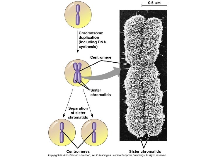
Dividing Cells • Once the c-some has been duplicated, there are now 2 sister chromatids which contain identical DNA molecules and are attached by proteins along their lengths. • The region where the sister chromatids appear to be pinched together is called the centromere.
Mitosis • During mitosis, the sister chromatids separate, the nucleus divides, and cytokinesis separates the cytoplasm of the cell. • Each new cell now contains one copy of DNA from the parent cell and the cycle repeats.
Meiosis • In a completely different form of cell division, meiosis yields cells that are not identical and contain only one set of c-somes. • Occurs in the testes and ovaries and results in a sperm or an egg. • During fertilization, the egg and the sperm fuse and restore the normal number of csomes of the organism.
Meiosis in Humans • Meiosis reduces the number of c-somes from 46 to 23. 22 autosomes and 1 sex c-some. • Fertilization then restores the c-some number back to 46. 44 autosomes and 2 sex c-somes.
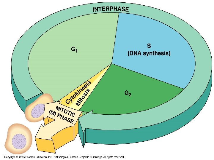
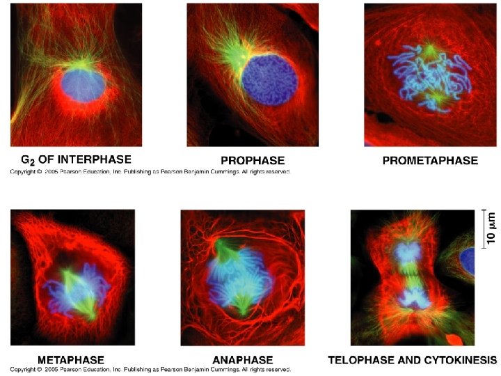
2 Main Phases of the Cell Cycle • Interphase which is broken into: • G 1 • S-Phase • G 2 • Mitosis (M-Phase) which is broken into: • Prophase • Prometaphase • Metaphase • Anaphase • Telophase • Cytokinesis
Mitosis Overview • Mitosis
Interphase • Interphase is the longest part of the cell cycle and is broken into: • G 1 phase is which is also called the 1 st gap phase or 1 st growth phase • S phase is where DNA is synthesized and the cell continues to grow. • G 2 phase is more growth and preparation for cell division.
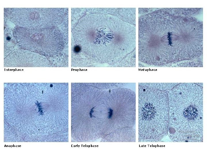
Mitosis-Prophase • Prophase is when the chromatin becomes tightly coiled. • Prometaphase is when each c-some has a distinct centromere.
Prophase • Prophase
Prometaphase • Prometaphase
Mitosis-Metaphase • Metaphase is when the c-somes align at the metaphase plate.
Metaphase • Metaphase
Mitosis-Anaphase • Anaphase occurs when sister chromatids begin to move apart.
Anaphase • As the proteins which bind the sister chromatids together become inactivated, anaphase begins as the sister chromatids are separated and begin to move to opposite ends of the cell. • Motor proteins walk the chromatids to the poles of the cell.
Anaphase • Anaphase
Mitosis-Telophase • Telophase occurs when the 2 daugher nuclei begin to form.
Telophase • Telophase
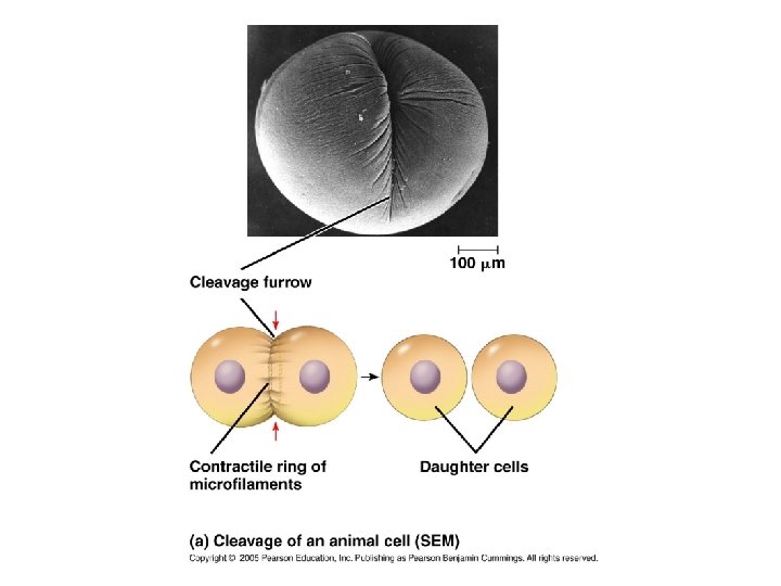
Mitosis-Cytokinesis • Cytokinesis is when a cleavage furrow forms which pinches the cell into two new daughter cells.
Cytokinesis • Cytokinesis
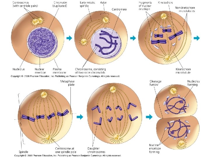
Interphase and Centrosomes • The centrosomes are nonmembranous organelles which organize the microtubules. • Sometimes the centrosomes are called the microtubule organizing center. • During interphase they duplicate and move to opposite ends of the cell.
Prophase and Centrosomes • As the centrosomes are moving to opposite ends of the cell (prophase and prometaphase are now occurring), microtubules grow out from them and attach to the kinetochore on each sister chromatid. • After attachment, each sister chromatid begins to move toward the pole from which the microtubule extends.
Centrosomes and Prophase • The actual movement of the chromatids is prevented because of the binding of the microtubule from the opposite end of the cell. • A “tug-of-war” now takes place until the c-somes are aligned at the metaphase plate.
Aster • As all of this is happening, a radial array of short microtubules (called the aster) is extending outward from each centromere (moving in the opposite direction) and attaches to the plasma membrane.
Mitotic Spindle • The mitotic spindle is an important part mitosis because is the organized array of microtubules that moves the chromosomes during cell division.
Mitotic Spindle • In addition to the asters sending microtubules outward, the microtubules which have grown and not attached to the kinetochores interact with other microtubules (from centrioles at the opposite end of the cell) that haven’t attached to the kinetochore forming the mitotic spindle.
Cytokinesis in Animals • During cytokinesis, a cleavage furrow forms and cleaves the cell into 2 new cells. • The cleavage furrow forms as a contractile ring of actin microfilaments interacts with myosin molecules on the cytoplasmic side of the plasma membrane. • When this interaction occurs, the ring contracts pinching the cell in two.
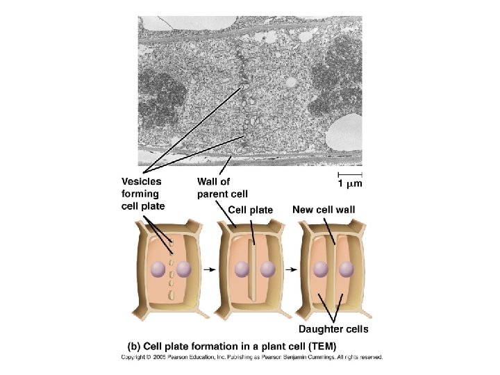
Cytokinesis in Plants • In plants, cytokinesis is much different. No cleavage furrow forms. Instead, vesicles which come from the Golgi migrate along microtubules to the center of the cell after division of the cytoplasmic contents. • The vesicles which collect at the center of the cell form a cell plate which eventually becomes a cell wall and two new cells are formed.
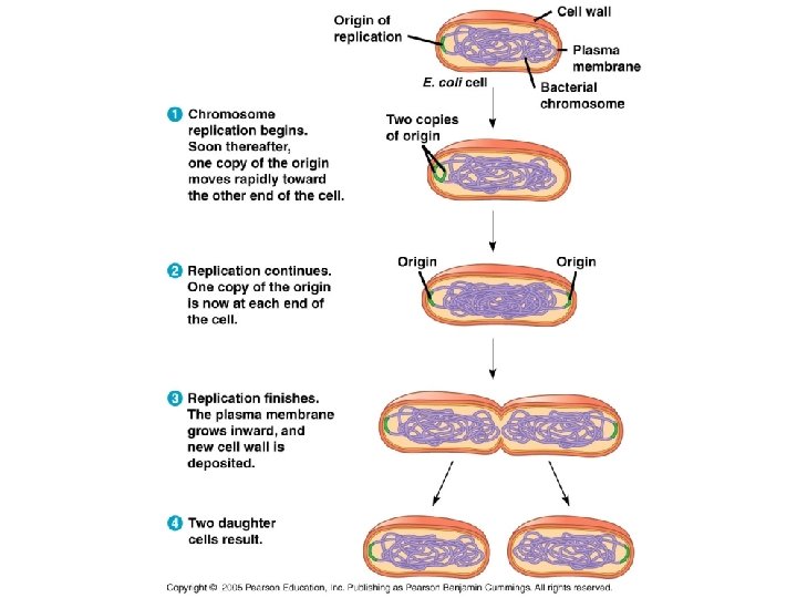
Bacterial Cell Division • Prokaryotic cells divide by a process called binary fission.
Stages of the Cell Cycle • The various stages of the cell cycle are determined by a variety of cytoplasmic signals. • Evidence comes from experiments performed in the early 1970’s. • When this happens, a cell replicates its DNA, the cell elongates and the plasma membrane grows inward dividing the cell in 2.
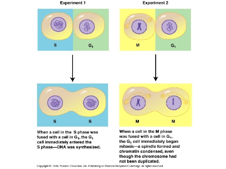
Cytoplasmic Signals--Evidence • Scientists grew cells in culture and harvested them at different stages of the cell cycle. • They then fused them together and found a variety of interesting things.
Results of Cell Fusion Experiments • When cells in the S phase were fused with those in the G 1 phase, the G 1 nuclei immediately entered the S phase. • Likewise, when M-phase cells were fused with cells in any other phase, the second cells immediately entered the Mphase.
What This Means… • All of the experiments lent support to the notion that cytoplasmic signals caused the various stages of the cell cycle and cell division. • They are sometimes called a “Cell Cycle Control System. ”
Cell Cycle Control System • This system cyclically operates and controls the key events in the cell cycle. • Each of the phases of the cell cycle seem to be controlled by a checkpoint which is where critical stop-and-go signals regulate the cell cycle.
Cell Cycle Control System • These signals shut down certain cell cycles and start them when key cellular processes have been completed. • The 3 major checkpoints are G 1, G 2 and M.
G 1 • For many cells, G 1 is important because cells receive a “go” signal here and will usually go through S, G 2 and M. • If no go signal is received, the cell enters G 0 which is a phase of nondivision.
G 0 • Most cells in the human body are in the G 0 phase and never divide (nerve and muscle cells). • Others are in G 0, but can be stimulated to divide when needed (liver).
Regulatory Proteins • Cyclins and Kinases are the two main types of regulatory molecules (proteins) found in cells.
Kinases • Kinases are enzymes that function by either activating or inactivating other proteins by phosphorylation. • Certain proteins give “go” signals at the G 1 and G 2 checkpoints.
Kinases • Kinases are usually present in constant concentration within the cell but are inactive. • They become active when attached to a cyclin protein. • These kinases are called Cd. Ks for cyclin dependent kinases.
Cyclin-Kinase Interactions • When cyclins and kinases interact, they initiate a variety of changes which result in the division of the cell.
Cancer Cells • Cancer cells don’t respond normally to the body’s control mechanisms. • They don’t heed the normal signals to regulate the cell cycle. • Others don’t need growth factors in their medium to grow and divide, some may even make their own.
Cancer Cells • Cancer cell also fails to exhibit what is known as density dependent inhibition which is where an external physical factor regarding cell division stops the division of crowded cells.
Cancer Cells • Normal cells usually divide 20 -50 x in the process of aging and then die. • Cancer cells are seemingly immortal because they will continue to divide forever if they are given a continuous supply of nutrients.
He-La Cells • Henrietta Lacks was a patient from whom some cancer cells were taken and cultured in 1951. These cells have been continually grown in culture since then. • The total number of cell divisions of these cells have far exceeded the normal 20 -50 cell cycles.