
Chapter 12 Neural Tissue 2012 Pearson Education Inc


- Slides: 66
Chapter 12 Neural Tissue © 2012 Pearson Education, Inc. 1
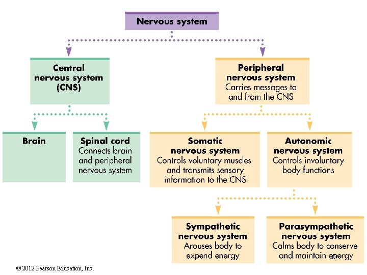
The Nervous System • Includes all neural tissue in the body • Neural tissue contains two kinds of CELLS 1. Neurons • Cells that send / receive signals 2. Neuroglia (glial cells) • Cells that support and protect neurons http: //iws. collin. edu/mweis/Images/Histology/2401%20 histology/Nervous%20 and%20 Speci al%20 Sense%20 Histo/nervous%20 and%20 ss%20 histo%20 labeled/NERVOUS_NEURON_S MEAR_labeled. png Organs of the Nervous System • Brain and spinal cord • Sensory receptors of sense organs (eyes, ears, etc. ) • Nerves connect nervous system with other systems Anatomical Divisions of the Nervous System Central nervous system (CNS) Peripheral nervous system (PNS) © 2012 Pearson Education, Inc. http: //www. mstrust. org. uk/information/public ations/msexplained/im ages/cns. gif 2
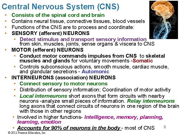
Central Nervous System (CNS) • • Consists of the spinal cord and brain Contains neural tissue, connective tissues, blood vessels Functions of the CNS are to process and coordinate: SENSORY (afferent) NEURONS • Detect stimulus and transport sensory information from skin, muscles, joints, sense organs & viscera to CNS • MOTOR (efferent) NEURONS • Conduct motor commands impulses from CNS to skeletal muscles and glands for voluntary movements -Somatic • Controls subconscious actions, smooth muscle, cardiac muscle, and glandular secretions - Automonic • INTERNEURONS (association) NEURONS • Connect sensory to motor neurons • Distribution of sensory information; Coordination of motor activity • Local interneurons short axons that form circuits with nearby neurons -analyze small pieces of information. Relay interneurons long axons that connect circuits of neurons in one region of the brain with those in other regions • Involved in higher functions- Intelligence, memory, planning, learning, emotion 3 • Accounts for 90% of neurons in the body - most of CNS © 2012 Pearson Education, Inc.
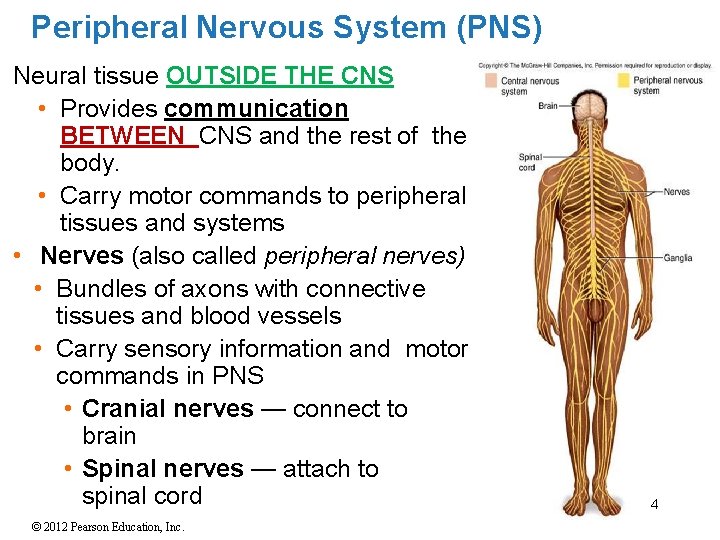
Peripheral Nervous System (PNS) Neural tissue OUTSIDE THE CNS • Provides communication BETWEEN CNS and the rest of the body. • Carry motor commands to peripheral tissues and systems • Nerves (also called peripheral nerves) • Bundles of axons with connective tissues and blood vessels • Carry sensory information and motor commands in PNS • Cranial nerves — connect to brain • Spinal nerves — attach to spinal cord © 2012 Pearson Education, Inc. 4
5 © 2012 Pearson Education, Inc.
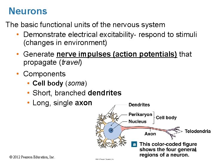
Neurons The basic functional units of the nervous system • Demonstrate electrical excitability- respond to stimuli (changes in environment) • Generate nerve impulses (action potentials) that propagate (travel) • Components • Cell body (soma) • Short, branched dendrites • Long, single axon 6 © 2012 Pearson Education, Inc.
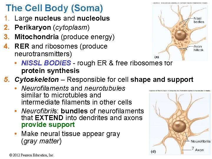
The Cell Body (Soma) 1. 2. 3. 4. Large nucleus and nucleolus Perikaryon (cytoplasm) Mitochondria (produce energy) RER and ribosomes (produce neurotransmitters) • NISSL BODIES - rough ER & free ribosomes for protein synthesis 5. Cytoskeleton – Responsible for cell shape and support • Neurofilaments and neurotubules similar to microtubles and intermediate filaments in other cells • Neurofibrils: bundles of neurofilaments that EXTEND into dendrites and axons provide support • Make neural tissue appear gray (gray matter) 7 © 2012 Pearson Education, Inc.
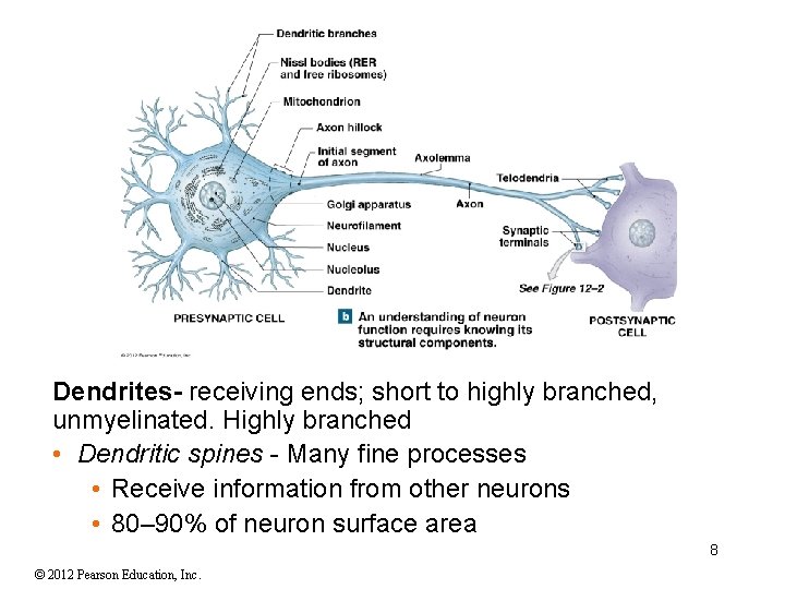
Dendrites- receiving ends; short to highly branched, unmyelinated. Highly branched • Dendritic spines - Many fine processes • Receive information from other neurons • 80– 90% of neuron surface area 8 © 2012 Pearson Education, Inc.
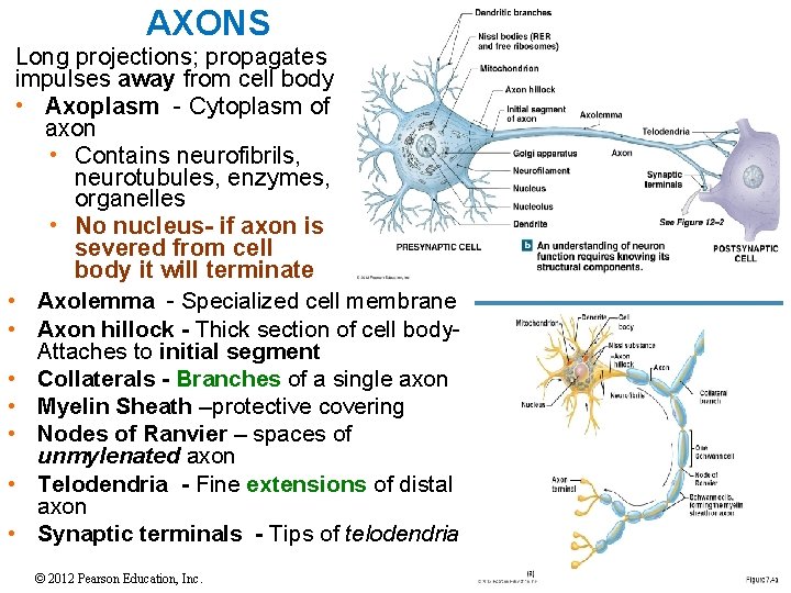
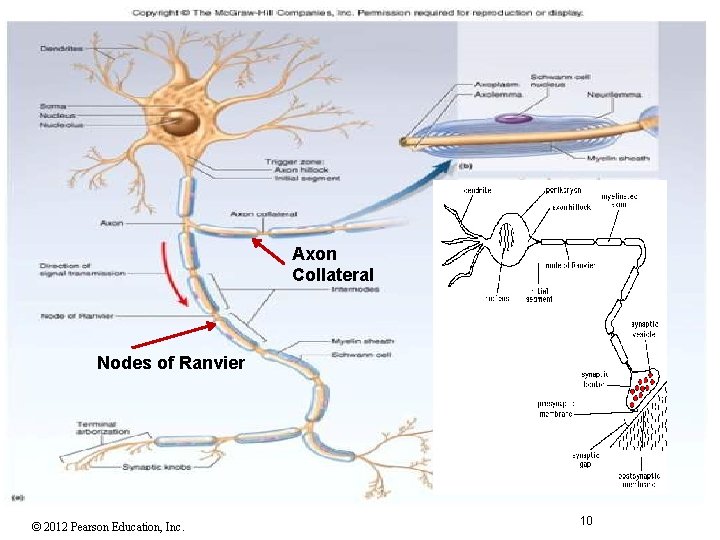
AXONS Long projections; propagates impulses away from cell body • Axoplasm - Cytoplasm of axon • Contains neurofibrils, neurotubules, enzymes, organelles • No nucleus- if axon is severed from cell body it will terminate • Axolemma - Specialized cell membrane • Axon hillock - Thick section of cell body. Attaches to initial segment • Collaterals - Branches of a single axon • Myelin Sheath –protective covering • Nodes of Ranvier – spaces of unmylenated axon • Telodendria - Fine extensions of distal axon • Synaptic terminals - Tips of telodendria © 2012 Pearson Education, Inc. 9
Axon Collateral Nodes of Ranvier © 2012 Pearson Education, Inc. 10
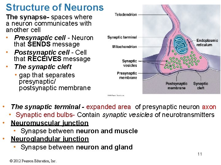
Structure of Neurons The synapse- spaces where a neuron communicates with another cell • Presynaptic cell - Neuron that SENDS message • Postsynaptic cell - Cell that RECEIVES message • The synaptic cleft • gap that separates presynaptic/ postsynaptic membrane • The synaptic terminal - expanded area of presynaptic neuron axon • Synaptic end bulbs- Contain synaptic vesicles of neurotransmitters • Neuromuscular junction • Synapse between neuron and muscle • Neuroglandular junction • Synapse between neuron and gland 11 © 2012 Pearson Education, Inc.
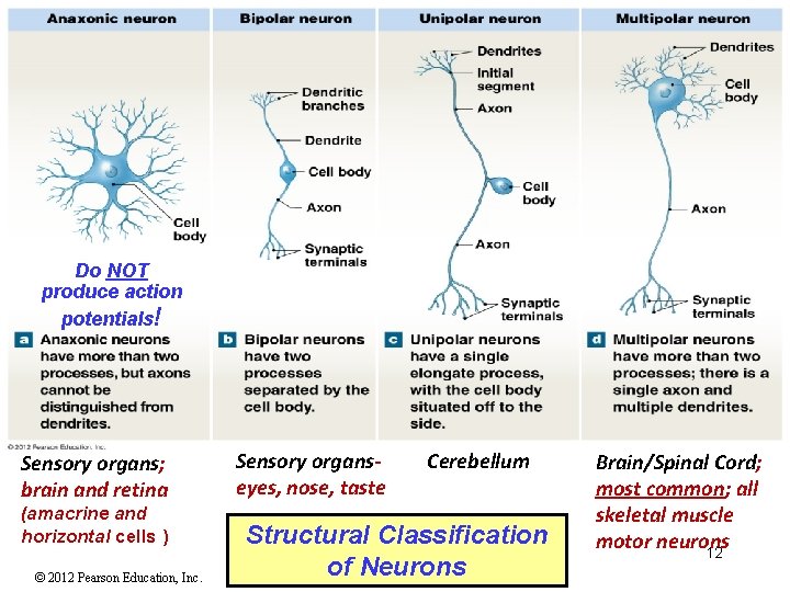
Do NOT produce action potentials! Sensory organs; brain and retina (amacrine and horizontal cells ) © 2012 Pearson Education, Inc. Sensory organseyes, nose, taste Cerebellum Structural Classification of Neurons Brain/Spinal Cord; most common; all skeletal muscle motor neurons 12
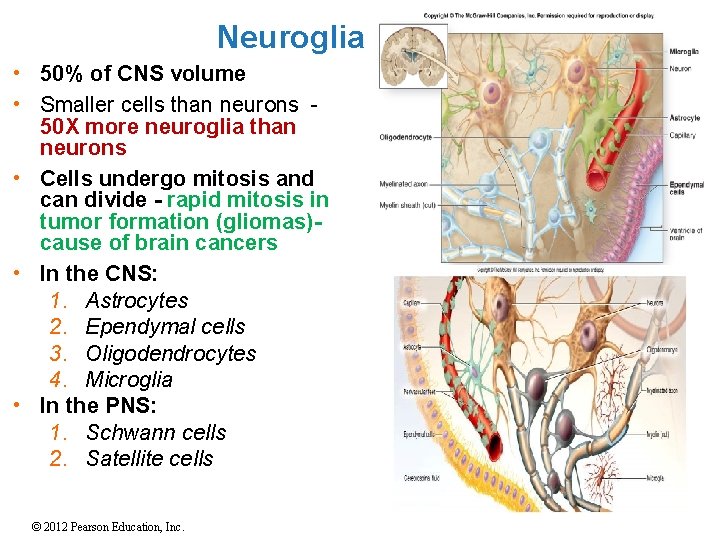
Neuroglia • 50% of CNS volume • Smaller cells than neurons 50 X more neuroglia than neurons • Cells undergo mitosis and can divide - rapid mitosis in tumor formation (gliomas)cause of brain cancers • In the CNS: 1. Astrocytes 2. Ependymal cells 3. Oligodendrocytes 4. Microglia • In the PNS: 1. Schwann cells 2. Satellite cells 13 © 2012 Pearson Education, Inc.
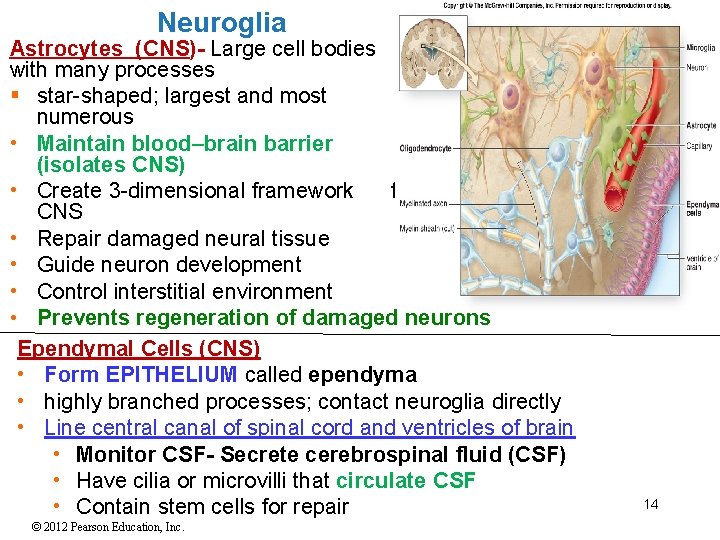
Neuroglia Astrocytes (CNS)- Large cell bodies with many processes § star-shaped; largest and most numerous • Maintain blood–brain barrier (isolates CNS) • Create 3 -dimensional framework for CNS • Repair damaged neural tissue • Guide neuron development • Control interstitial environment • Prevents regeneration of damaged neurons Ependymal Cells (CNS) • Form EPITHELIUM called ependyma • highly branched processes; contact neuroglia directly • Line central canal of spinal cord and ventricles of brain • Monitor CSF- Secrete cerebrospinal fluid (CSF) • Have cilia or microvilli that circulate CSF • Contain stem cells for repair © 2012 Pearson Education, Inc. 14
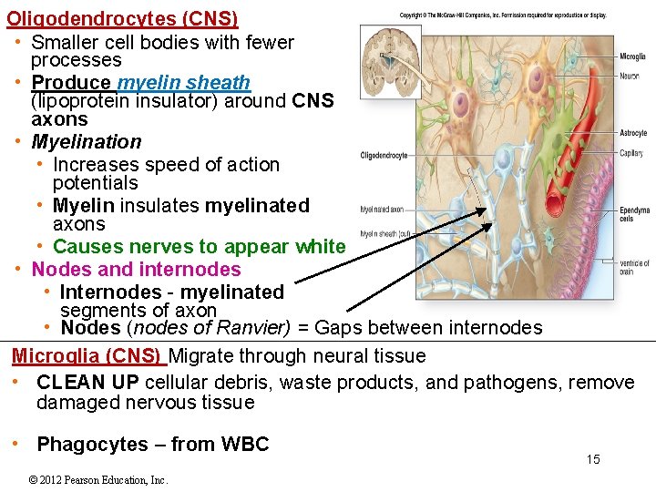
Oligodendrocytes (CNS) • Smaller cell bodies with fewer processes • Produce myelin sheath (lipoprotein insulator) around CNS axons • Myelination • Increases speed of action potentials • Myelin insulates myelinated axons • Causes nerves to appear white • Nodes and internodes • Internodes - myelinated segments of axon • Nodes (nodes of Ranvier) = Gaps between internodes Microglia (CNS) Migrate through neural tissue • CLEAN UP cellular debris, waste products, and pathogens, remove damaged nervous tissue • Phagocytes – from WBC © 2012 Pearson Education, Inc. 15
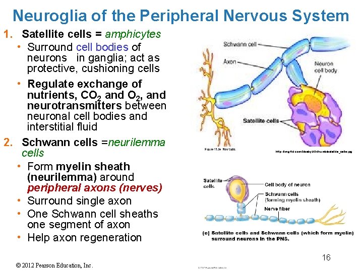
Neuroglia of the Peripheral Nervous System 1. Satellite cells = amphicytes • Surround cell bodies of neurons in ganglia; act as protective, cushioning cells • Regulate exchange of nutrients, CO 2 and O 2, and neurotransmitters between neuronal cell bodies and interstitial fluid 2. Schwann cells =neurilemma cells • Form myelin sheath (neurilemma) around peripheral axons (nerves) • Surround single axon • One Schwann cell sheaths one segment of axon • Help axon regeneration © 2012 Pearson Education, Inc. http: //img. tfd. com/Mosby. MD/thumb/satellite_cells. jpg 16 16
Electrical Potentials and Currents • All plasma (cell) membranes produce electrical signals by ion movements (ex: Na+). Membrane potential is particularly important to neurons. • Electrical potential – a difference in the concentration of charged particles between one point and another • **Cells use the changes in membrane potential as COMMUNICATION SIGNALS for receiving, integrating and sending information. • Electrical current – a flow of charged particles from one point to another; Movement of charges(ions) works to eliminate potential difference • currents occur by movement of ions, such as Na+ or K+ through gated channels in the plasma membrane • gated channels are opened or closed by various stimuli • The Electrochemical Gradient • The sum of chemical /electrical forces for a particular ion 17 © 2012 Pearson Education, Inc.
Membrane Processes Five Main Membrane Processes in Neural Activities 1. Resting potential • The membrane potential of resting cell 2. Graded potential - Caused by stimulus • Temporary, LOCALIZED CHANGE in resting potential 3. Action potential- is an electrical impulse • Produced by graded potential • Propagates along surface of axon to synapse 4. Synaptic activity –role of neurons to communicate a message to target cell via neurotransmitters • Releases neurotransmitters at presynaptic membrane • Produces graded potentials in postsynaptic membrane 5. Information processing • Response (integration of stimuli) of postsynaptic cell 18 © 2012 Pearson Education, Inc.
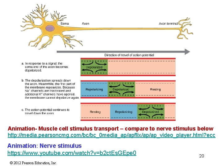
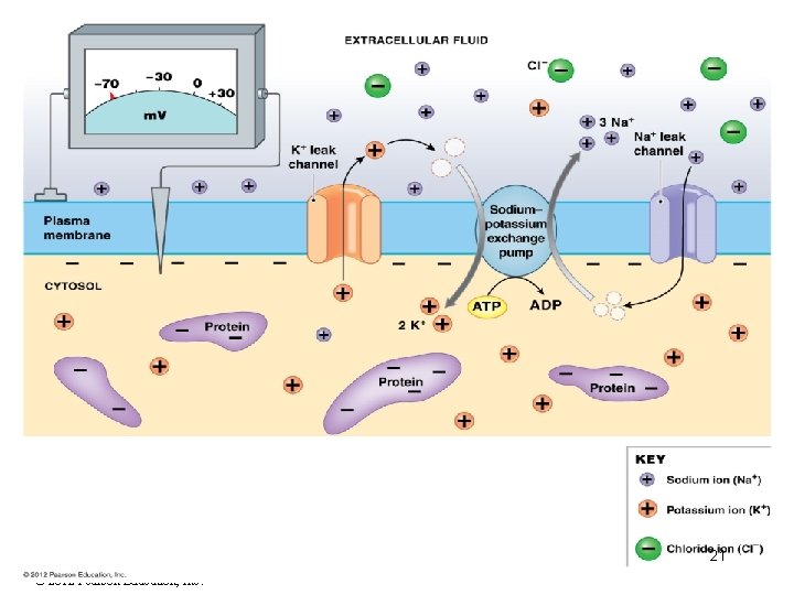
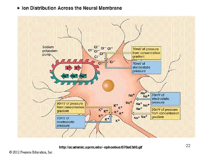
Resting Membrane Potential • Resting membrane potential (RMP) [Typically -70 mv ] is the charge difference across the plasma membrane due to unequal electrolyte distribution between extracellular (ECF) and intracellular fluid (ICF) Measured in millivolts. • Cells are said to be POLARIZED • Cells have selectively permeable membranes that separate charges of positive (+) and negative(-) ions resulting in potential difference • Membrane permeability varies by ion. Changes in membrane potential result from opening or closing specific membrane channels • Extracellular fluid higher concentration of Na+ ions than Cl- ions. Membrane has low permeability Na+ ions • Intracellular fluid contains K+ ions. Membrane highly permeable to potassium (K+) ions • Leakage K+ channels allow K+ to leak out resulting in net negative charge on the inside • Na+K+ exchange pump ejects 3 Na+ ions for each 2 K+ ions. This helps to maintains resting potential (– 70 m. V) • Intracellular fluid also contains negatively charged Cl- ions, phosphates (PO 43 - ), amino acids (ex: aspartic acid), and ADP. • Most negatively charged ions are NOT FREE to leave the neuron 19 © 2012 Pearson Education, Inc.
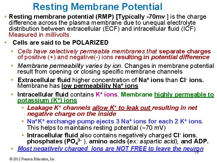
Animation- Muscle cell stimulus transport – compare to nerve stimulus below http: //media. pearsoncmg. com/bc/bc_0 media_ap/apflix/ap/ap_video_player. html? ecc Animation: Nerve stimulus https: //www. youtube. com/watch? v=b 2 ct. Es. GEpe 0 © 2012 Pearson Education, Inc. 20
21 © 2012 Pearson Education, Inc.
http: //academic. uprm. edu/~ephoebus/070 a 63 d 0. gif © 2012 Pearson Education, Inc. 22
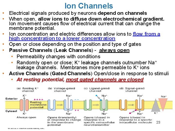
Ion Channels • Electrical signals produced by neurons depend on channels • When open, allow ions to diffuse down electrochemical gradient. Ion movement causes flow of electrical current that can change the membrane potential. • Ion concentration and electric differences allow ions to flow from a high concentration to a lower concentration • Open or close depending on the position and type of gates • Passive Channels (Leak Channels) - always open • Permeability changes with conditions • Randomly open or close; K+ leakage channels outnumber Na+ leakage channels. Membranes more permeable to K+ ions • Active Channels (Gated Channels) Open/close in response to stimuli • At resting potential, most gated channels are closed 23 © 2012 Pearson Education, Inc.
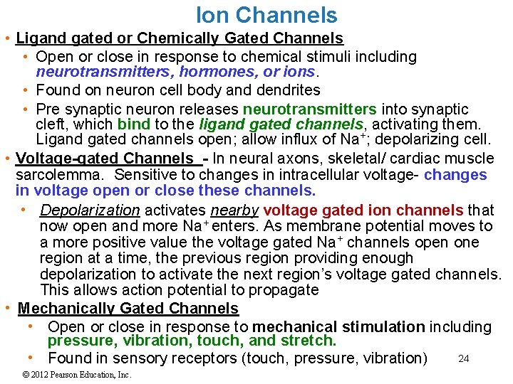
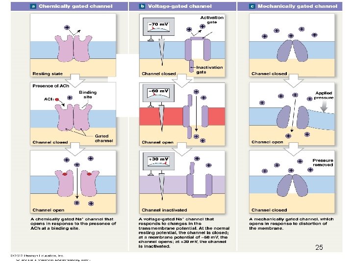
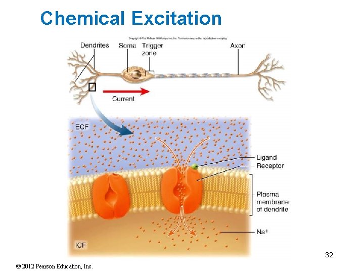
Ion Channels • Ligand gated or Chemically Gated Channels • Open or close in response to chemical stimuli including neurotransmitters, hormones, or ions. • Found on neuron cell body and dendrites • Pre synaptic neuron releases neurotransmitters into synaptic cleft, which bind to the ligand gated channels, activating them. Ligand gated channels open; allow influx of Na+; depolarizing cell. • Voltage-gated Channels - In neural axons, skeletal/ cardiac muscle sarcolemma. Sensitive to changes in intracellular voltage- changes in voltage open or close these channels. • Depolarization activates nearby voltage gated ion channels that now open and more Na+ enters. As membrane potential moves to a more positive value the voltage gated Na+ channels open one region at a time, the previous region providing enough depolarization to activate the next region’s voltage gated channels. This allows action potential to propagate • Mechanically Gated Channels • Open or close in response to mechanical stimulation including pressure, vibration, touch, and stretch. 24 • Found in sensory receptors (touch, pressure, vibration) © 2012 Pearson Education, Inc.
25 © 2012 Pearson Education, Inc.
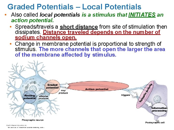
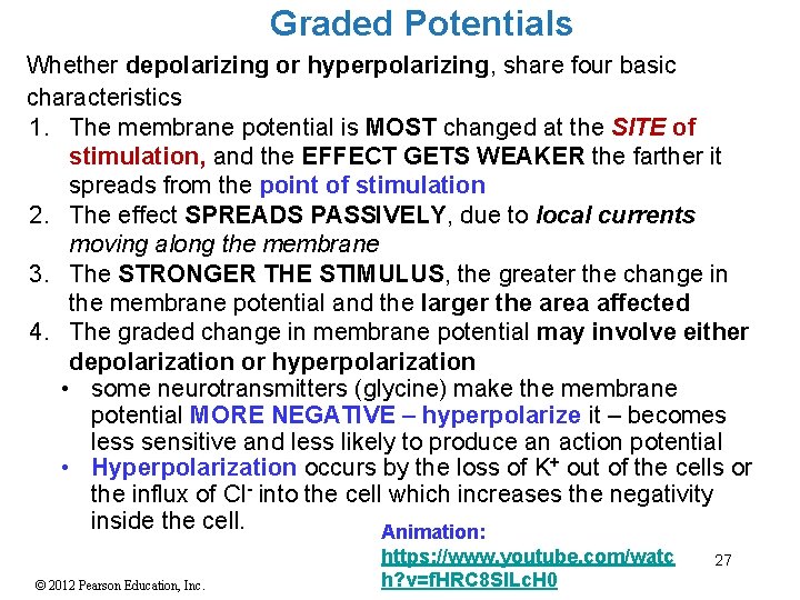
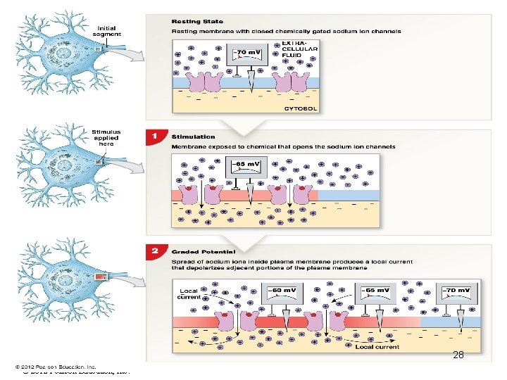
Graded Potentials – Local Potentials • Also called local potentials is a stimulus that INITIATES an action potential. • Spreads/travels a short distance from site of stimulation then dissipates. Distance traveled depends on the number of sodium channels open. • Change in membrane potential is proportional to strength of stimulus. The more channels that open the larger the area of the membrane affected by stimulus. 26 © 2012 Pearson Education, Inc.
Graded Potentials Whether depolarizing or hyperpolarizing, share four basic characteristics 1. The membrane potential is MOST changed at the SITE of stimulation, and the EFFECT GETS WEAKER the farther it spreads from the point of stimulation 2. The effect SPREADS PASSIVELY, due to local currents moving along the membrane 3. The STRONGER THE STIMULUS, the greater the change in the membrane potential and the larger the area affected 4. The graded change in membrane potential may involve either depolarization or hyperpolarization • some neurotransmitters (glycine) make the membrane potential MORE NEGATIVE – hyperpolarize it – becomes less sensitive and less likely to produce an action potential • Hyperpolarization occurs by the loss of K+ out of the cells or the influx of Cl- into the cell which increases the negativity inside the cell. Animation: © 2012 Pearson Education, Inc. https: //www. youtube. com/watc h? v=f. HRC 8 Sl. Lc. H 0 27
28 © 2012 Pearson Education, Inc.
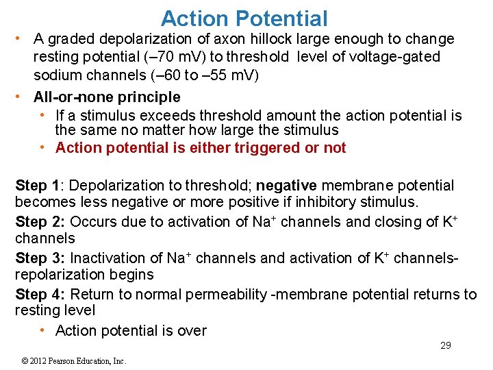
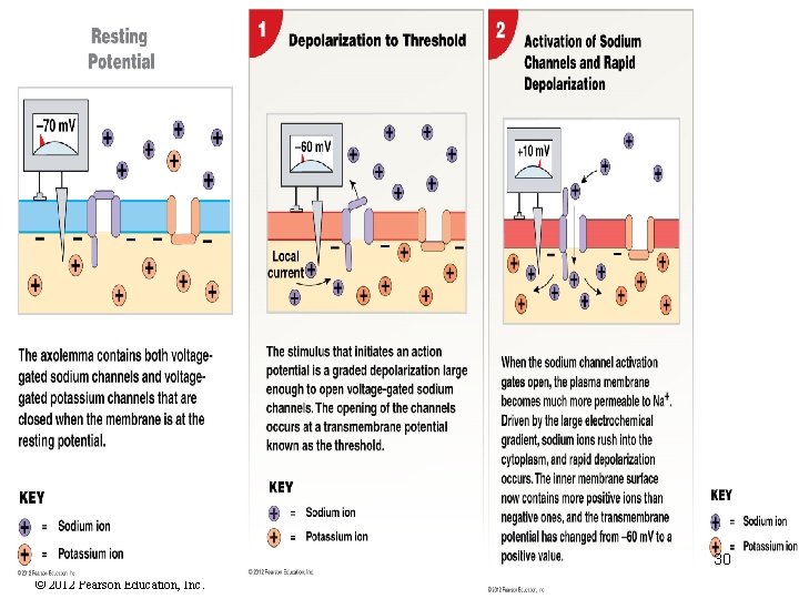
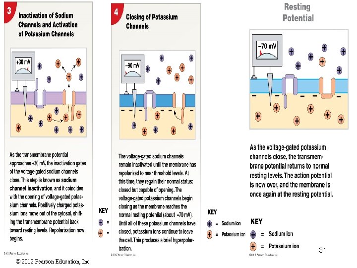
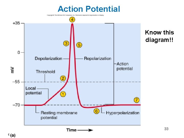
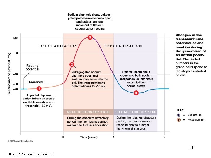
Action Potential • A graded depolarization of axon hillock large enough to change resting potential (– 70 m. V) to threshold level of voltage-gated sodium channels (– 60 to – 55 m. V) • All-or-none principle • If a stimulus exceeds threshold amount the action potential is the same no matter how large the stimulus • Action potential is either triggered or not Step 1: Depolarization to threshold; negative membrane potential becomes less negative or more positive if inhibitory stimulus. Step 2: Occurs due to activation of Na channels and closing of K channels Step 3: Inactivation of Na channels and activation of K channelsrepolarization begins Step 4: Return to normal permeability -membrane potential returns to resting level • Action potential is over 29 © 2012 Pearson Education, Inc.
30 © 2012 Pearson Education, Inc.
31 © 2012 Pearson Education, Inc.
Chemical Excitation 32 © 2012 Pearson Education, Inc.
Action Potential Know this diagram!! 33 © 2012 Pearson Education, Inc.
34 © 2012 Pearson Education, Inc.
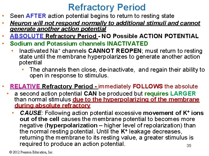
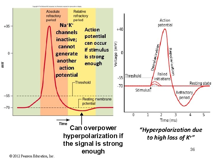
Refractory Period • Seen AFTER action potential begins to return to resting state • Neuron will not respond normally to additional stimuli and cannot generate another action potential • ABSOLUTE Refractory Period - NO Possible ACTION POTENTIAL • Sodium and Potassium channels INACTIVATED • Inactivated Na+ channels CANNOT REOPEN; must return to resting state until the membrane hyperpolarizes to generate another action potential • The channels then close, de-inactivate, and regain their ability to open in response to stimulus. • RELATIVE Refractory Period - immediately FOLLOWS the absolute • a second action potential CAN be produced but requires LARGER than normal stimulus due to the hyperpolarizing of the membrane during absolute refractory • CAUSE: Following action potential excessive movement of K+ ions out of the cell causes the membrane potential to becomes more negative (hyperpolarization – higher level of repolarization) than the normal resting potential. Until the K+ leakage decreases, returning the membrane to its resting value, a greater stimulus is required to produce an action potential. 35 © 2012 Pearson Education, Inc.
Na+K+ channels inactive; cannot generate another action potential © 2012 Pearson Education, Inc. Action potential can occur if stimulus is strong enough Can overpower hyperpolarization if the signal is strong enough “Hyperpolarization due to high loss of K+” 36
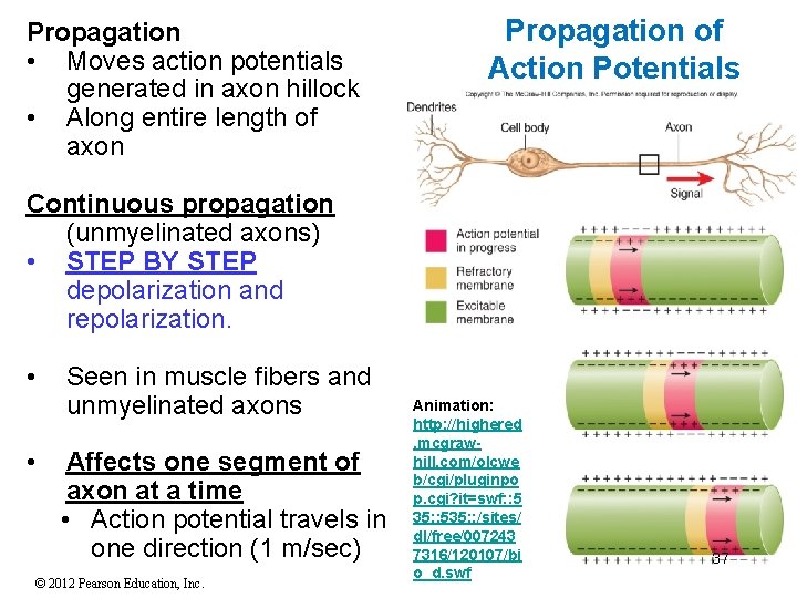
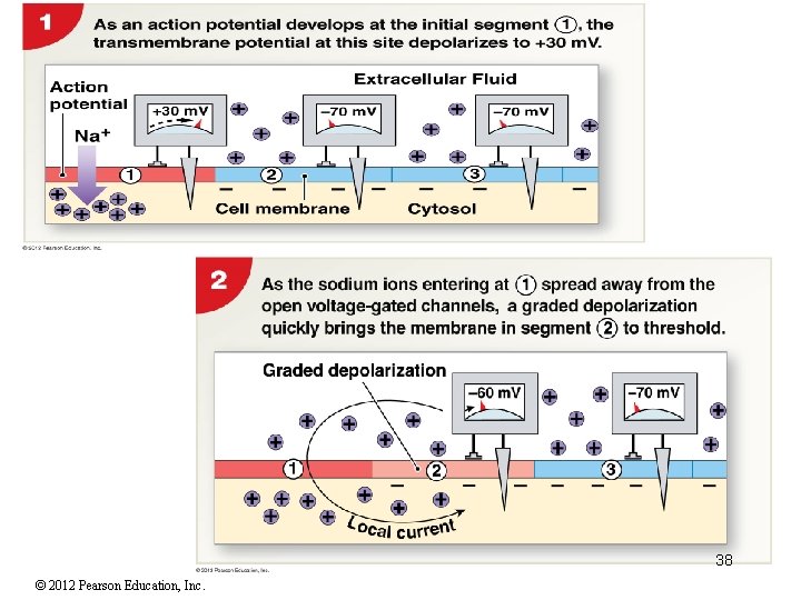
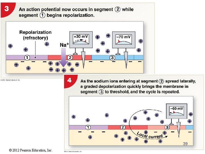
Propagation • Moves action potentials generated in axon hillock • Along entire length of axon Propagation of Action Potentials Continuous propagation (unmyelinated axons) • STEP BY STEP depolarization and repolarization. • • Seen in muscle fibers and unmyelinated axons Affects one segment of axon at a time • Action potential travels in one direction (1 m/sec) © 2012 Pearson Education, Inc. Animation: http: //highered. mcgrawhill. com/olcwe b/cgi/pluginpo p. cgi? it=swf: : 5 35: : 535: : /sites/ dl/free/007243 7316/120107/bi o_d. swf 37
38 © 2012 Pearson Education, Inc.
39 © 2012 Pearson Education, Inc.
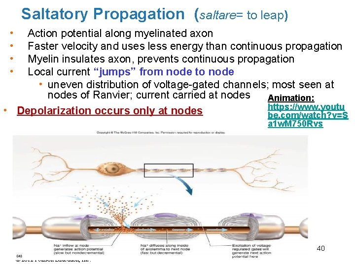
Saltatory Propagation (saltare= to leap) • • Action potential along myelinated axon Faster velocity and uses less energy than continuous propagation Myelin insulates axon, prevents continuous propagation Local current “jumps” from node to node • uneven distribution of voltage-gated channels; most seen at nodes of Ranvier; current carried at nodes Animation: https: //www. youtu • Depolarization occurs only at nodes be. com/watch? v=S a 1 w. M 750 Rvs 40 © 2012 Pearson Education, Inc.
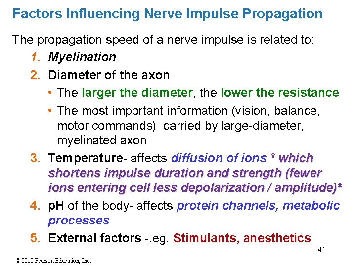
Factors Influencing Nerve Impulse Propagation The propagation speed of a nerve impulse is related to: 1. Myelination 2. Diameter of the axon • The larger the diameter, the lower the resistance • The most important information (vision, balance, motor commands) carried by large-diameter, myelinated axon 3. Temperature- affects diffusion of ions * which shortens impulse duration and strength (fewer ions entering cell less depolarization / amplitude)* 4. p. H of the body- affects protein channels, metabolic processes 5. External factors -. eg. Stimulants, anesthetics 41 © 2012 Pearson Education, Inc.
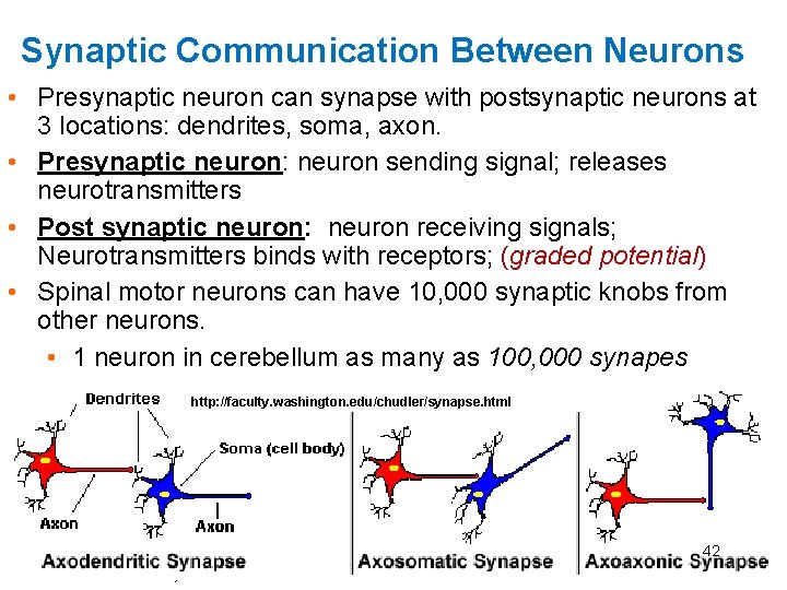
Synaptic Communication Between Neurons • Presynaptic neuron can synapse with postsynaptic neurons at 3 locations: dendrites, soma, axon. • Presynaptic neuron: neuron sending signal; releases neurotransmitters • Post synaptic neuron: neuron receiving signals; Neurotransmitters binds with receptors; (graded potential) • Spinal motor neurons can have 10, 000 synaptic knobs from other neurons. • 1 neuron in cerebellum as many as 100, 000 synapes http: //faculty. washington. edu/chudler/synapse. html 42 © 2012 Pearson Education, Inc.
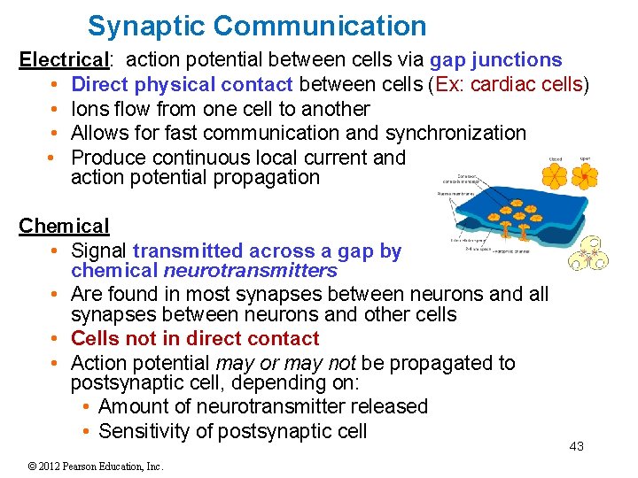
Synaptic Communication Electrical: action potential between cells via gap junctions • Direct physical contact between cells (Ex: cardiac cells) • Ions flow from one cell to another • Allows for fast communication and synchronization • Produce continuous local current and action potential propagation Chemical • Signal transmitted across a gap by chemical neurotransmitters • Are found in most synapses between neurons and all synapses between neurons and other cells • Cells not in direct contact • Action potential may or may not be propagated to postsynaptic cell, depending on: • Amount of neurotransmitter released • Sensitivity of postsynaptic cell © 2012 Pearson Education, Inc. 43
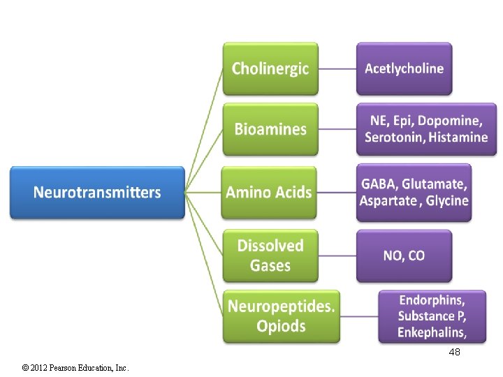
Chemical Synapse • The synaptic terminal releases a neurotransmitter that binds to the postsynaptic plasma membrane • Produces temporary, localized change in permeability or function of postsynaptic cell • Over 100 substances known as neurotransmitters • Types of neurotransmitters released by neurons • Cholinergic, Biogenic amines, Amino acids, Dissolved gases • Neuropeptides 1. EXCITATORY neurotransmitters • Cause depolarization of postsynaptic membranes • Promote action potentials 2. INHIBITORY neurotransmitters • Cause hyperpolarization of postsynaptic membranes • Suppress action potentials 44 © 2012 Pearson Education, Inc.
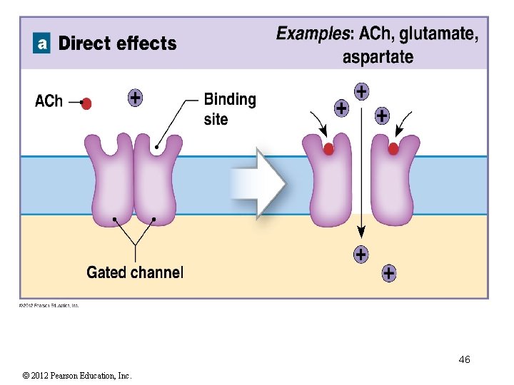
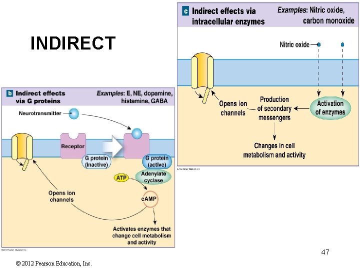
Function of Neurotransmitters and Neuromodulators Neurotransmitters promote the opening of channels. They are classified as either direct or indirect dependent on their method of activation of these channels 1. Direct effects open/close membrane channel • Ex: ACh, glycine, aspartate • Ionotropic effecs –alter ion movement 2. Indirect effects work through intermediate substances known as second messengers • G proteins – an enzyme complex coupled to the membrane receptor that binds GTP (high-energy compound) • Link between neurotransmitter (first messenger) and second messenger • Activate enzyme adenylate cyclase • Which produces second messenger cyclic-AMP (c. AMP) • Ex: E, NE, dopamine, histamine, GABA • Indirect effects via intracellular enzymes • For example, lipid-soluble gases (NO, CO) 45 © 2012 Pearson Education, Inc.
46 © 2012 Pearson Education, Inc.
INDIRECT 47 © 2012 Pearson Education, Inc.
48 © 2012 Pearson Education, Inc.
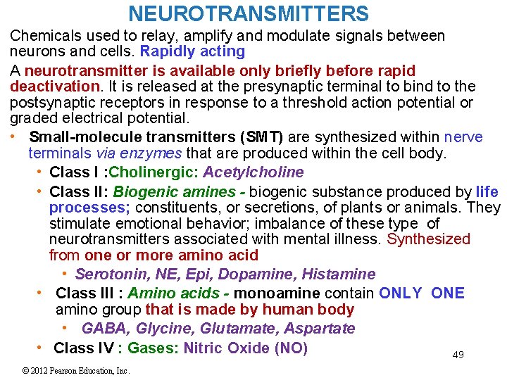
NEUROTRANSMITTERS Chemicals used to relay, amplify and modulate signals between neurons and cells. Rapidly acting A neurotransmitter is available only briefly before rapid deactivation. It is released at the presynaptic terminal to bind to the postsynaptic receptors in response to a threshold action potential or graded electrical potential. • Small-molecule transmitters (SMT) are synthesized within nerve terminals via enzymes that are produced within the cell body. • Class I : Cholinergic: Acetylcholine • Class II: Biogenic amines - biogenic substance produced by life processes; constituents, or secretions, of plants or animals. They stimulate emotional behavior; imbalance of these type of neurotransmitters associated with mental illness. Synthesized from one or more amino acid • Serotonin, NE, Epi, Dopamine, Histamine • Class III : Amino acids - monoamine contain ONLY ONE amino group that is made by human body • GABA, Glycine, Glutamate, Aspartate • Class IV : Gases: Nitric Oxide (NO) 49 © 2012 Pearson Education, Inc.
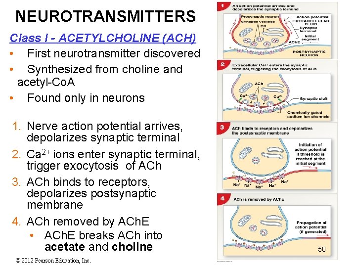
NEUROTRANSMITTERS Class I - ACETYLCHOLINE (ACH) • First neurotransmitter discovered • Synthesized from choline and acetyl-Co. A • Found only in neurons 1. Nerve action potential arrives, depolarizes synaptic terminal 2. Ca 2+ ions enter synaptic terminal, trigger exocytosis of ACh 3. ACh binds to receptors, depolarizes postsynaptic membrane 4. ACh removed by ACh. E • ACh. E breaks ACh into acetate and choline © 2012 Pearson Education, Inc. 50
Class II SMT Neurotransmitters - BIOGENIC AMINES NOREPINEPHRINE (NE) arousal and concentrated attention; and mood-feeling good [limbic system] • Indirect-2 nd messenger; Synthesized from amino acid tyrosine • Excitatory / inhibitory depending on receptors • Widely distributed in brain; portions of ANS only in neurons DOPAMINE: (Prolactin Inhibiting Hormone) emotional responses, pleasure experiences; skeletal muscle tone, cognitive attention; addictions; synthesized from amino acid tyrosine • Kidneys increase Na+ secretion; pancreas reduces insulin release • A CNS neurotransmitter- found only in neurons • Indirect- 2 nd messenger; May be excitatory or inhibitory • Involved in Parkinson’s disease • Cocaine and methamphetamine, act directly on increasing the levels of dopamine; Overproduction responsible for schizophrenia SEROTONIN: concentrated in neurons in the raphe nucleus. Involved in temperature regulation, mood and appetite, sleep, sexuality. Affects attention and emotional states • Generally inhibitory 51 • Synthesized from the amino acid TRYPTOPHAN © 2012 Pearson Education, Inc.
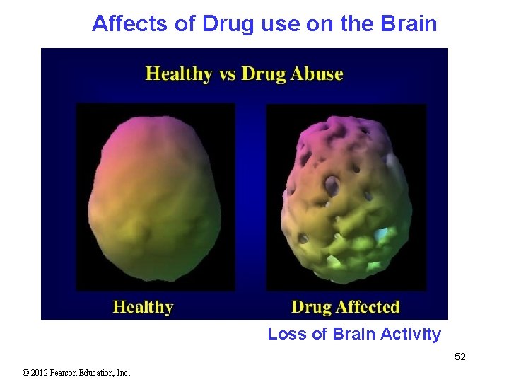
Affects of Drug use on the Brain Loss of Brain Activity 52 © 2012 Pearson Education, Inc.
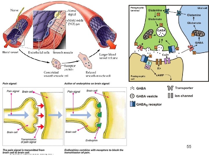
Small Molecule Transmitters - AMINO ACIDS § Gamma Aminobutyric Acid (GABA) cause hyperpolarization resulting in inhibitory; in chemical terms classified as amino acid. Primary inhibitory neurotransmitter in the CNS • Direct • Binds to chloride channels and allows Cl- ions in or K+ ions out of neuron, moving potential away from threshold to more negative levels and less likely to generate action potential • GABA found only in the CNS • Used to treat epilepsy; primarily responsible for muscle tone § Glycine- inhibitory • 2 nd messenger • Strychnine poison blocks glycine receptors resulting in uncontrolled convulsions and respiratory arrest § Glutamate- excitatory; seen in most excitatory neurons in the CNS--half of synapses in brain; inhibitory in night vision • Direct § Aspartate- excitatory stimulates glutamate receptors strengthens or weakens synaptic activity, has a role in memory function. 53 © 2012 Pearson Education, Inc.
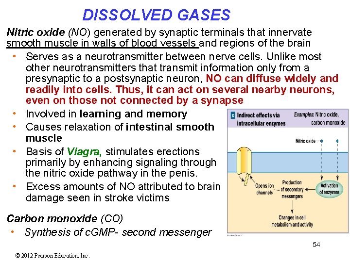
DISSOLVED GASES Nitric oxide (NO) generated by synaptic terminals that innervate smooth muscle in walls of blood vessels and regions of the brain • Serves as a neurotransmitter between nerve cells. Unlike most other neurotransmitters that transmit information only from a presynaptic to a postsynaptic neuron, NO can diffuse widely and readily into cells. Thus, it can act on several nearby neurons, even on those not connected by a synapse • Involved in learning and memory • Causes relaxation of intestinal smooth muscle • Basis of Viagra, stimulates erections primarily by enhancing signaling through the nitric oxide pathway in the penis. • Excess amounts of NO attributed to brain damage seen in stroke victims Carbon monoxide (CO) • Synthesis of c. GMP- second messenger 54 © 2012 Pearson Education, Inc.
55 © 2012 Pearson Education, Inc.
Neuropeptides NEUROPEPTIDES Similar in function to neurotransmitters • Neuropeptides are secreted primarily from neurons but also glia for some peptides and signal to neighboring cells (primarily neurons). • Released by nerve endings and their effect sometimes occurs quite far from the releasing neuron. • Not rapidly degraded or taken up, so their active time is not limited as in neurotransmitters. Much LARGER = 3 -40 amino acids in length synthesized within the cell body; transported along axon to nerve terminal within vesicles • Peptides are NOT RECYCLED back into the cell once secreted, unlike many conventional neurotransmitters • Neuromodulators bind to receptors; activate enzymes • Many work as hormones • Can be excitatory or inhibitory • involved in a wide range of brain functions, including analgesia, reward, food intake, metabolism, reproduction, social behaviors, learning and memory 56 © 2012 Pearson Education, Inc.
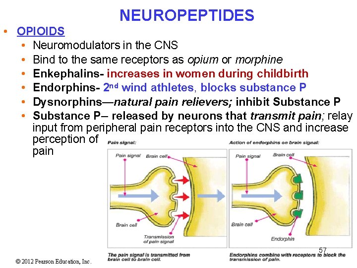
NEUROPEPTIDES • OPIOIDS • Neuromodulators in the CNS • Bind to the same receptors as opium or morphine • Enkephalins- increases in women during childbirth • Endorphins- 2 nd wind athletes, blocks substance P • Dysnorphins—natural pain relievers; inhibit Substance P • Substance P-- released by neurons that transmit pain; relay input from peripheral pain receptors into the CNS and increase perception of pain 57 © 2012 Pearson Education, Inc.
Information Processing Postsynaptic Potentials • Graded potentials developed in a postsynaptic cell • In response to neurotransmitters Two Types of Postsynaptic Potentials 1. Excitatory postsynaptic potential (EPSP) • Graded depolarization of postsynaptic membrane • any voltage change that shifts membrane potential closer to threshold and makes a neuron more likely to fire 2. Inhibitory postsynaptic potential (IPSP) • Graded hyperpolarization of postsynaptic membrane • any voltage change away from threshold that makes a neuron less likely to fire 58 © 2012 Pearson Education, Inc.
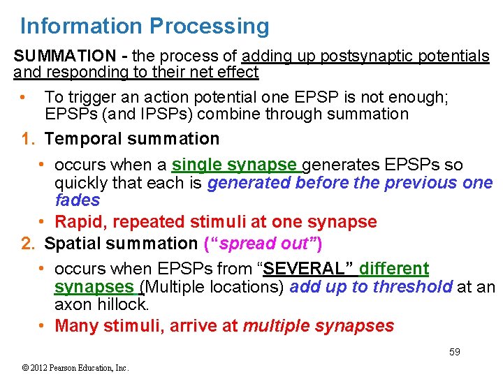
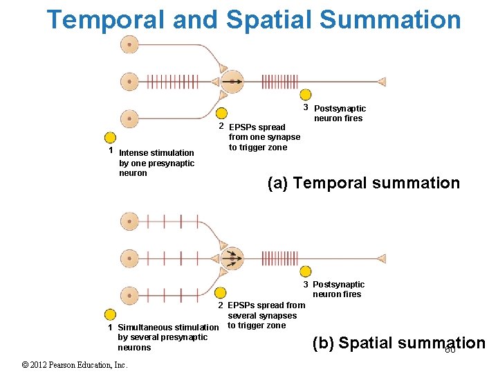
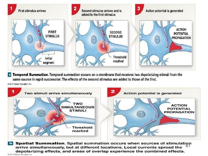
Information Processing SUMMATION - the process of adding up postsynaptic potentials and responding to their net effect • To trigger an action potential one EPSP is not enough; EPSPs (and IPSPs) combine through summation 1. Temporal summation • occurs when a single synapse generates EPSPs so quickly that each is generated before the previous one fades • Rapid, repeated stimuli at one synapse 2. Spatial summation (“spread out”) • occurs when EPSPs from “SEVERAL” different synapses (Multiple locations) add up to threshold at an axon hillock. • Many stimuli, arrive at multiple synapses 59 © 2012 Pearson Education, Inc.
Temporal and Spatial Summation 1 Intense stimulation by one presynaptic neuron 2 EPSPs spread from one synapse to trigger zone 3 Postsynaptic neuron fires (a) Temporal summation 3 Postsynaptic neuron fires 2 EPSPs spread from several synapses 1 Simultaneous stimulation to trigger zone by several presynaptic neurons (b) Spatial summation 60 © 2012 Pearson Education, Inc.
61 © 2012 Pearson Education, Inc.
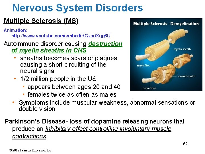
Nervous System Disorders Multiple Sclerosis (MS) Animation: http: //www. youtube. com/embed/KGzsri. Xqg 6 U Autoimmune disorder causing destruction of myelin sheaths in CNS • sheaths becomes scars or plaques causing a short circuiting of the neural signal • 1/2 million people in the US • appears between ages 20 and 40 • females twice as often as males • Symptoms include muscular weakness, abnormal sensations or double vision Parkinson’s Disease- loss of dopamine releasing neurons that produce an inhibitory effect controlling involuntary muscle contractions 62 © 2012 Pearson Education, Inc.
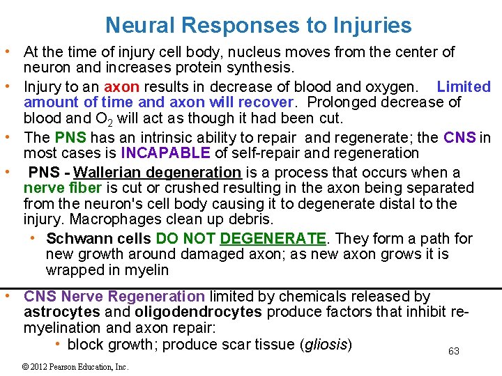
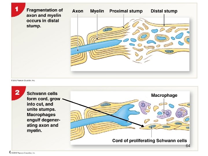
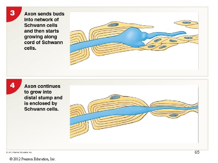
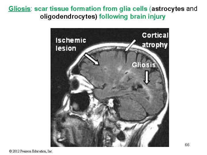
Neural Responses to Injuries • At the time of injury cell body, nucleus moves from the center of neuron and increases protein synthesis. • Injury to an axon results in decrease of blood and oxygen. Limited amount of time and axon will recover. Prolonged decrease of blood and O 2 will act as though it had been cut. • The PNS has an intrinsic ability to repair and regenerate; the CNS in most cases is INCAPABLE of self-repair and regeneration • PNS - Wallerian degeneration is a process that occurs when a nerve fiber is cut or crushed resulting in the axon being separated from the neuron's cell body causing it to degenerate distal to the injury. Macrophages clean up debris. • Schwann cells DO NOT DEGENERATE. They form a path for new growth around damaged axon; as new axon grows it is wrapped in myelin • CNS Nerve Regeneration limited by chemicals released by astrocytes and oligodendrocytes produce factors that inhibit remyelination and axon repair: • block growth; produce scar tissue (gliosis) 63 © 2012 Pearson Education, Inc.
64 © 2012 Pearson Education, Inc.
65 © 2012 Pearson Education, Inc.
Gliosis: scar tissue formation from glia cells (astrocytes and oligodendrocytes) following brain injury 66 © 2012 Pearson Education, Inc.