Chapter 12 Lecture Slides Copyright The Mc GrawHill


 • Myocardium: thick, middle layer composed")

 - right atrium")


: - AV valve between LA and LV - 2 cusps")


 that receive blood from lungs •")


- Slides: 58
Chapter 12 Lecture Slides Copyright © The Mc. Graw-Hill Companies, Inc. Permission required for reproduction or display.
Functions 1. 2. 3. 4. Regulates blood supply Generates blood pressure Routes blood Ensures 1 way blood flow
Heart Characteristics • Size: size of a fist and weighs less than 1 lb. • Location: between lungs in thoracic cavity • Orientation: apex (bottom) towards left side 3
Heart Coverings • Pericardium: double-layered sac that anchors and protects heart • Parietal pericardium: membrane around heart’s cavity • Visceral pericardium: membrane on heart’s surface • Pericardial cavity: space around heart 4
Heart Layers • Epicardium: surface of heart (outside) • Myocardium: thick, middle layer composed of cardiac muscle • Endocardium: smooth, inner surface 5
Cardiac Muscle • 1 centrally located nucleus • Branching cells • Rich in mitochondria • Striated (actin and myosin) • Ca 2+ and ATP used for contractions • Intercalated disks connect cells 6
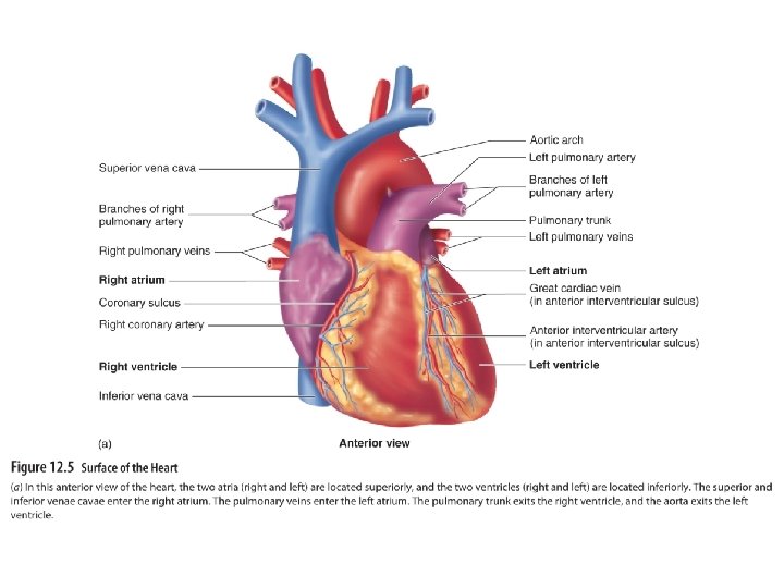
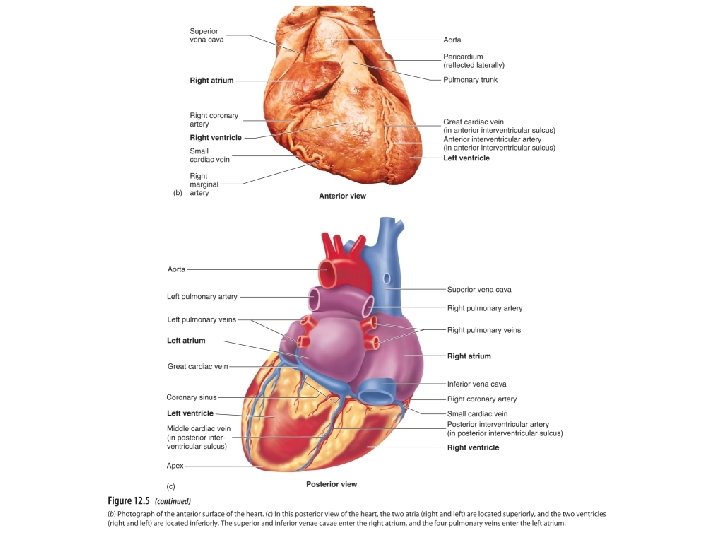
Chambers and Blood Vessels • 4 Chambers: - left atrium (LA) - right atrium (RA) - left ventricle (LV) - right ventricle (RV) • Coronary sulcus: separates atria from ventricles 7
Atria • • Upper portion Holding chambers Small, thin walled Contract minimally to push blood into ventricles • Interatrial septum: separates right and left atria 8
Ventricles • • Lower portion Pumping chambers Thick, strong walled Contract forcefully to propel blood out of heart • Interventricular septum: separates right and left ventricles 9
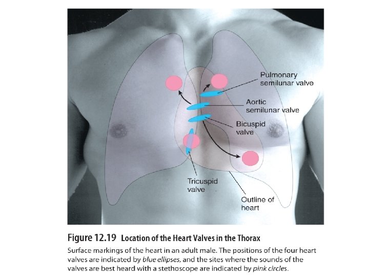
Valves • What are they? structures that ensure 1 way blood flow • Atrioventricular valves (AV): between atria and ventricles - Tricuspid valve: - AV valve between RA and RV - 3 cusps 11
- Bicuspid valve (mitral): - AV valve between LA and LV - 2 cusps • Chordae tendineae: - attached to AV valve flaps - support valves 12
• Semilunar valves: - Pulmonary: base of pulmonary trunk - Aortic: base of aorta 13
What happens when Bicuspid Valve is Open? • Blood flows from LA into LV. • Aortic semilunar valve is closed. • Tension on chordae tendineae is low. 15
What happens when Bicuspid Valve is Closed? • Blood flows from LV into aorta. • Aortic semilunar valve is open. • Tension on chordae tendineae is high. 16
Right Side of Heart • Pulmonary circuit: - carries blood from heart to lungs - blood is O 2 poor, CO 2 rich 18
• Right Atrium: - receives blood from 3 places: superior and inferior vena cava and coronary sinus - Superior vena cava: drains blood above diaphragm (head, neck, thorax, upper limbs) - Inferior vena cava: drains blood below diaphragm (abdominopelvic cavity and lower limbs) - coronary sinus: drains blood from myocardium 19
• Right Ventricle: - opens into pulmonary trunk - Pulmonary trunk: splits into right and left pulmonary arteries - Pulmonary arteries: carry blood away from heart to lungs 20
Left Side of Heart • Systemic circuit: - carries blood from heart to body - blood is O 2 rich, CO 2 poor 21
• Left Atrium: 4 openings (pulmonary veins) that receive blood from lungs • Left Ventricle: - opens into aorta - thicker, contracts more forcefully, higher blood pressure than right ventricle has to get to body • Aorta: carries blood from LV to body 22
Figure 12. 5 a
25
Blood Flow through Heart 1. RA 2. Tricuspid valve 3. RV 4. Pulmonary semilunar valve 5. Pulmonary trunk 6. Pulmonary arteries 7. Lungs 8. Pulmonary veins 9. LA 10. Bicuspid valve 11. LV 12. Aortic semilunar valve 13. Aorta 14. Body 26
27
Blood Supply to Heart • Coronary arteries: - supply blood to heart wall - originate from base of aorta (above aortic semilunar valve) • Left coronary artery: - has 3 branches - supply blood to anterior heart wall and left ventricle 28
• Right coronary artery: - originates on right side of aorta - supply blood to right ventricle 29
Figure 12. 11
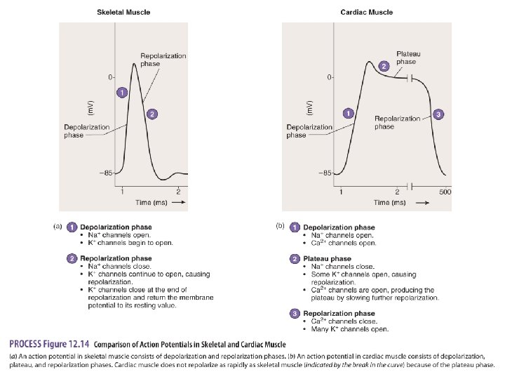
Action Potentials in Cardiac Muscle • Changes in membrane channels’ permeability are responsible for producing action potentials and is called pacemaker potential. 1. Depolarization phase: - Na+ channels open - Ca 2+ channels open 2. Plateau phase: - Na+ channels close - Some K+ channels open 31 - Ca 2+ channels remain open
3. Repolarization phase: - K+ channels are open - Ca 2+ channels close • Plateau phase prolongs action potential by keeping Ca 2+ channels open. • In skeletal muscle action potentials take 2 msec, in cardiac muscle they take 200 -500 msec. 32
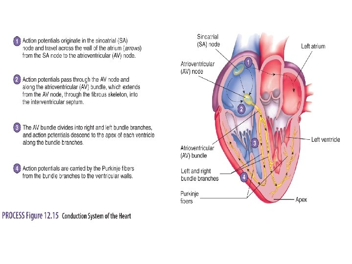
Conduction System of Heart • What is it? contraction of atria and ventricles by cardiac muscle cells • Sinoatrial node (SA node): - in RA - where action potential originates - functions as pacemaker - large number of Ca 2+ channels 34
Path of Action Potential through Heart 1. 2. 3. 4. 5. SA node AV node (atrioventricular) AV bundle Right and Left Bundle branches Purkinje fibers 35
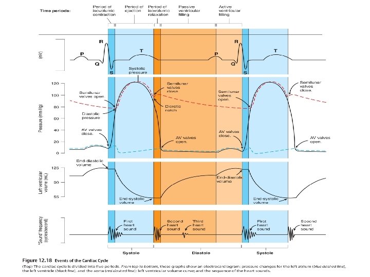
Electrocardiogram • What is it? - record of electrical events in heart - diagnoses cardiac abnormalities - uses electrodes - contains P wave, QRS complex, T wave 37
Components of ECG/EKG • P wave: depolarization of atria • QRS complex: - depolarization of ventricles - contains Q, R, S waves • T wave: repolarization of ventricles 38
Figure 12. 16
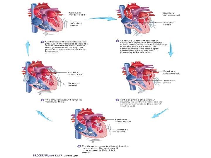
Cardiac Cycle • Heart is 2 side by side pumps: right and left • Atria: primers for pumps • Ventricles: power pumps • Cardiac Cycle: repetitive pumping action which includes contraction and relaxation 40
• Cardiac muscle contractions produce pressure changes within heart chambers. • Pressure changes are responsible for blood movement. • Blood moves from areas of high to low pressure. 41
• Atrial systole: contraction of atria • Ventricular systole: contraction of ventricles • Atrial diastole: relaxation of atria • Ventricular diastole: relaxation of ventricles 42
Heart Sounds • Stethoscope is used to hear lung and heart sounds • First sound is lubb, second is dupp • Sounds result from opening and closing valves • Murmurs are due to faulty valves 45
Regulation of Heart Function • Stroke Volume: - volume of blood pumped per ventricle per contraction - 70 ml/beat • Heart Rate: - number of heart beats in 1 min. - 72 beats/min. • Cardiac Output: - volume of blood pumped by a ventricle in 1 min. - 5 L/min. CO = SV x HR 47
Intrinsic Regulation of Heart • What is it? mechanisms contained within heart • Venous return: amt. of blood that returns to heart • Preload: degree ventricular walls are stretched at end of diastole
• Venous return, preload, stroke volume are related to each other • Starlings Law of the Heart: - relationship between preload and stroke volume - influences cardiac output - Ex. Exercise increases venous return, preload, stroke volume, and cardiac output • After load: pressure against which ventricles must pump blood
Extrinsic Regulation of Heart • What is it? - mechanisms external to heart - nervous or chemical regulation
Figure 12. 20
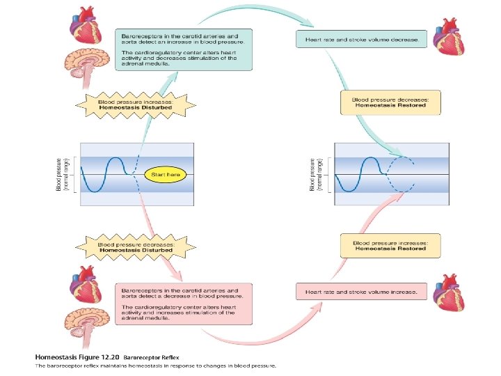
Nervous Regulation: Baroreceptor Reflex • What is it? - mechanism of nervous system which regulates heart function - keeps heart rate and stroke volume in normal range - baroreceptors monitor blood pressure in aorta and carotid arteries (carry blood to brain) - changes in blood pressure cause changes in frequency of action potentials 52 - involves medulla oblongata
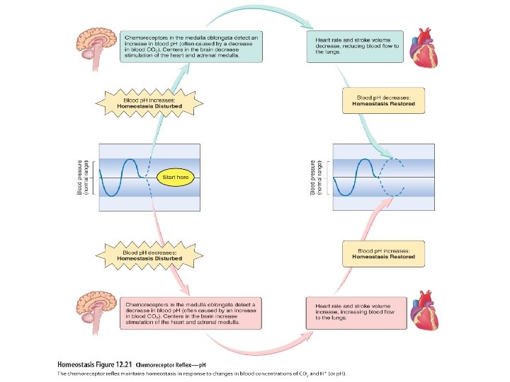
Chemical Regulation: Chemoreceptor Reflex • What is it? - chemicals can affect heart rate and stroke volume - epinephrine and norepinephrine from adrenal medulla can increase heart rate and stroke volume - excitement, anxiety, anger an increase cardiac output - depression can decrease cardiac output
- medulla oblongata has chemoreceptors for changes in p. H and CO 2 - K+, Ca 2+, and Na+ affect cardiac function
Heart Attack • Thrombus: - blood clot blocks coronary blood vessel causes heart attack - daily aspirin can prevent by thinning blood • Infarct: area that dies from lack of O 2
Heart Procedures • Angioplasty: procedure opens blocked blood vessels • Stent: structures inserted to keep vessels open • Bypass: procedure reroutes blood away from blocked arteries