Chapter 12 Cell Cycle Control Cancer Part 3
 AP Biology Ms. Day")
 l. Reproduce by a type")


 http: //www.")

 CYCLIN + ACTIVE FORM ATP CDK/CYCLIN COMPLEX Pi")

 l Type of protein & are substrates ¡Many different")
 of enzyme (a protein, too ) l Cyclin-dependent kinases (Cdks) ¡Many")

 cyclins? ¡ Proteolytic l Break enzymes (proteins) down/degrade cyclins cause")


 ¡ ¡ ¡ If cell doesn’t “pass” checkpoint, it goes")


 securin --| separase breaks down cohesins ¡ Also called")

, and")
 and external (outside the")


 Cells anchor to dish surface and divide (anchorage dependence). When cells have formed")


 ¡")


 and Metatasis http: //www. hhmi. org/biointeractive /media/angiogenesis-lg.")
- Slides: 40
Chapter 12 Cell Cycle Control & Cancer (Part 3) AP Biology Ms. Day
Another Type of Cell Division: Binary Fission ¡Prokaryotes (bacteria) l. Reproduce by a type of cell division called binary fission
In binary fission, ¡ The bacterial chromosome replicates ¡ The two daughter chromosomes move apart ¡ Origin of replication Cell wall Plasma Membrane E. coli cell 1 Chromosome replication begins. Soon thereafter, one copy of the origin moves rapidly toward the other end of the cell. 2 Replication continues. One copy of the origin is now at each end of the cell. 3 Replication finishes. The plasma membrane grows inward, and new cell wall is deposited. Figure 12. 11 4 Two daughter cells result. Two copies of origin Origin Bacterial Chromosome Origin
The cell cycle is HIGHLY regulated ¡ The frequency of cell division l Varies with the type of cell ¡ These cell cycle differences l Result from regulation at the molecular level l l http: //highered. mcgrawhill. com/sites/0072495855/student_view 0/chapter 2/animati on__how_the_cell_cycle_works. html https: //www. youtube. com/watch? v=p. Os. Ab. Ti 9 t. Hw
Cell Cycle Checkpoints ¡ The clock has specific checkpoints l l l ¡ a critical control point stop and “go-ahead” signals regulate cycle signals report whether crucial cellular processes up to that specific point have been completed and completed correctly There are 3 checkpoints l G 1 checkpoint l G 2 Checkpoint l M checkpoint
The Cell Cycle Control System G 1 checkpoint Control system S G 1 M Figure 12. 14 M checkpoint G 2 checkpoint
G 1 Checkpoint = most critical l called the “restriction point” G 0 G 1 checkpoint G 1 If a cell receives a go-ahead signal at the G 1 checkpoint, the cell continues on in cell cycle. G 1 If a cell does not receive a go-ahead signal at the G 1 checkpoint, cell exits the cell cycle and goes into G 0, a nondividing state.
• Chromosomes are lined up in the middle properly (replicated DNA) http: //www. cellsal ive. com/cell_cycl e. htm (original DNA) http: //highered. mcgrawhill. com/sites/0072495855/student_view 0/chapter 2/animation__control_of_the_cell_cycle. html
The Cell Cycle Clock: Cyclins and Cyclin-Dependent Kinases types of regulatory proteins in cytoplasm are involved in cell cycle control l Cyclins l cyclin-dependent kinases (Cdks) ¡ Two
INACTIVE FORM CYCLIN DEPENDENT KINASE (CDK) CYCLIN + ACTIVE FORM ATP CDK/CYCLIN COMPLEX Pi It gets PHOSPHORYLATED BY ATP!!!
Active vs. Inactive? ? ¡ What happens when cyclins and cdks are in the ACTIVE form? l ¡ can Cells pass through the cell cycle to the NEXT phase What happens when cyclins and cdks are in the INACTIVE form? l can NOT Cells pass through the cell cycle to the NEXT phase
Cyclins ¡ Cyclins (activate Cdk’s) l Type of protein & are substrates ¡Many different types! *Cyclin levels in the cell rise and fall (“cycle”) with the stages of the cell cycle.
Cyclin-dependent kinases (Cdks) of enzyme (a protein, too ) l Cyclin-dependent kinases (Cdks) ¡Many different types ¡ Type *Cdk levels remain constant but mostly inactive *Cdks add phosphate groups to variety of protein substrates that control processes in the cell cycle.
cyclin degrades & breaks apart “OFF” RED LIGHT cyclin degrades & breaks apart
What degrades (breaks down) cyclins? ¡ Proteolytic l Break enzymes (proteins) down/degrade cyclins cause them to fluctuate in [ ] l “PROTEO” means protein l “LYTIC” means break or lyse REMEMBER: l Cyclin concentration fluctuates (changes) l Cdk concentration stays the SAME
An Important Cyclin/Cdk to know… ¡ MPF “maturation-promoting factor” l think of M-Phase promoting factor l Allows cell to proceed from G 2 M ¡Cyclins increase during G 2 MPF (cyclin/cdk) forms ¡cell goes into M b/c MPF phosphorylates proteins in cycle l Ex: nuclear deconstruction/DNA condensation l In anaphase, MPF degrades it own cyclins and complex falls apart (turns “off”)
Nuclear reconstruction; DNA decondenses Nuclear destruction DNA condenses
The activity of cyclins and Cdks Fluctuates during the cell cycle http: //faculty. plattsburgh. edu/donald. slish/MPF. html
Programmed Cell Death (Apoptosis) ¡ ¡ ¡ If cell doesn’t “pass” checkpoint, it goes through apoptosis http: //www. dnatube. com/video/1188/Ap optosis-animation Cell signaling is involved in programmed cell death needed to maintain healthy tissues/ cell function http: //bioalive. com/categories/ apoptosis/apoptosis. h tm Figure 21. 17 2 µm
P 53: A Cell Cycle Inhibitor A TUMOR REPRESSOR GEN ¡ Creates protein 53 or tumor protein 53 ¡ p 53 has many anti-cancer mechanisms l Can recognized DNA damage l Can activate DNA repair proteins l Can initiate APOPTOSIS (programmed cell death) l P 53 protein binds damaged DNA cell cannot pass through to next stage. l Mutant p 53 protein not made no longer bind DNA CAN’T act as 'stop signal' for cell division tumors form
nn ¡ ¡ https: //highered. mheducation. com/sites/9834092339/student_view 0/chapter 1 0/how_tumor_suppressor_genes_block_cell_division. html http: //www. dnatube. com/video/224/p 53 -protects-us-from-getting-cancer
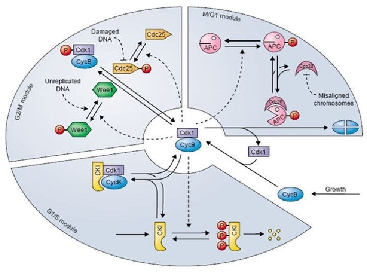
Another “helper” Anaphase-Promoting Complex (APC) securin --| separase breaks down cohesins ¡ Also called the cyclosome (often designated as APC/C) l triggers destruction of cohesins (proteins that make up centromere) allowing sister chromatids to separate l Cohesin breakdown is caused by a protease called separase l Separase is kept inactive until late metaphase by an inhibitory protein called securin l APC destroys securin allowing separase to break down the cohesins. l http: //faculty. plattsburgh. edu/donald. slish/Anaphase%20 transition. II. ht ml
https: //www. youtube. com/watch? v= Qh. Tbka. WQQAA
Cell Cycle Regulation The cell cycle is regulated by cyclins, cyclin-dependent kinases (CDKs), and cyclin-dependent kinase inhibitors (CDKIs) and is divided into 4 distinct phases (G 1, S, G 2, and M). G 0 represents exit from the cell cycle. The cell cycle is driven by CDKs, which are positively and negatively regulated by cyclins and CDKIs, respectively. The restriction point governs the transition point beyond which progression through the cell cycle is independent of external stimuli. Adapted from Shah and Schwartz. Clin Cancer Res. 2001; 7: 2168 -2181, with permission.
What CONTROLS the Checkpoints? ¡ Both internal (inside the cell) and external (outside the cell) signals
Internal and External Signals ¡Internal signals l DNA replication l Growth/Nutrition l CDK/Cyclins ¡External signals l Growth factors & Hormones l Density Dependent Inhibition l Anchorage Dependence
External Influences on Cell Division ¡ Growth factors l Stimulate other & hormones cells to divide ¡ Density-dependent inhibition l Crowded cells stop dividing ¡ Most animal cells exhibit anchorage dependence l Cells must be attached to a structure to divide ¡Ex: extracellular matrix of a tissue other protein or cells
(a) Cells anchor to dish surface and divide (anchorage dependence). When cells have formed a complete single layer, they stop dividing (density-dependent inhibition). Normal mammalian cells. **The availability of nutrients, growth factors, and a substratum for attachment limits cell density to a single layer. If some cells are scraped away, the remaining cells divide to fill the gap and then stop (density-dependent inhibition). Figure 12. 18 A 25 µm
Cancer cells l Exhibit neither density-dependent inhibition nor anchorage dependence l Immortal cells (if enough nutrients) Cancer cells usually continue to divide well beyond a single layer, forming a clump of overlapping cells. Figure 12. 18 B
Loss of Cell Cycle Controls in Cancer Cells ¡ Cancer cells l Do not “listen” to control mechanisms (internal and/or external) l CONTINUE TO DIVIDE l Form tumors ¡TUMOR= mass or group of abnormal dividing cells
Genes and Cancer ¡ Proto-oncogenes l Genes that create proteins that normally activates cell division growth factor genes ¡ become oncogenes (cancer-causing) when mutated ¡ ¡ Tumor-suppressor genes l l l normally inhibits (turns “off”) cell division if switched “OFF” can cause cancer example: p 53 gene
Why are Cancer cells IMMORTAL? ¡ Don’t need growth factors maybe they make their own growth factors ¡ Mutations in GENES!!! l Ex: cyclin or Cdk genes CANCER IS CAUSED BY A LOT OF MUTATIONS (AN ACCUMULATION OF MUTATIONS)
Cancer cells are “hungry”… http: //www. hhmi. org/biointeractive/angiogenesis ¡ Angiogenesis is the recruitment of blood vessels from the network of neighbouring vessels. Without blood and the nutrients it carries, a tumor would be unable to continue growing. l ¡
Loss of Cell Cycle Controls in Cancer Cells ¡ Cancer cells l Normal cell cancer cells using process of transformation l Form tumors ¡Benign “fine” l Clump of cells remain at orginal spot ¡Malignant “mean” “cancer” l Loose/destroy attachments to other cells they can spread!!!
Malignant tumors ¡ These tumors invade surrounding tissues and can metastasize l Tumors that can SPREAD and form secondary tumors l USE BLOOD STREAM and LYMPH VESSELS TO SPREAD!!!
Cancer Treatment ¡ Radiation l destroys DNA in cancer cells (so can’t divide) ¡ Chemotherapeutic l interfere drugs with specific steps in cell cycle (Ex: spindle formation or function) l also ¡Ex: effects normal cells hair loss
Pre-cancerous group of cells = polyp Tumor Lymph vessel Blood vessel Glandular tissue Cancer cell 1 A tumor grows from a single cancer cell. Figure 12. 19 2 Cancer cells invade neighboring tissue. 3 Cancer cells spread through lymph and blood vessels to other parts of the body. Metastatic Tumor 4 A small percentage of cancer cells may survive and establish a new tumor in another part of the body.
Cancer Movie ¡ ¡ http: //www. hhmi. org/biointeractive/using-p 53 -fight-cancer http: //www. cancerquest. org/index. cfm? page=3102&lang=en glish
Angiogenesis (the formation of new blood vessels) and Metatasis http: //www. hhmi. org/biointeractive /media/angiogenesis-lg. mov ¡ http: //www. hhmi. org/biointeractive /media/vegf-lg. mov ¡