Chapter 10 The Muscular System and Homeostasis Chapter


. For the")


 Anaerobic production of ATP by glycolysis occurs during strenuous exercise after")


 A simple")


. Relaxation is")


- Slides: 31
Chapter 10 The Muscular System and Homeostasis
Chapter 10 The Muscular System and Homeostasis 10. 1 Movement and Muscle Tissue 10. 2 Muscles, Health, and Homeostasis
Chapter 10 The Muscular System and Homeostasis In this chapter, you will learn: � There are three types of muscle tissue: skeletal muscle, smooth muscle, and cardiac muscle. � Skeletal muscle produces body movement, maintains body temperature, and provides support for the body. � Muscle fibres are filled with myofibrils that house thin (actin) and thick (myosin) contractile protein myofilaments. � Actin and myosin slide past each other during a muscle contraction. � Creatine phosphate, fermentation, and aerobic cellular respiration provide energy for muscle contractions. � Three types of skeletal muscle—slow-twitch, fast-twitch, and an intermediate type—are found in different parts of the body. � Muscles atrophy with inadequate stimulation and can hypertrophy with appropriate repeated stimulation. � The muscular system works with other body systems to maintain homeostasis.
10. 1 Movement and Muscle Tissue In this section, you will: �observe and compare three types of muscle tissue �describe, in general, the action of actin and myosin in muscle contraction and heat production �identify the sources of energy for muscle contraction
Muscle Cells
Muscle Function • Skeletal muscle supports the body. • Skeletal muscle makes the bones move. • Skeletal muscle helps to maintain a constant body temperature. • Skeletal muscle helps to protect the internal organs and stabilize the joints.
Muscle Fibres
Components of Skeletal Muscle Fibres Component Description Function Muscle fibre single muscle cell is responsible for muscle contractions myoglobin oxygen-binding pigment (similar to hemoglobin) in a skeletal muscle fibre stores oxygen for use during muscle contractions sarcolemma membrane of a muscle fibre surrounds the muscle fibre and regulates the entry and exit of materials sarcoplasm cytoplasm of a muscle fibre is the site of metabolic processes for normal cell activities; contains myoglobin and glycogen (which stores energy for muscle contractions) sarcoplasmic reticulum smooth endoplasmic reticulum in a muscle fibre stores calcium ions needed for muscle contractions Myofibrils organized bundles of myofilaments; cylindrical structures, as long as the muscle fibre itself contain myofilaments that are responsible for muscle contractions thick filament fine myofilament composed of bundles of protein called myosin (about 11 nm in diameter) binds to actin and causes muscle contractions thin filament fine myofilament composed of strands of protein called actin (about 5 nm in diameter) binds to myosin and causes muscle contractions
The Movement of Actin and Myosin
Sliding Filament Model
Calcium Controls Muscle Contractions http: //www. youtube. com/watch? v =Cbf. K 1 Gia. Ck&playnext=1&list=PLE 7 E 224845 4 B 7 C 3 E 9
animations �http: //www. youtube. com/watch? v=Y 1 tx 8 U 8 c 670 �http: //media. pearsoncmg. com/bc/bc_campbell_biolo gy_7/media/interactivemedia/activities/load. html? 49 &C �http: //www. mcgrawhill. ca/school/applets/abbio/quiz /ch 10/myofilament_contraction. swf �http: //www. mcgrawhill. ca/school/applets/abbio/quiz /ch 10/sarcomere_contraction. swf �http: //media. pearsoncmg. com/bc/bc_campbell_biolo gy_7/media/interactivemedia/activities/load. html? 49 &D
Crash Course �https: //www. youtube. com/watch? v=jqy 0 i 1 KXUO 4 Big Guns: The Muscular System - Crash. Course Biology #31
Energy for Muscle Contraction ATP required for contraction and is generated by THREE mechanisms.
Creatine phosphate builds up and is stored in resting muscle (purple box). For the muscle to contract (green area), it needs to acquire ATP. (A) When the muscle starts contracting, it breaks down stored creatine phosphate. This generates some ATP that is used immediately. (B) To continue contracting, the muscle carries out aerobic cellular respiration as long as oxygen is available. When the oxygen has been used up, the muscle can carry out fermentation for a limited period of time. Fermentation results in only a small amount of ATP compared with the amount produced by aerobic cellular respiration, and lactate builds up. Once the muscle resumes resting (purple box), creatine phosphate builds up again.
#1 Creatine Phosphate Breakdown Creatine phosphate quickly regenerates ATP by directly donating a phosphate (good for about 15 seconds, first ATP regeneration mechanism to kick in under anaerobic conditions)
#2 Cellular Respiration ATP is made in mitochondria by aerobic means - requires oxygen. Pyruvic acid is transported into mitochondria and oxidized to carbon dioxide and water.
“#3 Fermentation" (glycolysis) Anaerobic production of ATP by glycolysis occurs during strenuous exercise after CP stores are exhausted. Very inefficient, good for only 1 -2 minutes before muscle fatigue occurs. The end product of glycolysis, pyruvic acid, is converted to lactic acid if it isn't metabolized by cellular respiration fast enough to meet ATP needs of the muscle. Lactic acid builds up, the p. H drops, muscles fatigue and ache.
Oxygen Debt �Lactic acid is transported to the liver where it is converted back to pyruvic acid, which is then metabolized by cellular respiration, which of course requires oxygen. �The amount of oxygen required to finish metabolism of lactic acid is the oxygen debt - that’s the wind-sucking you do after going anaerobic.
10. 2 Muscles, Health, and Homeostasis In this section, you will: �explain how the skeletal muscles of the motor system support other body systems to maintain homeostasis �identify conditions that impair the healthy functioning of muscles and technologies that are used to treat or prevent these conditions �describe the benefits of exercise for maintaining the healthy structure and functioning of muscles
Muscular Atrophy, Sprains and Strains A sprain is an injury to a ligament, the tough, fibrous tissue that connects bones to other bone. Ligament injuries involve a stretching or a tearing of this tissue. A strain is an injury to either a muscle or a tendon, the tissue that connects muscles to bones. Depending on the severity of the injury, a strain may be a simple overstretch of the muscle or tendon, or it can result in a partial or complete tear.
Common Disorders & Ailments of Skeletal Muscles Condition Description muscular dystrophy a collective term for several hereditary conditions in which the skeletal muscles degenerate, lose strength, and are gradually replaced by fatty and fi brous tissue that impedes blood circulation; this, in turn, accelerates muscle degeneration in a fatal spiral of positive feedback botulism a potentially fatal muscular paralysis caused by a toxin produced by the bacterium Clostridium botulinum; the toxin prevents the release of a muscle-stimulating compound (acetylcholine) released by muscle-related cells of the nervous system, thus leading to paralysis cramps painful muscle spasms triggered by strenuous exercise, extreme cold, dehydration, salt (electrolyte) imbalance, low blood glucose, or reduced blood fl ow contracture abnormal muscle shortening not caused by nerve stimulation; can result from inability to remove calcium ions from the sarcoplasm or from the contraction of scar tissue (as in people who have experienced severe burns) fibromyalgia chronic muscular pain and tenderness often associated with fatigue and sleep disturbances; can be caused by infectious diseases, physical or emotional trauma, or medications crush syndrome a shock-like state following massive crushing of the muscles (as in, for example, the aftermath of an earthquake, the collapse of a building following an explosion, or a traffi c accident); associated with high fever, heart irregularities caused by potassium ions released from the muscles, and kidney failure caused by blockage of the renal tubules with myoglobin released by the traumatized muscles delayed onset muscle soreness pain, stiffness, and tenderness felt from several hours to a day after strenuous exercise; associated with trauma to the muscles, disruptions in the myofibrils and sarcolemma, and increased levels of myoglobin and muscle-fibre enzymes in the blood myositis muscle inflammation and weakness resulting from infection or an autoimmune disease
Myograms These graphs show the force of muscle contraction with time. (A) A simple muscle twitch has three periods: latent, contraction, and relaxation. (B) When a muscle is not allowed to relax completely between stimuli, the contraction gradually increases in intensity until it reaches a maximum, which is sustained until the muscle fatigues.
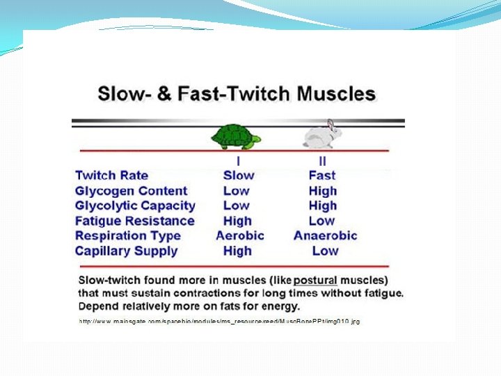
Types muscle fibres
Skeletal muscles have different proportions of fasttwitch and slow-twitch fibres. Thus, the force and response times of their contractions differ.
Chapter 10 Review �What body systems do muscles support? �How do muscles support these systems? �Explain the difference in muscle composition between a person who is not active versus someone who is very active? �Describe two different types of muscle injuries. Recommend appropriate treatments for each. �Explain how muscles change with strength training? With endurance training? With inactivity?
Concept Organizer
Chapter 10 Summary � All muscles do their work by contracting (shortening). Relaxation is the passive state of a muscle. There are three types of muscle cells: skeletal, smooth, and cardiac. � Skeletal muscle cells are attached to the bones of the skeleton, have many nuclei, are striated and tubular, and contract voluntarily. � Smooth muscle cells are found in the walls of internal organs, have one nucleus, are not striated, and contract involuntarily. � Cardiac muscle cells form the walls of the heart; have one nucleus, are striated, tubular, and branched; and contract involuntarily. � The fact that skeletal muscles can only either contract or relax means they can pull but not push. Therefore, they must work in pairs to move any part of the body—a relaxed muscle is only lengthened when the opposing muscle contracts to stretch it. Skeletal muscle contractions are explained by the sliding filament model.
Chapter 10 Summary �Skeletal muscle produces heat as well as movement and also supports and pads the body. Each muscle is made up of clusters of bundles of muscle fibres, which enclose bundles of myofibrils containing thin myofilaments of actin and thick myofilaments of myosin. Blood vessels supply nutrients and oxygen to the fibre bundles and remove wastes. The oxygen fuels the cellular respiration that supplies most of the energy muscles use. �Nerves trigger and control muscle contractions, which last for only a fraction of second in each muscle fibre. It is the wave of successive contractions, or twitches, that result in a muscle contraction. Skeletal muscles have both slow and fast-twitch fibres that are good for either endurance (slowtwitch) or intense (fast-twitch) activities. Using the skeletal muscles is the only way to maintain and build their function.
Additional Videos http: //www. youtube. com/watch? v=BXOTb 4 Zx. KDI Extraordinary Humans: Muscles (45 min) http: //www. youtube. com/watch? v=EKWa. J_7 PPXE Science of steroids National geographic (45 min)