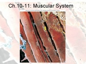
Chapter 10 Muscle Tissue and Organization 1 Muscles


- Slides: 31

Chapter 10 Muscle Tissue and Organization 1

Muscles • Skeletal muscles are voluntary muscles because they can be moved voluntarily • Heart and digestive muscles contract on their own

Muscles • Functions: – Body movement – Maintenance of posture – Temperature regulation (especially shivering when you are cold) – Storage and movement of materials • control of openings (sphincters) to gastrointestinal and urinary tracts – Support • abdominal cavity • floor of pelvic cavity

Table 10. 1 a • Muscles made up of fascicles – groups of muscle cells Muscle Fascicle Copyright © Mc. Graw-Hill Education. Permission required for reproduction or display.

Table 10. 1 a • Muscle cells are called muscle fibers • Muscle fibers contain protein strands called myofibrils Muscle Myofibril Muscle fiber Fascicle Copyright © Mc. Graw-Hill Education. Permission required for reproduction or display.

Table 10. 1 a • Myofibrils made up of proteins called myofilaments Thin filament Thick filament Actin molecules Heads of myosin molecules Myofibril Muscle fiber Fascicle Copyright © Mc. Graw-Hill Education. Permission required for reproduction or display.

Fig. 10. 1 Tendon Deep fascia Skeletal muscle • Tendons attach muscles to bones • Deep fascia covers muscle – – – dense irregular connective tissue separates individual muscles binds muscles with similar function helps distribute nerves and blood vessels fills spaces between muscles sits deep to superficial fascia (aka subcutaneous layer)

Fig. 10. 1 • Epimysium surrounds whole muscle (deep to deep fascia) Tendon Deep fascia Skeletal muscle (a) Muscle Epimysium

Fig. 10. 1 • Perimysium surrounds each fascicle – dense irregular connective tissue – contains neurovascular bundles Tendon Deep fascia Skeletal muscle Epimysium Perimysium Nuclei Muscle fiber Fascicle Vein Nerve. Artery (a) Muscle (b) Fascicle

Fig. 10. 1 • Endomysium surrounds each muscle fiber – areolar connective tissue – insulates each fiber from electrical charge of other fibers (each can contract individually) – contains reticular fibers that bind neighboring muscle fibers together Fascicle Muscle fiber Endomysium (a) Muscle Copyright © Mc. Graw-Hill Education. Permission required for reproduction or display.

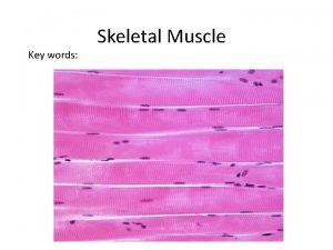
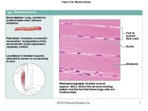
Endomysium Table 10. 1 c Myofibril Sarcoplasm Sarcolemma (plasma membrane) Satellite cell Muscle fiber Striations Nuclei (c) Muscle fiber

Fig. 10. 4 • Muscle fibers derive from myoblasts in embryo • Multiple myoblasts fuse into single cell with multiple nuclei • Satellite cells are myoblasts that didn’t fuse – assist in repair if muscle injured Myoblasts Satellite cell Myoblasts fuse to form a skeletal muscle fiber Muscle fiber

Fig. 10. 3 • Plasma membrane of muscle cell = sarcolemma (sarco = flesh) • Cytoplasm = sarcoplasm • Sarcoplasmic reticulum stores calcium ions needed for muscle contraction – Parts of SR extend deeper across cell, called transverse tubules Sarcolemma Sarcoplasm Mitochondria Myofibrils Myofilaments Transverse (T) Sarcoplasmic reticulum tubule Terminal cisternae Nucleus

Table 10. 1 d • Muscles move when myofilaments “walk” past each other • Myofibril shortens, contracting muscles • Muscles don’t “push”; They always pull Sarcomere Myofibril Myofilaments Thin filament Thick filament Actin molecules Heads of myosin molecules

Copyright © Mc. Graw-Hill Education. Permission required for reproduction or display. Muscle fiber Fig. 10. 6 Sarcomeres I band A band Z disc H zone I band Myofibril • Sections of fibers create striations (stripes) in muscle tissue Z disc Myofilaments M line Sarcomere Transverse sectional plane (a) Sarcomere Z disc Thick filament Z disc Thin filament Connectin M line Thick filaments and accessory proteins H zone I band A band I band (c) (b) Sarcomere TEM 400 x Z disc M line Z disc H zone I band (d) A band I band H zone Thick filaments Thin filaments Connectin Z disc Thin filaments Connectin and accessory proteins

Copyright © Mc. Graw-Hill Education. Permission required for reproduction or display. Fig. 10. 7 Thin filament • As muscles contract, striations change size M line Connectin Z disc H zone Thick filament Z disc I band M line Z disc I band A band I band Sarcomere M line I band A band Sarcomere A band I band Sarcomere (a) Relaxed muscle Sarcomere, I band, and H zone at a relaxed length. M line Z disc • https: //www. youtube. co m/watch? v=Cjx 3 v. Sm 54 N 8 Z disc I band H zone A band H zone Z disc M line Z disc I band A band M line A band I band Sarcomere Z disc H zone I band Sarcomere (b) Partially contracted muscle Thick and thin filaments start to slide past one another. The sarcomere, I band, and H zone are narrower and shorter. M line Z disc M line A band Z disc Sarcomere (c) Fully contracted muscle The H zone and I band disappear, and the sarcomere is at its shortest length. Remember the lengths of the thick and thin filaments do not change. a-c: © Dr. H. E. Huxley Sarcomere A band

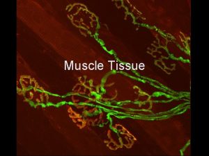
Fig. 10 • Motor unit is a motor neuron and all the muscle fibers it controls • One motor unit controls some fibers in a muscle • Smaller motor units provide finer control (ex. eye muscles) Spinal cord Motor neuron 1 Motor neuron 2 Neuromuscular junctions Muscle fibers innervated by motor neuron 1

Fig. 10 • all-or-none principle means Spinal cord fibers contract completely or not at all – Force exerted depends on number of motor units activated Neuromuscular junctions Motor neuron 1 Motor neuron 2 Muscle fibers innervated by motor neuron 1

Fig. 10. 8 Neuromuscular junction Axon of a motor neuron Synaptic knob Skeletal muscle fiber LM 500 x

Fig. 10. 8 Neuromuscular junction Axon of a motor neuron Path of nerve impulse Synaptic knob Skeletal muscle fiber Endomysium Sarcolemma Synaptic cleft Acetylcholine (ACh) ACh receptor Acetylcholinesterase (ACh. E) Synaptic knob Motor end plate Synaptic vesicles Sarcolemma Sarcoplasm

Fig. 10. 2 Muscle attachment • At end of muscle, connective tissue layers merge to form tendon – attaches muscle to bone, Tendon skin, or another muscle • thin, flattened sheet of tendon is called aponeurosis Copyright © Mc. Graw-Hill Education. Permission required for reproduction or display.

Fig. 10. 2 Muscle attachment Origin • Less mobile point of attachment of muscle is origin Relaxed • More mobile point of muscle attachment is insertion • Insertion usually moves Tendon toward origin • In limbs, origin is proximal to insertion Contracted muscle Movement of insertion of muscle Insertion Copyright © Mc. Graw-Hill Education. Permission required for reproduction or display.

Actions of Skeletal Muscles • Agonist is a muscle that contracts to produce a particular movement • Antagonist is a muscle whose actions oppose agonist – ex. agonist extends; antagonist flexes • Synergist assists the agonist – stabilizes point of origin or contributes to tension at point of insertion – ex. biceps brachii and brachialis muscles

Naming of Skeletal Muscles • Muscle action – Some names reflect function or movement of muscle -flexor -extensor -pronator • Specific body region – anterior, posterior – superficialis or externus are visible from body surface – profundus or internus are deeper or more internal

Naming of Skeletal Muscles • Muscle attachment – Named for origin, insertion, or prominent attachment – first part of name indicates origin and second part indicates insertion – ex. sternocleidomastoid originates on sternum and clavicle and inserts on mastoid • Orientation of muscle fibers – rectus = straight – oblique = oblique angle to longitudinal axis of body

Naming of Skeletal Muscles • Shape and size – – – deltoid = shaped like a triangle (delta, Δ) orbicularis = shaped like a circle (orbit) trapezius = shaped like a trapezoid brevis = short longus or longissimus = long teres = long and round

Naming of Skeletal Muscles • Shape and size – magnus = large, big – major = bigger – maximus = biggest – minor = small – minimus = smallest

Naming of Skeletal Muscles • Heads/tendons of origin – biceps = two tendons of origin – triceps = three tendons of origin – quadriceps = four tendons of origin • ex. quadriceps femoris = thigh muscle with four heads/tendons of origin

Cardiac muscle Fig. 10. 15 • Striated; bands visible under microscope • One nucleus per cell • Autorhythmic: individual cells can generate contractions by themselves Cardiac muscle cell (cardiocyte) Endomysium Z discs Intercalated disc Centrally located nucleus Intercalated discs I band Endomysium A band Gap junctions Desmosomes Mitochondrion (a) Sarcolemma Nucleus (b) Copyright © Mc. Graw-Hill Education. Permission required for reproduction or display. Cardiac muscle cell

Cardiac Muscle Intercalated discs

Fig. 10. 16 Smooth muscle • Not striated; each cell tapered at each end • Contractions slow, resistant to fatigue • Present in gut, blood vessel walls, etc. ; under involuntary control
 Muscles and muscle tissue chapter 9
 John wiley
 Chapter 4 the tissue level of organization
 Chapter 4 the tissue level of organization
 Chapter 4 the tissue level of organization
 Muscle and nervous tissue
 How is aerolar tissue different than aerenchyma tissue?
 Is skeletal muscle an organ
 Muscle tissue
 Muscle tissue
 Smooth muscle gap junctions
 Tropomyosin muscle contraction
 Classification of muscle tissue
 Skeletal muscle
 Epithelium location
 Muscle tissue
 Skeletal muscle tissue 40x
 Muscle tissue
 Summation anatomy
 Muscle tissue parts
 Inner quads
 Axial muscles
 Types of muscle
 Tissue that connects muscle to bone
 Structural proteins in muscle
 Myalgia is abnormal softening of muscle tissue
 Skeletal muscle tissue structure
 Bone cells
 Muscle tissue parts
 Cardiac muscle tissue parts
 Skeletal muscle location
 Muscle cells
