Chapter 10 Molecular Biology of the Gene Power

Chapter 10 Molecular Biology of the Gene Power. Point Lectures for Biology: Concepts & Connections, Sixth Edition Campbell, Reece, Taylor, Simon, and Dickey Lecture by Mary C. Colavito Copyright © 2009 Pearson Education, Inc.

THE STRUCTURE OF THE GENETIC MATERIAL Copyright © 2009 Pearson Education, Inc.

10. 2 DNA and RNA are polymers of nucleotides § The monomer unit of DNA and RNA is the nucleotide, containing – Nitrogenous base – 5 -carbon sugar – Phosphate group Copyright © 2009 Pearson Education, Inc.

§ DNA and RNA are polymers called polynucleotides – A sugar-phosphate backbone is formed by covalent bonding between the phosphate of one nucleotide and the sugar of the next nucleotide – Nitrogenous bases extend from the sugar-phosphate backbone Animation: DNA and RNA Structure Copyright © 2009 Pearson Education, Inc.
")
Sugar-phosphate backbone Phosphate group Nitrogenous base Sugar Nitrogenous base (A, G, C, or T) DNA nucleotide Phosphate group Thymine (T) Sugar (deoxyribose) DNA nucleotide DNA polynucleotide
 Phosphate group Thymine (T) Sugar (deoxyribose)")
Nitrogenous base (A, G, C, or T) Phosphate group Thymine (T) Sugar (deoxyribose)
 Cytosine (C) Pyrimidines Adenine (A) Guanine (G) Purines")
Thymine (T) Cytosine (C) Pyrimidines Adenine (A) Guanine (G) Purines
 Phosphate group Uracil (U) Sugar (ribose)")
Nitrogenous base (A, G, C, or U) Phosphate group Uracil (U) Sugar (ribose)

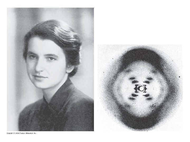
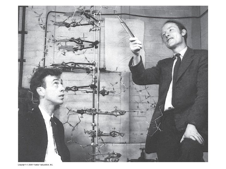
10. 3 DNA is a double-stranded helix § James D. Watson and Francis Crick deduced the secondary structure of DNA, with X-ray crystallography data from Rosalind Franklin and Maurice Wilkins Copyright © 2009 Pearson Education, Inc.

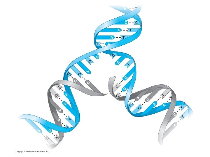
§ DNA is composed of two polynucleotide chains joined together by hydrogen bonding between bases, twisted into a helical shape – The sugar-phosphate backbone is on the outside – The nitrogenous bases are perpendicular to the backbone in the interior – Specific pairs of bases give the helix a uniform shape – A pairs with T, forming two hydrogen bonds – G pairs with C, forming three hydrogen bonds Animation: DNA Double Helix Copyright © 2009 Pearson Education, Inc.

Twist

Hydrogen bond Base pair Ribbon model Partial chemical structure Computer model

DNA REPLICATION Copyright © 2009 Pearson Education, Inc.

10. 4 DNA replication depends on specific base pairing § DNA replication follows a semiconservative model – The two DNA strands separate – Each strand is used as a pattern to produce a complementary strand, using specific base pairing – Each new DNA helix has one old strand with one new strand Animation: DNA Replication Overview Copyright © 2009 Pearson Education, Inc.

Parental molecule of DNA

Nucleotides Parental molecule of DNA Both parental strands serve as templates

Nucleotides Parental molecule of DNA Both parental strands serve as templates Two identical daughter molecules of DNA

10. 5 DNA replication proceeds in two directions at many sites simultaneously § DNA replication begins at the origins of replication – DNA unwinds at the origin to produce a “bubble” – Replication proceeds in both directions from the origin – Replication ends when products from the bubbles merge with each other § DNA replication occurs in the 5’ 3’ direction – Replication is continuous on the 3’ – Replication is discontinuous on the 5’ forming short segments Copyright © 2009 Pearson Education, Inc. 5’ template 3’ template,

10. 5 DNA replication proceeds in two directions at many sites simultaneously § Proteins involved in DNA replication – DNA polymerase adds nucleotides to a growing chain – DNA ligase joins small fragments into a continuous chain Animation: Origins of Replication Animation: Leading Strand Animation: Lagging Strand Animation: DNA Replication Review Copyright © 2009 Pearson Education, Inc.

Origin of replication Parental strand Daughter strand Bubble Two daughter DNA molecules

5 end P 4 3 P 5 2 1 3 end 2 3 1 4 5 P P P 3 end 5 end

DNA polymerase molecule 5 3 3 5 Parental DNA 3 5 5 3 DNA ligase Overall direction of replication Daughter strand synthesized continuously Daughter strand synthesized in pieces

THE FLOW OF GENETIC INFORMATION FROM DNA TO RNA TO PROTEIN Copyright © 2009 Pearson Education, Inc.

10. 6 The DNA genotype is expressed as proteins, which provide the molecular basis for phenotypic traits § A gene is a sequence of DNA that directs the synthesis of a specific protein – DNA is transcribed into RNA – RNA is translated into protein § The presence and action of proteins determine the phenotype of an organism Copyright © 2009 Pearson Education, Inc.

DNA Nucleus Cytoplasm

DNA Transcription RNA Nucleus Cytoplasm

DNA Transcription RNA Nucleus Cytoplasm Translation Protein

DNA strand Transcription RNA Codon Translation Polypeptide Amino acid

10. 8 The genetic code is the Rosetta stone of life § Characteristics of the genetic code – Triplet: Three nucleotides specify one amino acid – 61 codons correspond to amino acids – AUG codes for methionine and signals the start of transcription – 3 “stop” codons signal the end of translation Copyright © 2009 Pearson Education, Inc.

10. 8 The genetic code is the Rosetta stone of life – Redundant: More than one codon for some amino acids – Unambiguous: Any codon for one amino acid does not code for any other amino acid – Does not contain spacers or punctuation: Codons are adjacent to each other with no gaps in between – Nearly universal Copyright © 2009 Pearson Education, Inc.

Third base First base Second base

Strand to be transcribed DNA

Strand to be transcribed DNA Transcription RNA Start codon Stop codon

Strand to be transcribed DNA Transcription RNA Start codon Polypeptide Met Translation Lys Phe Stop codon

10. 9 Transcription produces genetic messages in the form of RNA § Overview of transcription – The two DNA strands separate – One strand is used as a pattern to produce an RNA chain, using specific base pairing – For A in DNA, U is placed in RNA – RNA polymerase catalyzes the reaction Copyright © 2009 Pearson Education, Inc.

RNA nucleotides RNA polymerase Direction of transcription Newly made RNA Template strand of DNA

RNA polymerase DNA of gene Promoter DNA Terminator DNA 1 Initiation 2 Elongation 3 Termination Completed RNA Area shown in Figure 10. 9 A Growing RNA polymerase

10. 11 Transfer RNA molecules serve as interpreters during translation § Transfer RNA (t. RNA) molecules match an amino acid to its corresponding m. RNA codon – t. RNA structure allows it to convert one language to the other – An amino acid attachment site allows each t. RNA to carry a specific amino acid – An anticodon allows the t. RNA to bind to a specific m. RNA codon, complementary in sequence – A pairs with U, G pairs with C Copyright © 2009 Pearson Education, Inc.

Amino acid attachment site Hydrogen bond RNA polynucleotide chain Anticodon

10. 12 Ribosomes build polypeptides § Translation occurs on the surface of the ribosome – Ribosomes have two subunits: small and large – Each subunit is composed of ribosomal RNAs and proteins – Ribosomal subunits come together during translation – Ribosomes have binding sites for m. RNA and t. RNAs Copyright © 2009 Pearson Education, Inc.

t. RNA molecules Growing polypeptide Large subunit m. RNA Small subunit

t. RNA-binding sites Large subunit m. RNA binding site Small subunit

Next amino acid to be added to polypeptide Growing polypeptide t. RNA m. RNA Codons

10. 13 An initiation codon marks the start of an m. RNA message § Initiation brings together the components needed to begin RNA synthesis § Initiation occurs in two steps 1. m. RNA binds to a small ribosomal subunit, and the first t. RNA binds to m. RNA at the start codon – The start codon reads AUG and codes for methionine – The first t. RNA has the anticodon UAC 2. A large ribosomal subunit joins the small subunit, allowing the ribosome to function – The first t. RNA occupies the P site, which will hold the growing peptide chain – The A site is available to receive the next t. RNA Copyright © 2009 Pearson Education, Inc.

Start of genetic message End

Met Initiator t. RNA P site 1 m. RNA Start codon Small ribosomal subunit 2 Large ribosomal subunit A site

10. 14 Elongation adds amino acids to the polypeptide chain until a stop codon terminates translation § Elongation is the addition of amino acids to the polypeptide chain § Each cycle of elongation has three steps 1. Codon recognition: next t. RNA binds to the m. RNA at the A site 2. Peptide bond formation: joining of the new amino acid to the chain – Amino acids on the t. RNA at the P site are attached by a covalent bond to the amino acid on the t. RNA at the A site Copyright © 2009 Pearson Education, Inc.

10. 14 Elongation adds amino acids to the polypeptide chain until a stop codon terminates translation 3. Translocation: t. RNA is released from the P site and the ribosome moves t. RNA from the A site into the P site Copyright © 2009 Pearson Education, Inc.

10. 14 Elongation adds amino acids to the polypeptide chain until a stop codon terminates translation § Elongation continues until the ribosome reaches a stop codon § Applying Your Knowledge How many cycles of elongation are required to produce a protein with 100 amino acids? § Termination – The completed polypeptide is released – The ribosomal subunits separate – m. RNA is released and can be translated again Animation: Translation Copyright © 2009 Pearson Education, Inc.

Amino acid Polypeptide A site P site Anticodon m. RNA Codons 1 Codon recognition

Amino acid Polypeptide A site P site Anticodon m. RNA Codons 1 Codon recognition 2 Peptide bond formation

Amino acid Polypeptide A site P site Anticodon m. RNA Codons 1 Codon recognition 2 Peptide bond formation New peptide bond 3 Translocation

Amino acid Polypeptide A site P site Anticodon m. RNA Codons 1 Codon recognition m. RNA movement Stop codon 2 Peptide bond formation New peptide bond 3 Translocation

10. 15 Review: The flow of genetic information in the cell is DNA RNA protein § Does translation represent: – DNA RNA or RNA protein? § Where does the information for producing a protein originate: – DNA or RNA? § Which one has a linear sequence of codons: – r. RNA, m. RNA, or t. RNA? § Which one directly influences the phenotype: – DNA, RNA, or protein? Copyright © 2009 Pearson Education, Inc.

10. 16 Mutations can change the meaning of genes § A mutation is a change in the nucleotide sequence of DNA – Base substitutions: replacement of one nucleotide with another – Effect depends on whethere is an amino acid change that alters the function of the protein – Deletions or insertions – Alter the reading frame of the m. RNA, so that nucleotides are grouped into different codons – Lead to significant changes in amino acid sequence downstream of mutation – Cause a nonfunctional polypeptide to be produced Copyright © 2009 Pearson Education, Inc.

10. 16 Mutations can change the meaning of genes § Mutations can be – Spontaneous: due to errors in DNA replication or recombination – Induced by mutagens – High-energy radiation – Chemicals Copyright © 2009 Pearson Education, Inc.

Normal hemoglobin DNA Mutant hemoglobin DNA m. RNA Normal hemoglobin Sickle-cell hemoglobin Glu Val

Normal gene m. RNA Protein Met Lys Phe Gly Ala Lys Phe Ser Ala Base substitution Met Base deletion Met Missing Lys Leu Ala His
- Slides: 61