Chapter 10 Integumentary System Skin o Skin 3

 o Skin: 3 Layers n 1. Epidermis o Thickness varies based")
 o Skin: 3 Layers n 2. Dermis / Corium o o")
 o Skin: 3 Layers n 3. Sub Q / hypodermis o")


 n skin layers and")
- Slides: 13
Chapter 10
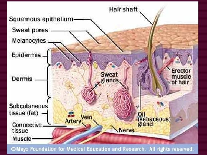
Integumentary System (Skin) o Skin: 3 Layers n 1. Epidermis o Thickness varies based on region o Stratified Squamous epithelium n Many flat layers of cells o Basal (Bottom Layer) produces new cells, oldest cells on top layer of skin. Dead skin cells are “shed” (dander) o Keratin= water proofing quality o Melanin (coloring), Albinism = absence of coloring
Integumentary System (Skin) o Skin: 3 Layers n 2. Dermis / Corium o o Blood and lymph vessels Nerve fibers (pain!) Accessory organs Connecting Tissue n Fibroblasts, collagen, histiocytes, mast cells, histamine, heparin, perception
Integumentary System (Skin) o Skin: 3 Layers n 3. Sub Q / hypodermis o Connective tissue o Fat n Adipocytes = fat cells that produce lipids
Skin: Appendages o o o o Glands Hair, wool, fur Feathers Scales Claws, hooves, nails Beaks Horns/ Antlers n Horns: grow continuously n Antlers: Shed each season n Cornification: Skin-> horns/keratin
Vestigial Appendages o Vestigial: rudimentary structures, seldom used for survival, “left overs” from evolution o Dewclaws: rudimentary bones n Dogs: Digits 2 and 4 o Front and back legs. Back dew claw normally removes o Chestnuts: medial surface of (horse) legs o Ergots: tuft of hair on the fetlock joint
Common Procedures o Biopsy: removal of LIVING tissue for examination n Incisional n Excisional: removal of entire tumor o Culture: growing microbes in a predetermined media for study o Skin Scrape: microscopic examination of the skin for presence of mites o Intradermal Testing: injection of test substances under the skin to test for body reaction (allergies)
Common Procedures o Cauterization: destruction of tissue using electrical current/heat/ chemicals o Lance: open to allow for drainage
Common Ailments o Abrasion: superficial cut o Abscess: localized collection of pus o Alopecia: abnormal hair loss(bald spots) n Shedding: normal hair loss o Contusion: Bruise o Dermatitis: inflammation n Allergies, fleas o Mange: skin disease caused by mites o Ulcers (decubical): sores , bed sores
Lesions o Surface Lesions n Raised and discolored n Papule, macule, scale, wheal, plaque, patch, crust o Fluid-Filled lesions n Cyst, pustule, vesicle, bulla o Erosive n Ulcer, fissure
Needle Activity : Wednesday
Activities o Review o Depict (YOU MUST DRAW IT) n skin layers and anatomy n Include a Hair follicle it must be colored and labeled (like the on this ppt) o Types of Hair n Give examples of/ on animals n Example: What type of hair are whiskers? o Draw examples of the types of injections n Intramuscular, Sub. Q, Intravenous, Cyntesis n Know what/when they are used