Chapter 10 Cell Division General Biology I BSC

Chapter 10 Cell Division General Biology I BSC 2010 Download for free at http: //cnx. org/contents/185 cbf 87 -c 72 e-48 f 5 -b 51 e-f 14 f 21 b 5 eabd@10. 61

CELL DIVISION • The cell is a basic unit of life. • All living organisms are composed of one of more cells. • Cells comes from pre-existing cells. • Cell division in unicellular organisms is for reproduction. • Cell division in multicellular organisms is for growth, development, regeneration, and repair.

Cell division • A sea urchin begins life as a single cell that • divides to form two cells, visible by scanning electron microscopy. After four rounds of cell division, • there are 16 cells, as seen in this SEM image. After many rounds of cell division, the individual develops into a complex, multicellular organism, as seen in this • mature sea urchin. Download for free at http: //cnx. org/contents/185 cbf 87 -c 72 e-48 f 5 -b 51 e-f 14 f 21 b 5 eabd@10. 61

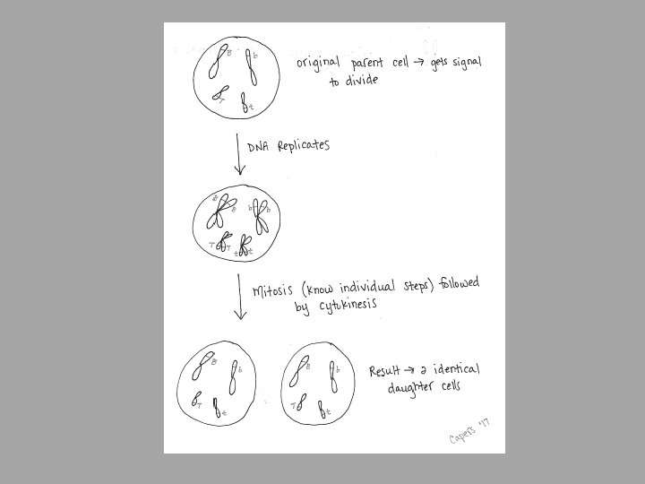
• Before division can happen, the cell must get the proper signal and the DNA has to replicate to make a replicate chromosome

Prokaryotic chromosome • Prokaryotes, including bacteria and archaea, have a single, circular chromosome located in a central region called the nucleoid. Download for free at http: //cnx. org/contents/185 cbf 87 -c 72 e-48 f 5 -b 51 e-f 14 f 21 b 5 eabd@10. 61

Bacterial Binary Fission – since bacteria only have 1 circular chromosome, their division is less complicated than ours Caption: Binary Fission (c) Ecoddington 14, Public domain

• Eukaryotic cells have multiple chromosomes • Eukaryotic cells have homologous pairs • Eukaryotic cells have linear chromosomes • Depending on the species, the number can range from a couple to hundreds • Humans have 46 chromosomes (usually defined as 22 autologous pairs and 1 pair of sex chromosomes) • Therefore, division is more complicated – have to keep track of the chromosomes so they end up in the right place • 2 types of division with eukaryotes we will talk about: • Mitosis – growth and repair – identical DNA • Meiosis – to make gametes – shuffle DNA

Human chromosomes • Humans have 46 chromosomes in 23 nearly identical pairs • Every species can have a different number of chromosomes Download for free at http: //cnx. org/contents/185 cbf 87 -c 72 e-48 f 5 -b 51 e-f 14 f 21 b 5 eabd@10. 61

Human chromosomes • Particular array of chromosomes in an individual organism is called karyotype. • Humans are diploid (2 n) • 2 complete sets of chromosomes • 46 total chromosomes • Haploid (n): 1 set of chromosomes • Gametes, some fungi, and some algae exist in this form • Pairs of chromosomes are homologous • Have the same genes, just might have different versions (alleles) of that gene • Example: the two chromosome #3’s are homologous, or the two chromosome #18’s are homologous • (each pair is 99+% identical; thus differences are less than 1%)

Chromosomes Composition • Over 2 meters of DNA inside a diploid human nucleus. • • When the cell is going about its normal function, the DNA is not condensed When the cell is getting ready to divide, it will STOP its normal function and condense its DNA (making chromosomes visible under the light microscope) • Chromosomes are composed of chromatin – complex of TIGHTLY PACKED DNA and protein • Typical human chromosome 140 million nucleotides long

Chromosomes organization Levels of Chromatin Organization: • Chromatin is organized to help form the proper structure • From naked DNA to Chromosomes 1) Naked DNA – form that is copied and transcribed 2) Nucleosome – ball of DNA wrapped around histone proteins – form that is stored and protected 3) Chromatin Loops – loops away from chromatin for gene expression 4) Chromosome – package of many genes

Download for free at http: //cnx. org/contents/185 cbf 87 -c 72 e-48 f 5 -b 51 e-f 14 f 21 b 5 eabd@10. 61

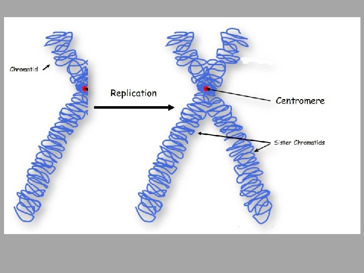
REPLICATION OF CHROMOSOMES • Before replication: each chromosome composed of a single DNA molecule • DNA must make a copy of itself • After replication: each chromosome composed of 2 identical DNA molecules • Identical copies are held together by cohesin proteins • Visible as 2 strands held together as chromosome becomes more condensed • One chromosome composed of 2 sister chromatids joined at centromere

The cell cycle • The cell cycle consists of 2 stages: interphase and the mitotic phase. • During interphase, the cell grows and the nuclear DNA is duplicated. • Interphase is followed by the mitotic phase. • During the mitotic phase, the duplicated chromosomes are segregated and distributed into daughter nuclei. • The cytoplasm is usually divided as well (cytokinesis), resulting in two daughter cells.

Eukaryotic Cell Division The eukaryotic cell cycle has two phases: 1. Interphase (Between cell divisions) • Cell is being a cell, performing its usual functions • Cell nucleus is visible • Cell metabolic functions, including DNA replication, occur • Begins after cytokinesis and ends when mitosis starts 2. Mitosis/cytokinesis: • Normal cell function stops because migrating DNA safely is priority • Eukaryotic nuclear division (mitosis) and cell division (cytokinesis)
 • Primary growth phase, longest")
Eukaryotic Cell Cycle 1. G 1 (gap phase 1) • Primary growth phase, longest phase • Replication of DNA 2. S (synthesis) 3. G 2 (gap phase 2) • Interphase Organelles replicate, microtubules organize; prepare for mitosis 4. M (mitosis) – Nuclear division • Subdivided into 5 phases • Separation of 2 new cells 5. C (cytokinesis) – Cytoplasmic division

Duration of Cell Cycle • Most variation in the length of the cell cycle between organisms or cell types occurs in G 1 • Cells often pause in G 1 before DNA replication and enter a resting state called G 0 • Resting phase G 0 – cells spend more or less time here before resuming cell division. • This is when a cell is being a cell • Can range from hours to decades depending on cell type • Most nerve cells remain there permanently – that is why damage to nerve cells is permanent • Liver cells can resume G 1 phase in response to factors released during injury

THE CELL CYCLE - MITOSIS • Mitosis produces genetically-identical daughter nuclei • Mitosis is followed by cytokinesis which splits the cytoplasm into two daughter cells • 4 main stages of mitosis: • Prophase – chromosomes condense; nuclear membrane dissolves • Prometaphase – centrosomes push away from each other; attach to centromeres • Metaphase – chromosomes align • Anaphase – chromosomes separate and migrate • Telophase – after nuclear membrane reforms, chromosomes relax

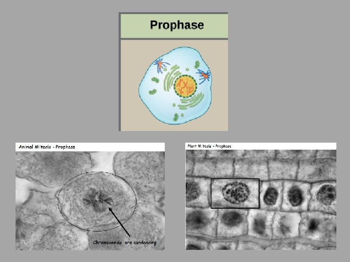
The Cell Cycle - Mitosis Prophase: • Chromosomes condense and become visible • Chromosomes appear as two sister chromatids held together at the centromere • Nuclear envelope breaks down • Mitotic spindle begins to form – 2 centrioles move to opposite poles forming spindle apparatus (no centrioles in plants) – Asters – radial array of microtubules in animals (not plants)

• During prometaphase, mitotic spindle microtubules from opposite poles attach to each sister chromatid at the kinetochore. • Because sister chromatids are still attached to each other, the result is a tug of war between the two poles at the “equator” • In anaphase, the connection between the sister chromatids breaks down, and the microtubules pull the chromosomes toward opposite poles. Download for free at http: //cnx. org/contents/185 cbf 87 -c 72 e-48 f 5 -b 51 e-f 14 f 21 b 5 eabd@10. 61

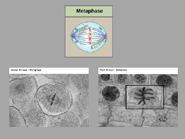
THE CELL CYCLE - MITOSIS • Metaphase: Alignment of chromosomes along metaphase plate • Not an actual structure; it is the midway between two centrioles pulling equally at chromosomes • Future axis of cell division

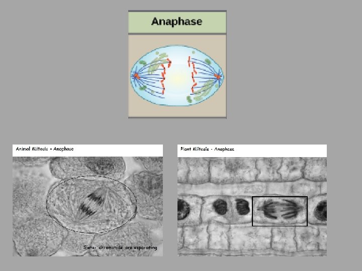
THE CELL CYCLE - MITOSIS • Anaphase: • Begins when centromeres split • Removal of cohesin proteins from all chromosomes • Sister chromatids pulled to opposite poles

THE CELL CYCLE - MITOSIS • Telophase: • Chromosomes are clustered at opposite poles • Nuclear envelopes re-forms around chromosomes • Chromosomes begin to uncoil and decondense once nucleus is there to trap them • Nucleolus reappears in each new nucleus • Golgi complex and ER re-form
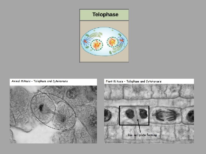

THE CELL CYCLE - CYTOKINESIS • Often immediately follows telophase or begins at the same time as telophase • Cytokinesis is the stage in which two daughter cells are formed from the original one • After cytokinesis, cells re-enter interphase. • Animals: • Proteins pinch the original cell into two new cells

THE CELL CYCLE - CYTOKINESIS • Cytokinesis in Plants: • Starts with vesicles forming the cell plate (segments of cell wall within vesicles) • This results in a new cell wall being formed between the cells forming daughter cells. • The cell wall is made from cellulose • Vesicles of cell plate fuse with each other and with membrane at equator and effectively split the cells

Control of the Cell Cycle • • • Cell division is a tightly controlled process Normal cells halt at checkpoints Proteins survey the condition of the cell Cell must pass the survey to proceed with cell division 3 checkpoints: G 1, G 2, and metaphase

Control of the Cell Cycle

Control of the Cell Cycle 3 Checkpoints: 1. G 1/S checkpoint • • Cell “decides” whether or not to divide Enough nutrients? Mitotic signals? • • Cell makes a commitment to mitosis Assesses success of DNA replication; fixes errors if necessary Can stall the cycle if DNA has not been accurately replicated. 2. G 2/M checkpoint • 3. Late metaphase (spindle) checkpoint • • Cell ensures that all chromosomes are attached to the spindle Or else, both sister chromatids could go the same way – genetic imbalance – PROBLEMS!

Control of the Cell Cycle • There are two groups of intracellular molecules that regulate the cell cycle. • These molecules can either promote progress of the cell to the next phase (positive regulation) or halt the cell cycle (negative regulation). • These molecules can either act individually or in partnership with other molecules. • Deficiency or non-functioning of a single regulator may not affect the control of cell cycle. • Many redundancies exist to ensure mutation does not prevent cell cycle progression or its regulation!
 are protein kinases that when fully")
• POSITIVE REGULATION • Cyclin-dependent kinases (Cdk’s) are protein kinases that when fully activated, can phosphorylate and thus activate other proteins that advance the cell cycle past a checkpoint. • To become fully activated, a Cdk must bind to a cyclin protein and then be phosphorylated by another kinase.
, p")
Negative regulation – checkpoint inhibitors • The best understood are retinoblastoma protein (Rb), p 53 and p 21. • Rb, p 53, and p 21 act primarily at the G 1 check point. . • P 53 detects damage DNA and recruits enzymes to repair DNA. If DNA cannot be repaired, it will trigger apoptosis (cell death) hence preventing duplication of damaged DNA. • Increasing of p 53 results in production of p 21 which in turn binds and inhibits the CDK/cyclin complexes. Ultimately, the cell cycle will not proceed.

Cancer and the cell cycle • Proto-oncogenes: genes that code for the positive cell cycle control proteins • When proto-oncogenes mutate, they become oncogenes • Their proteins no longer properly regulate cell division • They usually overstimulate cell division even though it is not supposed to.

CANCER AND THE CELL CYCLE • Tumor suppressor genes: genes for proteins that stop cell division if conditions are not favorable (negative regulator) • Rb, p 53, and p 21 are tumor suppressor genes • When mutated, can allow cells to override checkpoints

Control of the Cell Cycle
- Slides: 40