
CH5232 Villigen Switzerland The Paul Scherrer Institute PSI
- Slides: 1
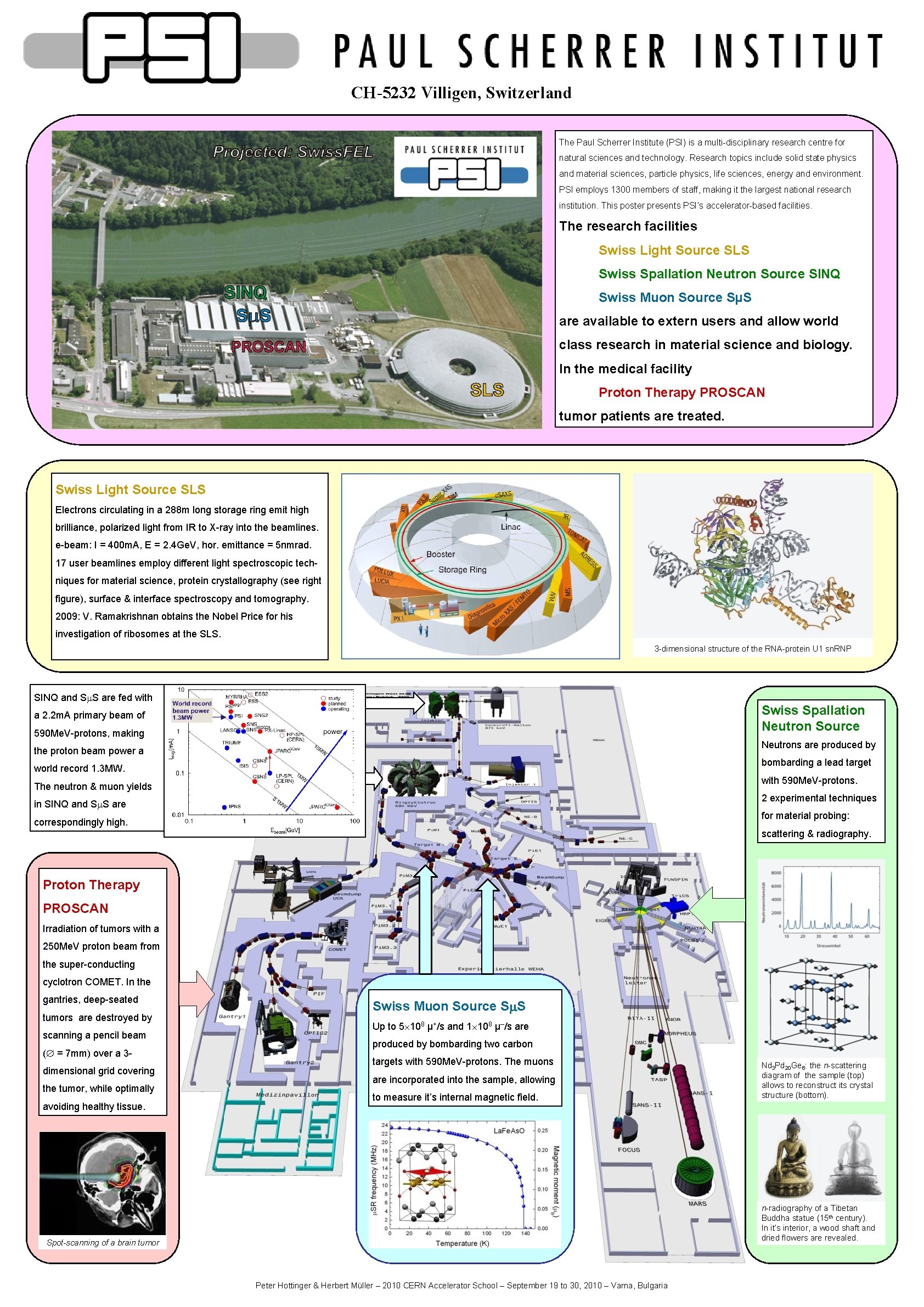
CH-5232 Villigen, Switzerland The Paul Scherrer Institute (PSI) is a multi-disciplinary research centre for natural sciences and technology. Research topics include solid state physics and material sciences, particle physics, life sciences, energy and environment. PSI employs 1300 members of staff, making it the largest national research institution. This poster presents PSI’s accelerator-based facilities. The research facilities Swiss Light Source SLS Swiss Spallation Neutron Source SINQ Swiss Muon Source SµS are available to extern users and allow world class research in material science and biology. In the medical facility Proton Therapy PROSCAN tumor patients are treated. Swiss Light Source SLS Electrons circulating in a 288 m long storage ring emit high brilliance, polarized light from IR to X-ray into the beamlines. e-beam: I = 400 m. A, E = 2. 4 Ge. V, hor. emittance = 5 nmrad. 17 user beamlines employ different light spectroscopic techniques for material science, protein crystallography (see right figure), surface & interface spectroscopy and tomography. 2009: V. Ramakrishnan obtains the Nobel Price for his investigation of ribosomes at the SLS. 3 -dimensional structure of the RNA-protein U 1 sn. RNP SINQ and Sm. S are fed with Swiss Spallation Neutron Source a 2. 2 m. A primary beam of 590 Me. V-protons, making Neutrons are produced by the proton beam power a bombarding a lead target world record 1. 3 MW. with 590 Me. V-protons. The neutron & muon yields 2 experimental techniques in SINQ and Sm. S are for material probing: correspondingly high. scattering & radiography. Proton Therapy PROSCAN Irradiation of tumors with a 250 Me. V proton beam from the super-conducting cyclotron COMET. In the gantries, deep-seated tumors are destroyed by scanning a pencil beam ( = 7 mm) over a 3 dimensional grid covering the tumor, while optimally avoiding healthy tissue. Swiss Muon Source Sm. S Up to 5 108 µ+/s and 1 108 µ–/s are produced by bombarding two carbon targets with 590 Me. V-protons. The muons are incorporated into the sample, allowing to measure it’s internal magnetic field. Nd 3 Pd 20 Ge 6: the n-scattering diagram of the sample (top) allows to reconstruct its crystal structure (bottom). n-radiography of a Tibetan Buddha statue (15 th century). In it’s interior, a wood shaft and dried flowers are revealed. Spot-scanning of a brain tumor Peter Hottinger & Herbert Müller – 2010 CERN Accelerator School – September 19 to 30, 2010 – Varna, Bulgaria