CH 5 Integumentary System General Characteristics The integumentary


")


")

- Slides: 42
CH 5 Integumentary System
General Characteristics • The integumentary system includes the skin and its accessory organs. • The skin includes the epidermis, dermis and hypodermis (subcutaneous layer). • The skin is the largest organ of the body – Approximately 22 ft 2 – Approximately 16% of total body weight – 0. 5 mm-4 mm thick • Thickest regions= hands and feet • Thinnest regions= eyelids and scrotum
THE SKIN Epidermis Dermis Subcutaneous (Hypodermis)
Skin Functions • Thermoregulation: regulation of body temperature. • Most important function • Excess heat is carried to the skin by the blood vessels. • Skin absorbs heat and transfers it to the surrounding air. • Protection: acts as a barrier against physical trauma, chemical and biological substances. • Works the best as a barrier when it is intact- no cuts, scrapes, etc. . • Approximately 50 layers of cells.
Skin Functions • Sense organ: provides the body with cutaneous sensations. • Sensations of touch, vibration and pain • Collects information from the outside world and sends it to the brain • What part of the body is the most sensitive? • Lips • Genitals • Hands/Feet
Skin Functions • Secretion and absorption: allowing certain materials to pass into or out of the skin. • Secretion: • Secretes water and oils through glands. • Can remove some waste like salts, urea, carbon dioxide and heat. • Absorption: • Taking materials into the body through the skin. • Most absorbed- fat soluble vitamins (A, D, E, K) • Some toxins are absorbed- rubbing alcohol, acetone, chlorine
Skin Functions • Vitamin D production: a precursor to the vitamin D molecule can be found within the skin. • When exposed to sunlight, the precursor becomes vitamin D because the light energy transforms its shapes. • It takes hours of sun exposure to produce the amount of vitamin D in a glass of milk. Is it worth it?
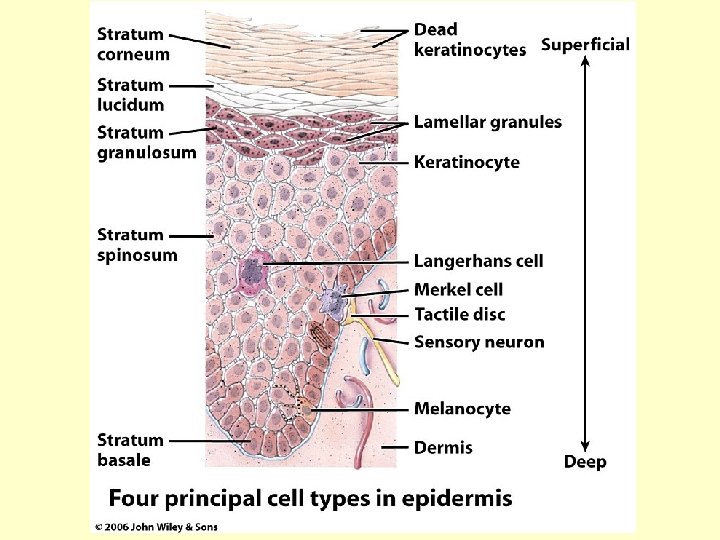
General Characteristics and Function of the Epidermis- outer layer of skin • 40 -50 layers of stratified squamous epithelial tissue. • Avascular • Designed to be shed and replaced frequently • Waterproof • Function: Protection
Cells of the Epidermis • Keratinocytes- cells that produce keratin. – These cells make up 90% of the epidermis – Keratin is a tough, fibrous, water-proof protein – Most keratinocytes are dead – They don’t really become flattened until they reach the top layers due to pressures from above and below as the cells move up the epidermis. – Keratinization: the process by which the cytoplasm and organelles of a keratinocyte are replaced by keratin. • Happens as cells move up through the epidermis • This is what eventually kills the keratinocytes
Cells of the Epidermis • Melanocytes- cells that produce melanin. – Function to protect the skin from UV radiation – Produce melanin- a brown, black pigment • It is produced by positive feedback. The more exposure- the more melanin produced- which means the more the skin can be exposed to the sun. – We all have the same # of melanocytes per square inch of the skin. Our melanocytes just produce a varying amount of melanin. • Langerhans cells- immune cells – Function to protect the body from foreign particles – These are phagocytes- engulf and destroy
Cells of the Epidermis • Merkel cells- touch cells. – Function to gather information (touch, temperature, vibration, and pain) and send it to the brain to be interpreted. – Melanocytes, Langerhans cells and Merkel cells account for the remaining 10% of the epidermis.
Layers of the Epidermis- from the bottom to top • Stratum Basale- deepest layer • Simple cuboidal layer • Lies above the dermis and blood vessels in the dermis supply this layer with blood (by diffusion) • Here is where cell division takes place, producing new skin cells and pushing older cells toward the surface. • Stratum Spinosum- superficial to the stratum basale. • 8 -10 layers • Cells have small projections “spiny” that help lock the cells together like velcro.
Layers of the Epidermis- from the bottom to top • Stratum Granulosum- superficial to the stratum spinosum. • 3 -5 layers • Cells are starting to look more squamous because of pressure from the cells above and below. • Layer where keratinization starts and cells produce keratin in large quantities. • Can see granules of keratin in the cells. • The transition layer between living and dead cells.
Layers of the Epidermis- from the bottom to top • Stratum Lucidum- superficial to the stratum granulosum. • 3 -5 layers • Only found on the soles and palms • Shock absorbers • Stratum Corneum- the most superficial layer of the epidermis • • 25 -30 layers of squamous keratinocytes Spines start to break off which causes the cells to fall off Waterproofs the skin Takes 5 weeks for cells to reach this layer from the stratum basale
LAYERS OF EPIDERMIS
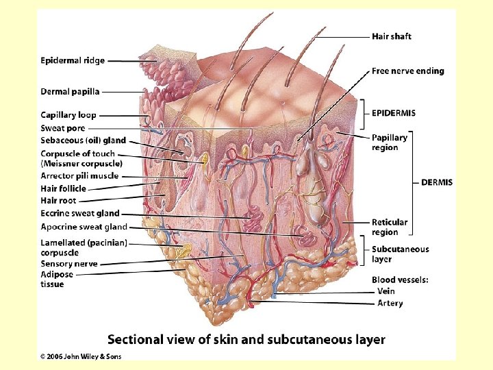
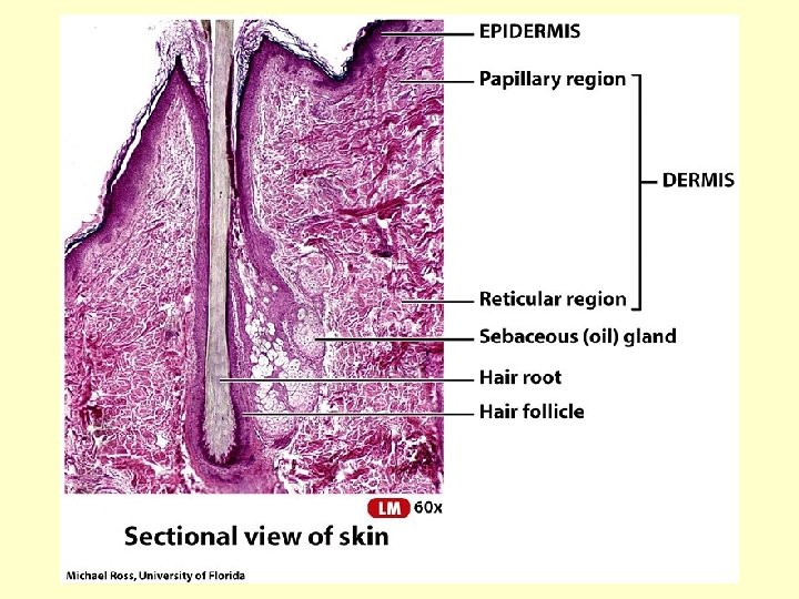
Dermis- Inner Layer of Skin • General Characteristics: – Thicker than the epidermis – Composed of connective tissue – Highly vascular – Contains all accessory organs necessary for skin function (nerves, blood vessels, glands, hair follicles) • That’s why burns that extend into the dermis are much more severe. They interfere with skin’s ability to function.
Dermis continued…. • Function: – Supports the epidermis by keeping the dividing cells of the stratum basale healthy – Regulates body temperature – Provides skin with its strength and flexibility
Dermis continued…. • Layers of the dermis: – Papillary region: lies deep to the stratum basale of the epidermis • 20% of the thickness of the dermis • Composed of areolar connective tissue with thin collagen and fine elastic fibers • Small capillary (tiny blood vessel) loops to supply nutrients to the epidermis • Contains dermal papillae
Dermis continued… • Dermal papillae: small projections that anchor the epidermis to the dermis – Prevent the epidermis from laterally sliding off of the dermis • Epidermal ridges: the ridges that form the fingerprints – Larger, downward projections of the epidermis that force some of the dermal papillae sideways – Function to lock the epidermis in place
Dermal papillae and epidermal ridges
What type are you? 65% 30% 5%
• Reticular layer: lies deep to the papillary region – Includes the remaining 80% of the dermis – Composed of dense irregular tissue – Collagen fibers run in various directions which adds strength – Functions to house the accessory organs of the skin and provide the skin with strength and flexibility
ACT-UP • Knowing what you know about the skin, answer the following questions: • 1) Why would a person with tattoos not need to have them retouched every five weeks? • 2) What would cause them to fade over time?
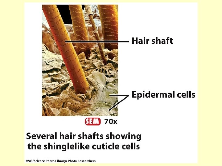
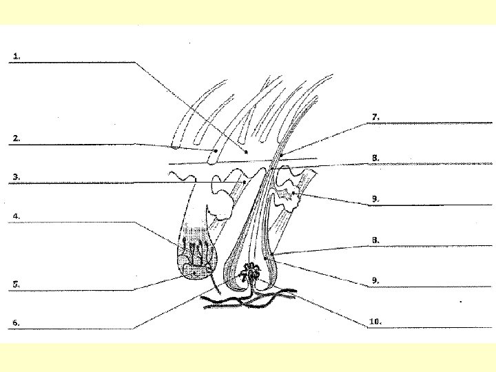
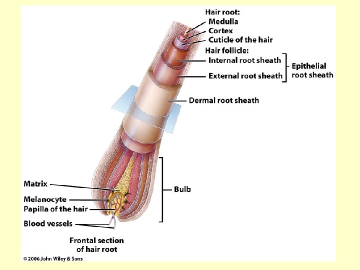
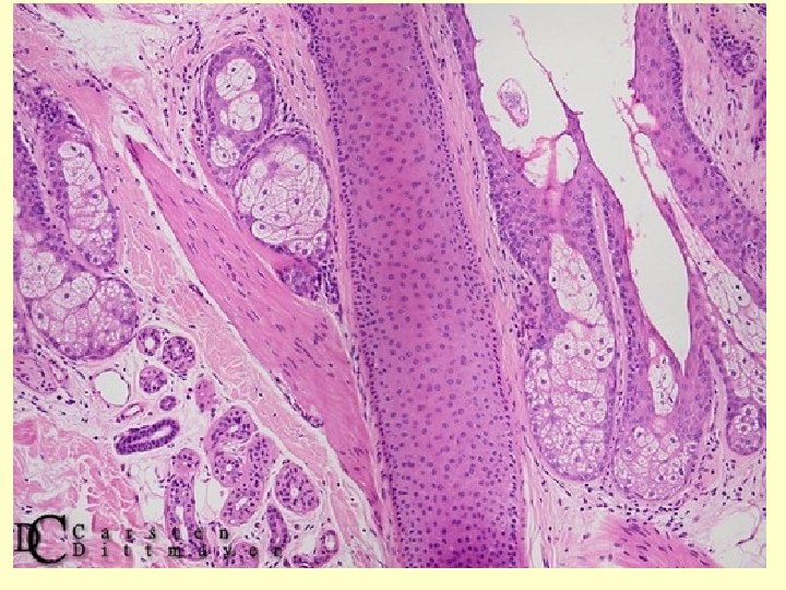
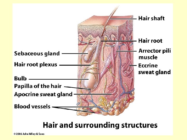
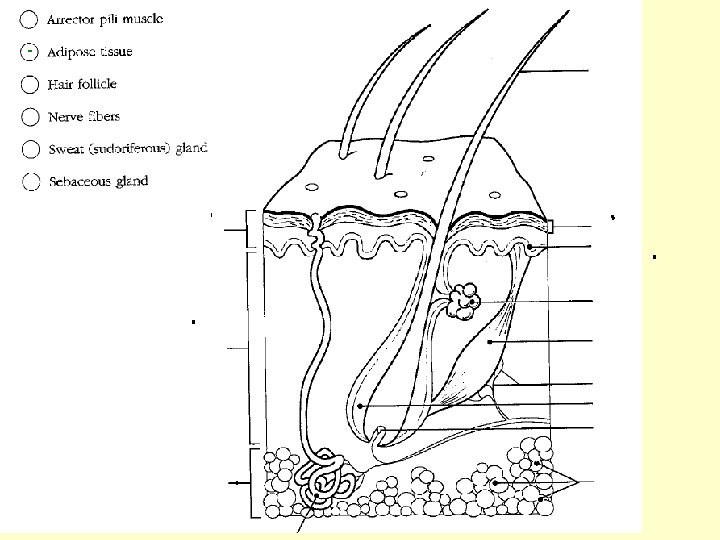
Accessory Structures of the Skin • Hair- present on most surfaces of the skin – Shaft is the hair above the skin (dead keratinocytes) – Root is the hair below the skin – Follicle- Layers that surround and protect the hair root – Matrix- The dividing cells of the hair root that cause hair growth – Papillae- Small projections that anchor each hair. • Contain blood vessels that keep the matrix alive.
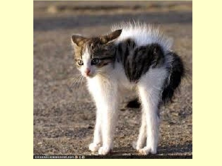
Accessory Structures of the Skin • Hair function: – Insulates against heat loss – Protects from UV radiation • Arrector pili muscle: A small muscle attached to the bulb of a hair. – Attaches to the follicle sheath – Contracts under physiological or emotional stress, which pulls the hair shafts perpendicular to the skin surface.
Glands- Present in different concentrations in all areas of the skin • Sebaceous glands: oil glands – Associated with hair follicles – Function to produce oil (sebum) • Keeps hair soft and flexible • Keeps skin around the hair soft and flexible • Destroys some bacteria
• Sudoriferous glands: sweat glands – Function to release a watery secretion (sweat) to cool the body. – Two types: • Eccrine- Respond to elevated body temperatures • Apocrine- Respond to stress (adrenaline rush) – These release sweat along with some proteins and fat that bacteria LOVE! As bacteria feed on this thick sweat, they produce a stinky by-product = B. O. !!
• Ceruminous glands: modified sweat glands that are only found within the external ear canal – Function to produce cerumen (wax) that protects the eardrum by trapping substances. – It’s important to keep this production at a low level- so water doesn’t get trapped that will support the breeding of bacteria.