Ch 18 Urinary System Urinary System Organs 1


 2. Regulate blood volume, electrolytes,")


- Slides: 38
Ch 18 Urinary System
Urinary System • Organs 1. 2 Kidneys 2. 2 Ureters 3. Urinary bladder 4. Urethra
Urinary System
Anatomy of Urinary System
Urinary System • Functions 1. Excrete waste products (urine) 2. Regulate blood volume, electrolytes, blood p. H (acid-base balance), tissue fluid 3. RBC production 4. Vit. D synthesis-kidneys along with skin and liver
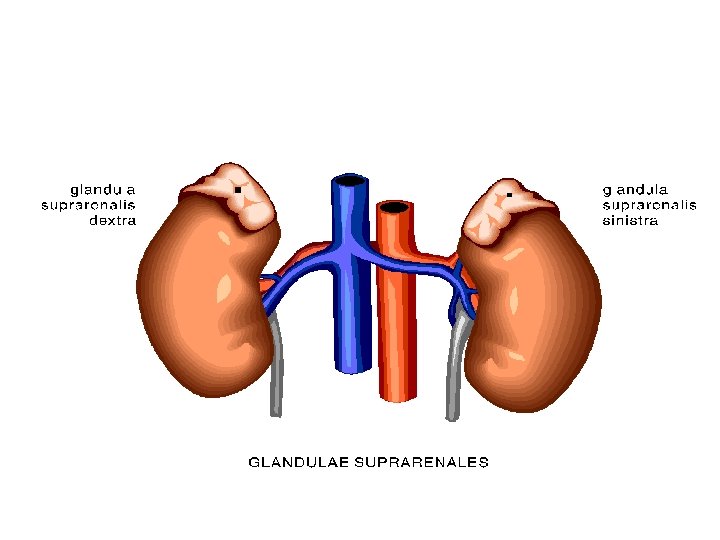
Kidneys • Structure 1. Bean-shaped 2. Retroperitoneal (upper abdominal c. , on either side of vertebral column) 3. Each covered by renal fascia (renal capsule) 4. Medial side indentation called hilus (site where blood vessels, nerves & ureters merge)
Kidneys • Internal Structure 1. 2 tissue portion renal cortex – outer renal medulla – inner 2. Renal medulla divided 3 areas: - renal pyramids - renal pelvis - calyces
renal columns renal cortex renal pyramids renal calyces renal vein renal artery renal pelvis ureter
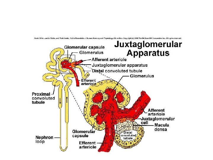
Kidneys • Internal Structure 3. Nephron = functional unit of kidney, consist of 2 major components - renal corpuscle - renal tubules
Kidneys • Nephron Structure 1. Renal corpuscle = consists of - glomerulus (tuft of capillaries- porous) - renal capsule surrounds glomerulus (very porous) - also called Bowman’s capsule
Kidneys • Nephron Structure 2. Renal tubules = continuous w/ Bowman’s capsule Tubules listed in order: - proximal convoluted tubule - loop of Henle (ascending & descending limbs) - distal convoluted tubule - collecting duct
Nephron-functional unit of kidney
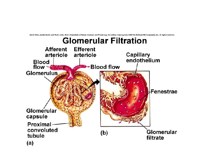
Renal Corpusle Bowman’s capsule is the enlarged end of the nephron. Outer layer simple squamous epi and inner layer podocytes Blood flows into the glomerulus from the afferent arteriole and leaves the glomerulus through the efferent arteriole
Filtration membrane Podocytes of Bowman’s capsule surround the porous glomerular capillaries Filtration Membrane consists of capillary endothelium, basement membrane and podocytes
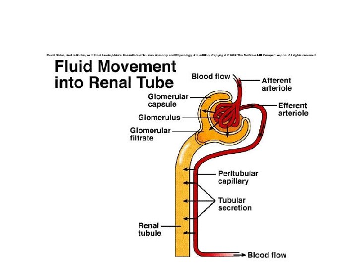
Kidneys • Nephron Structure 3. Peritubular capillaries = surround renal tubules to secrete/reabsorb materials to/from tubules
Blood flow Circulation through arteries capillaries and veins of the kidney and nephrons
Kidneys • Formation of urine: 1. 3 major processes - glomerular filtration - tubular reabsorption - tubular secretion
Urine formation 1. filtration- movement across filtation membrane into bowman’s capsule 2. Solutes are reabsorbed across the wall of the nephron by active transport and cotransport, water follows 3. Solutes are secreted across the wall of the nephron into the filtrate
Kidneys • Formation of urine: 1. Glomerular filtration= b. p. forces plasma, dissolved substances wastes and small molecules out glomerulus to renal capsule = is now called filtrate about 19 -20% of of the tt volume of blood that flows through the glomerular capillaries (no blood cells) – Rate = about 100 -125 ml/min (180 L/ day)
Kidneys • Formation of urine: 2. Tubular reabsorption = takes place between renal tubules & peritubular capillaries. Average urine output = 1 -2 L/day. • 99% filtrate is reabsorbed back into blood of peritubular capillaries 1 % becomes urine • Proteins, AA, glucose, fructose, Na+, K+, Ca++, HCO 3, Cl- are all reabsorbed
About 65 % of filtrate reabsorbed in proximal tubule, 15% in descending limb and 19% in distal tubule and collecting duct
Water reabsorption from filtrate
Kidneys • Formation of urine: 3. Tubular secretion = substances H+ wastes/ammonia, medications, are actively secreted from blood of peritubular capillaries into renal tubules
Regulation of Na+ & water in blood and other ECF Inc osmolality affects hypothalamic neurons and decreased BP affects baroreceptors inc water reabsorption Dec BP A II aldosterone inc BP Inc BP in the rt atrium inc secretion of atrial natriuretic hormone inc Na+ excretion and water loss in the form of urine
Kidneys • Elimination of urine: 1. Involves ureters, urinary bladder & urethra – ureters = muscular tube extends from hilus to posterior inferior portion of urinary bladder, peristaltic movements propel urine to bladder
Ureters and Bladder
Kidneys • Elimination of urine: 2. urinary bladder – trigone shaped muscular sac • women below uterus • men above prostate • temporarily stores urine (400 ml)
Urinary bladder
Kidneys • Elimination of urine: 3. urethra – tube carries urine from bladder to exterior. – women = 1 -1. 5 in – men = 7 -8 in. & also extends through prostate where it carries semen - micturition – expulsion of urine from urinary bladder
ureters Urinary bladder urethra
Hormonal regulation of blood volume and effects on urine volume and concentration
Regulation of K+ levels in blood and ECF
Regulation of Calcium Ions in the Blood
Regulation of Acid-Base Balance