
Cesarean Section and Simultaneous Transabdominal Repair Of Small
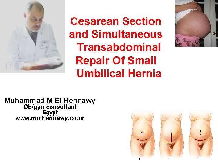
Cesarean Section and Simultaneous Transabdominal Repair Of Small Umbilical Hernia Muhammad M El Hennawy Ob/gyn consultant Egypt www. mmhennawy. co. nr
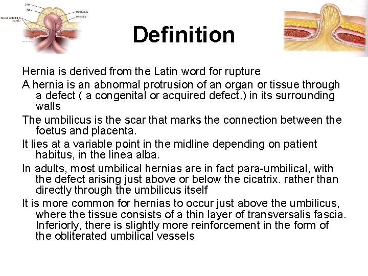
Definition Hernia is derived from the Latin word for rupture A hernia is an abnormal protrusion of an organ or tissue through a defect ( a congenital or acquired defect. ) in its surrounding walls The umbilicus is the scar that marks the connection between the foetus and placenta. It lies at a variable point in the midline depending on patient habitus, in the linea alba. In adults, most umbilical hernias are in fact para-umbilical, with the defect arising just above or below the cicatrix. rather than directly through the umbilicus itself It is more common for hernias to occur just above the umbilicus, where the tissue consists of a thin layer of transversalis fascia. Inferiorly, there is slightly more reinforcement in the form of the obliterated umbilical vessels
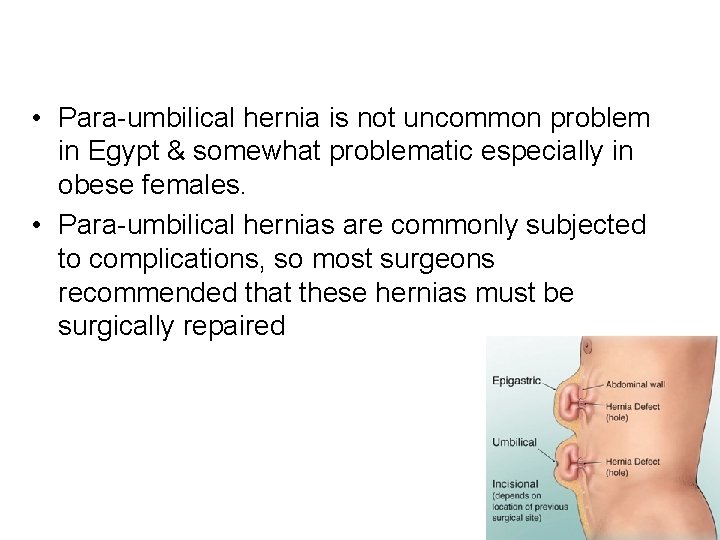
• Para-umbilical hernia is not uncommon problem in Egypt & somewhat problematic especially in obese females. • Para-umbilical hernias are commonly subjected to complications, so most surgeons recommended that these hernias must be surgically repaired

Incidence Umbilical hernias account for 6% of all abdominal wall hernias in adults The typical patient with an umbilical hernia is an overweight multiparous female between the ages of 35 and 50. Women are affected with umbilical hernias 3 to 5 times more frequently than men Ascites may be a contributing factor and makes the hernia more difficult to treat.
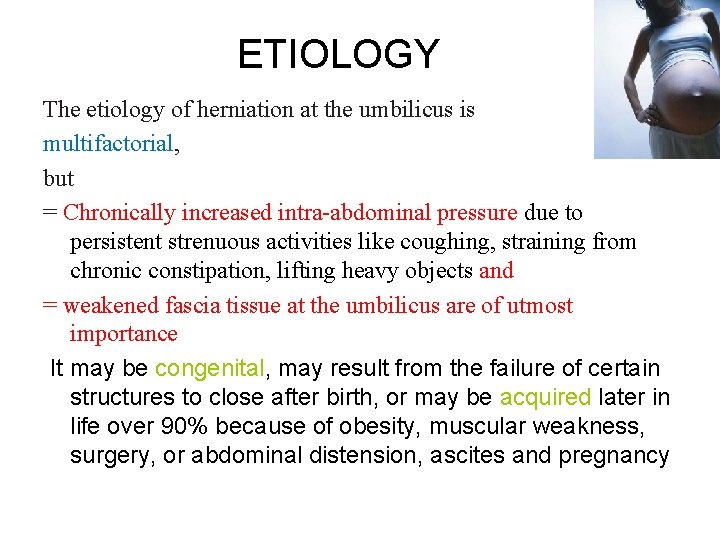
ETIOLOGY The etiology of herniation at the umbilicus is multifactorial, but = Chronically increased intra-abdominal pressure due to persistent strenuous activities like coughing, straining from chronic constipation, lifting heavy objects and = weakened fascia tissue at the umbilicus are of utmost importance It may be congenital, may result from the failure of certain structures to close after birth, or may be acquired later in life over 90% because of obesity, muscular weakness, surgery, or abdominal distension, ascites and pregnancy
Size Of Hernias • • The hernias can be Quite large, with fascial defects of 10 to 15 cm, Moderate Size with fascial defects of 5 -10 cm but most are smaller than 5 cm in diameter.
Content Of Hernias Omentum, colon, and small bowel can all be encountered within the umbilical hernia sac the presence of omentum alone or in combination with small or large bowel in 60% of patients. Small bowel alone 4%and large bowel were found in 7%, Adhesions from the omentum and bowel to the sac and the relatively small size of the fascial defect compared with the large amount of sac contents make these hernias prone to incarceration incarcerated umbilical hernia, most often containing herniated omentum or preperitoneal fat 1
CLINICAL PRESENTATION Although 39% of patients are asymptomatic at the time the hernia is discovered, 61% have experienced pain, pressure, nausea, or vomiting. or a lump at the umbilicus Of these, pain is the most common complaint, occurring in 44% of patients, followed by pressure in 20% and nausea and vomiting in 9%. The pain can be described as a “dragging” sensation or can be quite sharp and acute in nature when associated with coughing, straining, or incarceration of abdominal contents.
DIAGNOSIS The diagnosis of umbilical hernia is usually made by obtaining a history of pain or a lump at the umbilicus, Which is usually confirmed on physical examination. The appearance of an “outie” instead of an “innie” of the umbilicus in an adult suggests an umbilical hernia. This is confirmed by palpation of the incarcerated sac or protrusion of the sac through the fascial ring with straining maneuvers. Occasionally, for morbidly obese patients on whom it is difficult to perform an adequate abdominal physical examination, the diagnosis can be confirmed by a computed tomographic scan of the abdomen
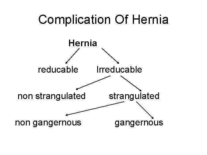
Complication Of Hernia reducable Irreducable non strangulated non gangernous
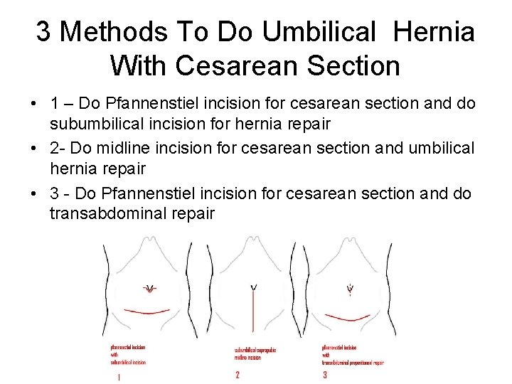
3 Methods To Do Umbilical Hernia With Cesarean Section • 1 – Do Pfannenstiel incision for cesarean section and do subumbilical incision for hernia repair • 2 - Do midline incision for cesarean section and umbilical hernia repair • 3 - Do Pfannenstiel incision for cesarean section and do transabdominal repair
1 -Do Pfannenstiel incision for cesarean section and do subumbilical incision for hernia repair For Large Hernia more than 10 cm • A-by closing the defect under tension with sutures ((herniorrhaphy)) • B-by reinforcement of the defect with a mesh (hernioplasty)-- prefacsial, retromuscular or preperitoneal) For small hernia less than 5 cm • C- tension free repair - (intraperitoneal) with a Ventralex hernia patch
Incision and dissection With a scalpel, make a curve-linear incision over the hernia. This can be extended either side as required Hold the skin edges either side using Littlewoods or Allis clamps Deepen the incision through sub-cutaneous fat and aponeurosis Insert a self-retaining retractor such as Travers or West’s to help Take care not to enter the hernial sac If iatrogenic peritoneal defects occur, make note and close them later with an absorbable suture such as 3’ 0 vicryl Continue dissection to identify the margins of the hernial defect circumferentially, down to the hernial neck If the hernia is small, preserve the umbilical skin by dissecting the sac off it In cases of large hernias, it may be necessary to excise the umbilicus and associated skin Skeletalise the sac from fatty tissue and clear an area of rectus sheath (the white, tough tissue) around it Use a finger to sweep the peritoneum away from the under surface of the defect, taking care not to tear into the peritoneum Inspect the contents of the sac. If freely reducible and there is no suspicion of ischaemia, proceed with repair
Repair Close any peritoneal defects with a continuous suture, using an absorbable material such as 3’ 0 vicryl Primary repair This should be performed on only the smallest of defects (<3 cm)- Place interrupted sutures transversely across the defect using a non-absorbable material such as 0 prolene - Instead of tying each stitch immediately, place both ends in a clip Place further sutures at evenly spaced points along the defect, again placing the ends of each in clips Use the first suture placed to lift up the sheath away from the peritoneal contents. This helps to avoid iatrogenic bowel injuries that can occur When all sutures are placed, tie them off Mayo’s ‘vest over pants’ repair The aim here is to overlap the upper and lower edges of the defect using a large non-absorbable suture such as 0’ prolene This is used to place interrupted horizontal mattress sutures transversely across the defect Starting on the lower edge, take full-thickness bite of the sheath 1 cm from its edge Then pass the needle under and through the upper leaf, bringing it out about 4 cm from the edge Now reverse the needle and take a bite from the upper leaf, about 2 cm from the edge Now pass the needle under the edge of the lower leaf, bringing it out close to the original entry point Again, you may find it easier to clip the suture and use it to lift the sheath as you place all the other stitches before tying them off individually at the end As the stitches are tied, the lower edge (‘pants’) is pulled under the upper edge (‘vest’), hence the name ‘vest over pants When all the stitches are tied, a further layer of continuous sutures is placed between the edge of the upper and lower sheath Mesh repair During dissection, ensure adequate exposure of the rectus sheath all around Take a bite of the sheath with your suture, then pass it through the mesh Do not tie immediately, but instead place the ends of the suture in a clip, which can then be used to lift the sheath away from the peritoneal contents below Place all the sutures in a similar fashion around the mesh, taking good bites of tissue - Tie off all the sutures
Closure Ensure adequate haemostasis The placement of ‘fat stitches’ is optional, but in large patients they can be used to close the potential space between sheath and skin. If placed, use an absorbable suture such as 3’ 0 vicryl Securing the umbilicus to the sheath is another optional step Skin closure is with an absorbable subcuticular suture such as 3’ 0 monocryl It is not usual practice to leave a drain
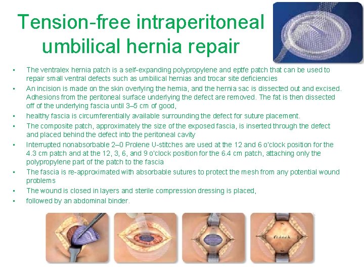
Tension-free intraperitoneal umbilical hernia repair • • The ventralex hernia patch is a self-expanding polypropylene and eptfe patch that can be used to repair small ventral defects such as umbilical hernias and trocar site deficiencies An incision is made on the skin overlying the hernia, and the hernia sac is dissected out and excised. Adhesions from the peritoneal surface underlying the defect are removed. The fat is then dissected off of the underlying fascia until 3– 5 cm of good, healthy fascia is circumferentially available surrounding the defect for suture placement. The composite patch, approximately the size of the exposed fascia, is inserted through the defect and placed behind the defect into the peritoneal cavity Interrupted nonabsorbable 2– 0 Prolene U-stitches are used at the 12 and 6 o’clock position for the 4. 3 cm patch and at the 12, 3, 6, and 9 o’clock position for the 6. 4 cm patch, attaching only the polypropylene part of the patch to the fascia The fascia is re-approximated with absorbable sutures to protect the mesh from any potential wound problems The wound is closed in layers and sterile compression dressing is placed, followed by an abdominal binder.
2 - Do midline incision for cesarean section and umbilical hernia repair • For Large Hernia more than 10 cm • A-by closing the defect under tension with sutures B-by reinforcement of the defect with a mesh -- prefacsial, retromuscular or preperitoneal)
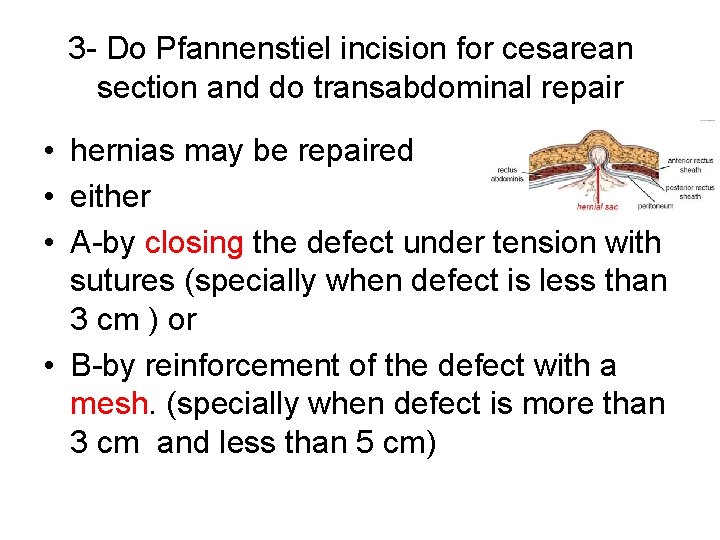
3 - Do Pfannenstiel incision for cesarean section and do transabdominal repair • hernias may be repaired • either • A-by closing the defect under tension with sutures (specially when defect is less than 3 cm ) or • B-by reinforcement of the defect with a mesh. (specially when defect is more than 3 cm and less than 5 cm)
Preoperative • Patient received standardized preoperative prophylactic antibiotic (ceftriaxone sodium, 1 g intravenously or intravenous cefotaxime sodium 1 gram half an hour before surgery and after placental extraction)
• a Pfannenstiel incision • Lower segment cesarean section (LSCS) was done • after the uterus was closed. • Lax abdominal wall was easily everted
A-closing the defect under tension with sutures It is relaitvely easy to close a small ( less than 3 cm) paraumbilical or umbilical hernia from within the abdomen by using the same technique of withdrawing of sac and closing ring with a purse-string suturea (primary suture from the inside)
B-by reinforcement of the defect with a mesh a defect more than 3 centimeters and less than 5 cm, repair was performed by inside mesh hernioplasty fixed to posterior rectus sheath by non-absorpable polypropylene suture
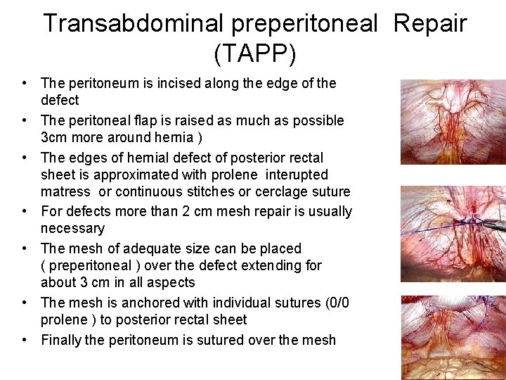
Transabdominal preperitoneal Repair (TAPP) • The peritoneum is incised along the edge of the defect • The peritoneal flap is raised as much as possible 3 cm more around hernia ) • The edges of hernial defect of posterior rectal sheet is approximated with prolene interupted matress or continuous stitches or cerclage suture • For defects more than 2 cm mesh repair is usually necessary • The mesh of adequate size can be placed ( preperitoneal ) over the defect extending for about 3 cm in all aspects • The mesh is anchored with individual sutures (0/0 prolene ) to posterior rectal sheet • Finally the peritoneum is sutured over the mesh
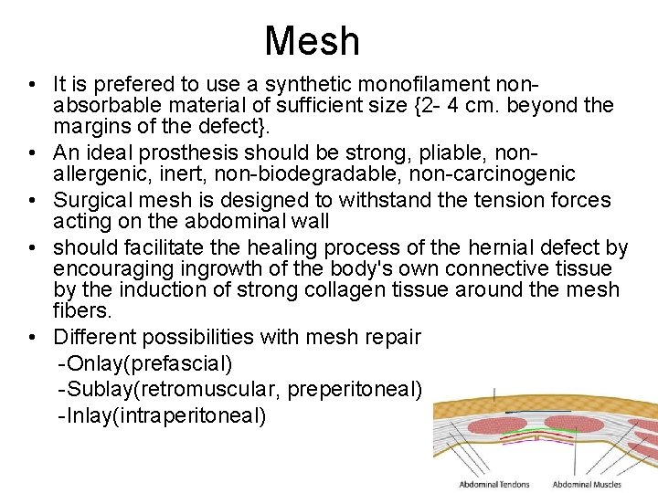
Mesh • It is prefered to use a synthetic monofilament nonabsorbable material of sufficient size {2 - 4 cm. beyond the margins of the defect}. • An ideal prosthesis should be strong, pliable, nonallergenic, inert, non-biodegradable, non-carcinogenic • Surgical mesh is designed to withstand the tension forces acting on the abdominal wall • should facilitate the healing process of the hernial defect by encouraging ingrowth of the body's own connective tissue by the induction of strong collagen tissue around the mesh fibers. • Different possibilities with mesh repair -Onlay(prefascial) -Sublay(retromuscular, preperitoneal) -Inlay(intraperitoneal)
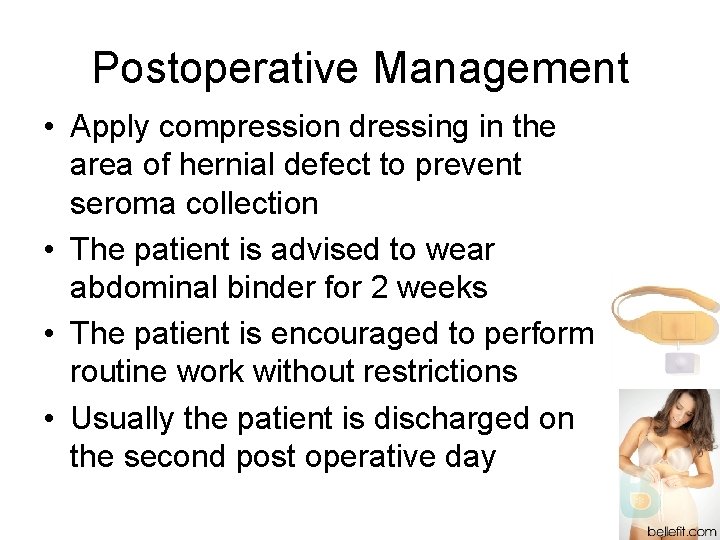
Postoperative Management • Apply compression dressing in the area of hernial defect to prevent seroma collection • The patient is advised to wear abdominal binder for 2 weeks • The patient is encouraged to perform routine work without restrictions • Usually the patient is discharged on the second post operative day
Complications Of Operation • • Pain Seroma Mesh Infection Recurrence
Pain • Some patients have pain • This usually subsides with conservative management like NSAIDs and injection of local anesthetics
Seroma • The fluid accumilation in the retained hernial sac is common and usually self limiting • Most of these fluid collections resolve with conservative management • Aspiration of the seroma can be performed if it is continuously enlarging in size • This should be done under strict aseptic precausions to avoid introduction of infection
Mesh Infection • The incidence of mesh infection is very low ( about 1% ) when compared to 10 -15 % incidence in the conventional hernia surgeries • All aseptic precautions should be taken to avoid this complication • As managing this complication is very difficult • The infected mesh usually need removal for effective healing of the wound
Recurrence • Recurrence was found lateral to mesh • Small size of the mesh was probably the cause of recurrence • Size of defect ( more than 4 cm = 40%recurrence ) • Ineffective anchoring of the mesh • Scar tissue reaction • Infection • Transfascial sutures not employed • Diabetes, obesity
Advantages cesarean section and hernia repair, With a single incision or two single anesthesia, and single hospital stay performed in one session, avoids need for readmission to hospital, is safe, effective, and well accepted. It neither increases the complication rate nor prolongs the hospital stay, valuable advantages for both patient and hospital in time, cost, and convenience, , the combined procedure confers not to mention avoiding the separation of mother from newborn entailed by reoperation. There were no major complications. Wound healing was delayed, without infection. Blood loss, opiate use, and hospital stay did not differ significantly
- Slides: 32